The importance of histological examination for modern medicine
The role of histology in modern medicine cannot be underestimated. Especially in the diagnosis of malignant neoplasms (cancer), because a correctly established diagnosis (correctly identified type of tumor) is already 50% of success in treatment.
Histological studies are used by almost all doctors of various specialties (gynecologists, oncologists, pathologists, gastroenterologists, surgeons, urologists, etc.).
Examination of the cervix and endometrium
Histological studies of the endometrium in gynecology make it possible to correctly assess the functioning of the ovaries and promptly identify any diseases. If the menstrual cycle continues, then diagnostic curettage is performed approximately 3 days before the start of menstruation. If the patient has dysfunctional bleeding, then the scraping must be taken precisely during bleeding. Biological tissues for research are stained using eosin or hematoxylin, as well as the Van Gieson method. After this, all the features of the endometrium, the structure of the glands and stroma are determined by analysis. Healthy glands during the luteal period of the menstrual cycle are slightly expanded and have a sawtooth shape. At the same time, the cells of the glandular epithelium themselves have pale nuclei and light cytoplasm, and the glands necessarily contain secretions.
Histological studies of the cervix are also carried out by collecting a small amount of tissue from the patient. If during the analysis a minor pathological change was detected in them, this means the presence of an inflammatory disease or a benign formation. If many cells with signs of pathological changes were identified, then we can talk about a precancerous condition or a malignant tumor.
The essence of histological examination
Any doctor you see can prescribe a histology test. Why is such an analysis prescribed? Of course, histology analysis is not prescribed for everyone and not in all cases; there must be certain indications for this.
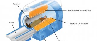
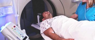
If, for example, studies (x-ray, ultrasound, CT, MRI, etc.) do not provide an accurate answer to the questions posed by the doctor and it is impossible to make an accurate diagnosis. And without an accurate diagnosis, it is unlikely that you will receive high-quality treatment for your disease.
The essence of histological examination is a correct diagnosis! In Russia, the histological method can be called the gold standard in diagnosis!
What will histological analysis tell you?
Histological examination requires strict indications. It is impossible to undergo it as a preventive screening, since it checks a specific organ, and not the general state of health. The analysis is carried out for patients who have certain health problems, namely tumors, the nature of which can be malignant.
Indications for histology are:
- clarification of the nature of the developing neoplasm;
- clarifying the reasons why pregnancy does not occur;
- analysis of the health of the female genitourinary system;
- determination of inflammation in the stomach and intestines.
Almost half of the cases when a diagnosis such as histology is prescribed is a suspicion of a cancerous tumor in the human body. Moreover, not only the formed tumors are studied, but also the tissues of organs in which neoplasms could potentially arise. This analysis is also effective for diagnosing the condition of the digestive organs, reproductive and reproductive systems of men and women.
Collection of biomaterial from the bronchi for histological analysis
What is the purpose of histological examination?
Histology analysis will help differentiate the diagnosis in unclear situations. There are often cases when it is not entirely clear what kind of process is happening in an organ (tissue). Some diseases are similar to each other in their manifestations and clinical course. For example, a round formation in the lung can be both a tumor and tuberculoma, etc. When the doctor cannot say with one hundred percent accuracy what it is, then, of course, a histological examination is necessary.
There are situations when, even during surgical interventions, surgeons discover some formations, which are also sent for histological examination.
What to do after receiving the result
After the patient receives the result from the laboratory, it is worth waiting for a consultation with the attending physician. There is no point in deciphering the received data yourself , since only an experienced attending physician will be able to correctly interpret them, based on previously obtained results of other tests and the patient’s medical history.
If the specialist has doubts, he will refer the patient for re-examination. And if the patient himself has doubts and uncertainty about his doctor, he can turn to another doctor with all his tests. In the same way, you can send your material for study to another laboratory.
There is no need to panic when receiving a referral for a histological examination. Histology will help identify and prevent the development of cancer.
This is a study that allows an accurate diagnosis to be made at the earliest stages of the disease, and even in a precancerous state. In this case, it is very important to find out about the disease as early as possible. In addition, histology will help solve the problem of infertility and miscarriage. Now this is especially true for many dozens of families.
Article design: Mila Friedan
How to obtain material for histological examination
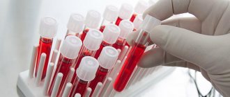
Materials for histological examination are organ tissues or entire organs (if surgically removed). Organ tissue is obtained through an invasive procedure - biopsy (plucking off a small piece). A piece of organ (tissue) in this case is called a biopsy.
When collecting material for research, they use the principle of collecting the most changed areas of the organ. Before proceeding with the biopsy itself, other additional studies may be performed (CT, MRI, ultrasound, endoscopic studies, etc.).
What methods can be used to obtain biopsy material?
- During a surgical operation (part of the affected organ or the entire removed organ);
- during an endoscopic examination (special tweezers are attached to the endoscope, which, in fact, extract the material during the examination);
- puncture with a thick needle (puncture biopsy) - sampling is done through the skin of nearby organs, for example, liver tissue can be obtained;
- suction of the material with a small tube (aspiration biopsy) - thus, material is obtained from the thyroid gland and the uterine cavity;
- scraping and smear methods are also used mainly in gynecology.
What can a histology analysis show?
Using a histological examination , the doctor can examine the tissue or organ at the cellular level.
This method makes it possible to detect the following processes in the body:
- Inflammatory processes in acute or chronic form.
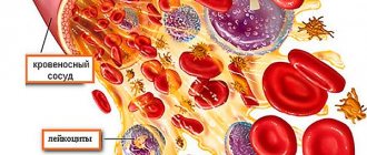
- Find hemorrhages, blood clots and other disorders associated with the circulatory system.
- Find new formations, find out what they are: benign or malignant.
- Determine the level of malignancy if doctors notice a cancerous tumor.
Various processes can be detected.
Thanks to such an examination, one can find out whether foreign bacteria, parasites and other foreign bodies are present in the tissue. This is just a small list of the capabilities histology has.
Very often, examination of a scraping of the surface of the uterus allows you to determine the presence of any changes on different days of menstruation. The results obtained during such examinations make it possible to find out how the ovaries work. And this, in turn, is very important data that makes it possible to cure many diseases, for example, female infertility.
When examining the cervix with this method, it is possible to detect diseases that have just begun to develop, if, of course, conservative treatment does not bring positive results.
Execution technique
Any histological specimen is studied under a light or electronic microscope, it depends on the equipment of the laboratory. But basically, of course, the specimen (biopsy) is studied under a light microscope.
A histologist or pathologist examines and gives an opinion (a pathologist is not only a mortuary worker, doctors of this specialty must master the technique of microscopic examination of a specimen!). Laboratory assistants prepare drugs for testing.
To examine tissue histologically, special preparations are required.
- The very first thing that happens is the collection of material (biopsy) , initially a piece of the organ must be placed in a special solution (formalin or alcohol), this is done to fix the tissue and to prevent various putrefactive processes.
- Then the biopsy is filled with paraffin (you will get a dense square).
- The next thing is that thin sections are made using a special device so that the layers of cells do not overlap each other, this improves the quality of the preparation.
- Staining of preparations. In order to study various structures or changed cells, special dyes are used.
- Well, in the end, in order for the drug to last longer, it undergoes further special processing.
Material collection rules
The reliability of the results of histological examination largely depends on compliance with the rules for taking a biopsy sample.
General standards for the collection and storage of biomaterial:
- Biopsy procedures are performed with a sharp instrument to avoid deformation of the material.
- A biopsy sample from a pathological formation is taken from the center and at the border with healthy tissue.
- Recommendations for the excised specimen: small size, flat shape, thickness 3-5 mm. These conditions are necessary for uniform fixation of the biopsy specimen.
- After collecting the material, it is immediately placed in a container with neutral formaldehyde (10%). The ratio of the volume of biomaterial and fixative should be 1:10.
- Do not place multiple biopsy samples in one container.
- The container with formaldehyde is tightly closed to prevent evaporation of the contents and drying out of the biopsy specimen.
- Dividing biomaterial into parts for sending to several laboratories is strictly prohibited. Pathological changes can be detected in only one part of the tissue being examined. This may lead to conflicting results.
- The sample is labeled (name of the patient, where the tissue was taken from, date) and sent to the pathohistology laboratory.
Interpretation of histological examination results
The histologist receives the material and begins with a description of the macroscopic picture. That is, at the beginning he describes the appearance (color, density, visible changes of the organ or piece of tissue received by him). Then the drug is prepared (information about this is presented above) and studied directly under a microscope. The microscopic picture is studied, which the histologist describes and finally makes a diagnosis.
In a referral for histology, the attending physician often indicates a preliminary diagnosis or a questionable diagnosis, which, in fact, can be confirmed or not.
Decoding the results
The pathologist draws up a conclusion with a detailed description of the detected morphological changes in the tissues and makes a preliminary diagnosis.
The patient is given a report form with a detailed description of the results of the pathological examination. It is not recommended to independently interpret the data obtained. Interpretation of histology results, making a final diagnosis and prescribing treatment is solely within the competence of a qualified physician.
The results of such studies contain virtually no errors and provide comprehensive information for making a diagnosis. Except in cases where an insufficient volume of biomaterial has been collected or changes in tissue are insignificant. Errors are also possible if the biopsy sample is taken and stored incorrectly.
Are there errors in histology results?
Unfortunately, mistakes happen everywhere! Therefore, the study is sometimes carried out several times. It is not always a matter of the doctor’s dishonesty; there are situations when the material was collected incorrectly from the very beginning. Therefore, the biomaterial should be taken exactly from the changed tissue (parts of the organ).
Also in the laboratory, improper preparation of material for research, poor equipment and expired reagents can affect the result.
Therefore, all stages from collecting material to examination by a histologist must be observed with particular precision and care.
Time frame for histological analysis
The duration of the study depends on many factors. The more outdated the equipment is in the laboratory, the longer it will take to wait for a histological answer. But no clinic will perform the analysis earlier than 7 days in advance. On average, histology takes up to 10 days.
Factors such as the professionalism of the staff, the speed of delivery of the sample being studied to the laboratory, compliance with the rules for its transportation, and the quality of the reagents used may also affect the deadlines. If the clinic where the biomaterial is collected has its own laboratory, then the time required to complete the study is significantly reduced. If the diagnosis is performed in another city (usually a regional or district center), then the time period increases by several more days.
Slide with cells for histological analysis
But the shortest method of histology is express diagnostics. It is performed during surgery, helping to make decisions about the course of surgery. Usually, ultrasound and computed tomography help to guess the nature of the tumor that has arisen, and therefore doctors already assume how the operation will go. But often, when tissue is dissected, the detected tumor does not correspond to predictions. Express diagnostics helps to quickly determine whether there are cancer cells in tissues. If they are detected, the area of surgical intervention will be expanded, since all tissues with which the tumor had contact will be removed.
In which branches of medicine is histology of great importance?
Histological studies are prescribed by almost all doctors. But this method is most common in oncology. Oncology dispensaries have a special department of pathological anatomy, where materials are sent and an accurate diagnosis is made.
For cancer patients, this research method is most important, firstly, in order to answer the question - what kind of tumor it is, benign or malignant, and secondly, to track the dynamics of treatment.
Gynecology
In gynecology, methods of collecting material such as smears, curettage (curettage), and examination of a whole or part of an organ are used. A large percentage of diseases of the female reproductive system implies that some materials are sent for histology.
If a disease is suspected, the material can be sent for histology? This:
- cancer of the uterus (cervix or body);
- ovarian cancer;
- breast cancer;
- uterine dysplasia;
- erosion;
- abortion, etc.
These are not all diseases, but only a small number of them. Recently, the increase in diseases among women (specifically female pathology) has been increasing, especially cancer.
Gastroenterology
In gastroenterology, most often the collection of test material occurs during an endoscopic examination (FGDS), that is, you can evaluate the appearance of the changed area and, in addition, take a biopsy sample. In this way, gastritis, ulcers, tumors are determined; in a biopsy specimen, the causative agent of peptic ulcer disease, the bacterium Helicobacter pylori, can also be determined (histochemical examination).
It should be remembered that peptic ulcers and erosive gastritis are precancerous conditions, so such patients should undergo an examination (endoscopic) of their gastrointestinal tract at least once a year.
Oncology
I repeat once again - this is the largest group of diseases (oncological diseases) that require this particular research method. A diagnosis that is confirmed histologically is called a verified diagnosis.
Why is it necessary to send material for research in cases of cancer? There are many different types of tumors, which may be similar to each other macroscopically, but differ upon detailed microscopic examination. Treatment of a particular tumor depends on its structure and the type of tissue from which it originates, so an accurate diagnosis is very important in this case.
Other areas of research
Histological studies are widely used in endocrinology (studies of the thyroid gland), especially in cases of autoimmune lesions, when hormone tests do not provide a complete and accurate picture.
Examination of the prostate gland is also widespread and is associated with frequent diseases in men after 40 years of age.
Histological analysis is also important in hematology; it is used both to determine tumor processes (chronic and acute leukemia) and to determine the nature of anemia (for example, B12-deficiency anemia, bone marrow puncture is used).
The list can be endless. Simply put, any organ and tissue can be subjected to histological analysis. Since each tissue has a certain structure and certain cells in its composition, if there are changes, the structural features change and pathology can already be suspected.
What is histology?
This science is considered very important as it studies all the tissues of living organisms. Tissue is a collection of cells and structures between cells, combined into one unified system. This system performs certain vital functions. The human body consists of 5 main types of tissues:
- Epithelial
- Muscular
- Nervous
- Connective
- Blood
Each type has its own characteristics of structure and growth during the life process. The science of histology allows doctors to properly study the structure of the listed tissues, and therefore it is considered mandatory knowledge for every doctor of any field.
Pathological histology is a special science that studies tissue during various diseases and conditions that are not normal. Deep knowledge of the normal state of every human cell and organ enables doctors and pathologists to detect pathology and then make the necessary diagnosis.
Important Analysis
Today in medicine, this science is considered one of the most important among other examination methods. There are laboratories that perform histological examinations in almost any medical center.
Histology examinations are often carried out in surgical departments, obstetrics, and gynecology. It is considered mandatory to examine tissues using this method after the death of the person himself in order to establish the cause of death and determine the presence of diseases.
In addition, the histological examination method helps to establish a reliable diagnosis if the death is related to crime. Using this method, the exact time of the injuries is determined. The limitation period for wounds is established. Such processes are carried out in special laboratories with people who have the appropriate education. The data obtained helps law enforcement agencies investigate a particular crime.
Erroneous data
Many patients are interested in the question of whether erroneous results can be obtained during histological examination. This usually happens if the doctor collected the biomaterial incorrectly. For example, he took a lot of healthy tissue, but almost completely missed the affected area of the organ. Also, the cause of the error may be incorrect storage conditions for the patient’s tissues or serious violations committed during their preparation for storage.
In addition, to obtain a reliable result, the number of sections is of great importance - the more there are, the better, because if there are not enough sections, the affected area of tissue can be skipped, in which case a thorough study will not be carried out.
Often, errors in such diagnostics are explained by the insufficient qualifications of the histologist and the lack of mutual understanding between him and the doctor treating the patient.