A medical examination for preventive purposes must be completed every year.
A mandatory procedure during the examination is fluorography, which makes it possible to identify lung pathologies in the early stages. A sure sign of pathological transformations in the lung tissue is darkening on a fluorographic image. The causes of darkening can be a variety of factors, so the doctor sends the patient for additional examinations. A fluorogram does not establish a reliable diagnosis, but only reveals the presence of a disease.
What are the types of blackouts?
First you need to understand what types of dark spots there are and what their origin is.
There are several types of darkening in the lungs on X-ray:
- focal;
- focal;
- segmental;
- equity;
- containing liquid;
- indefinite form.
What is meant by focal opacities? These are small spots in the form of nodules. They can manifest themselves in tumors and inflammation, vascular disorders. But one cannot draw a conclusion about the disease from just one image. It is necessary to undergo an additional full examination. This includes: x-ray, computed tomography, blood and urine tests,.
Many of our readers actively use the Monastic Collection of Father George to treat coughs and improve their condition with bronchitis, pneumonia, bronchial asthma, and tuberculosis. It contains 16 medicinal plants that are extremely effective in the treatment of chronic COUGH, bronchitis and cough caused by smoking.
Sometimes blood tests are normal, but a patient with such dark spots complains of weakness, lack of appetite, and severe cough. This may be a sign of the development of focal tuberculosis. Also, focal darkening in the lungs on fluorography can be a manifestation of oncological processes in the lung and many other diseases.
Focal shadows are dark spots of a round shape, having a diameter of more than ten millimeters. Their presence indicates many diseases, for example:
- pneumonia;
- bronchial asthma;
- a cyst filled with air;
- tuberculoma;
- abscess.
The presence of tumors can also be assumed. Sometimes this phenomenon indicates a rib fracture.
Segmental darkening in the lungs on fluorography indicates that this is:
- pneumonia;
- the presence of a foreign body in the lung;
- tuberculosis;
- metastases in other organs.
It all depends on how many such segments there are and what shape they are. Often segmental darkening in the lungs on fluorography appears in the form of a triangle. Sometimes children inhale small parts of toys, and this can be seen on a fluorographic image as a segmental spot on the lungs.
With lobar darkening, the outlines are clearly visible. They come in different shapes: convex, concave, rectilinear, etc. This phenomenon may indicate:
- possible chronic pulmonary disease;
- cirrhosis;
- bronchiectasis;
- purulent inflammation;
- tumors.
If the darkening contains liquid, this means that it is developing. It comes in two types:
A dark spot in the lungs of an indeterminate shape indicates development. This may also indicate a pulmonary infarction, edema, tumor, hemorrhage, accumulation of pleural fluid and other diseases that need to be confirmed with a full examination.
It must be said that there are not only dark spots in the pictures; with emphysema, for example, a white spot in the lungs can be seen on an x-ray. White spots also occur when foreign bodies enter the respiratory tract.
Key Features
In most situations, pulmonary diseases are accompanied by the appearance of compactions. Such problems occur due to a decrease in diameter or blockage of air passages at specific locations on the surface of the lung, and radiologists see dark spots on the fluorogram.
Symptoms of this kind are confirmation of the occurrence or development of pathologies in the lungs themselves or surrounding cells.
Shadows indicating pulmonary diseases often have varying intensity, clarity and dimensions. Such areas are evidence of the following health problems:
- Inflammation and tissue compaction;
- nodular neoplasms (tumors);
- clogged air passages;
- development of tuberculosis processes;
- fluid filling of the lung pleura (the layer of membrane that covers and protects every organ in the sternum);
- inflammation of the pleura;
- pustular abscesses.
Fluorographic images often contain dark spots that appear due to defects of any organ behind the ribs. Such symptoms are confirmation of such problems:
- Enlarged lymph nodes.
- Tumors on the vertebrae/ribs.
- Diseases of the esophagus, etc.
Traditional methods of treatment
Warming procedures. The bathhouse is popular among adults, as it helps increase blood circulation and defeat the disease at the very beginning. In children, a similar effect can be achieved by applying warming ointments, mustard plasters, and using pepper plaster. Compresses have a good effect. Treatment of bronchitis with propolis in adults or how to treat bronchitis with badger fat will help increase the body's defenses and get rid of the cause. Herbal tinctures and teas
When coughing, it is important to use products that have an antispasmodic and expectorant effect. Medicinal plants such as marshmallow, coltsfoot, thyme, and plantain have proven themselves well.
Inhalations. Moisturizing the mucous membrane of the respiratory tract, even with ordinary mineral water or saline, can reduce the inflammatory process and significantly speed up recovery.
How to evaluate x-rays for lung cancer
Compaction syndrome. Infiltrate. Cancer.
Lung cancer gives clear radiographic signs that are easy to compare with pathology
High-quality x-rays and increased attention from the doctor help diagnose a formation larger than 5 mm in the image. Unfortunately, at the very early stage, when the tumor is just forming, it is still indistinguishable on X-rays.
If the doctor suspects cancer even without a visible nodular neoplasm, he may send the patient for further examination. Using computed tomography, a malignant tumor measuring 2 mm in diameter can be diagnosed.
When receiving an X-ray with suspected lung cancer, doctors pay special attention to the following parameters:
- the presence of a peripheral shadow with a vague, bumpy contour - such signs can be caused by adenocarcinoma or squamous cell carcinoma;
- if dark grooves are found along the darkened contour, then this is a sign of growth of a carcinomatous node into the bronchus;
- “rising sun syndrome” is a typical manifestation of central lung cancer in the image, as evidenced by additional intense shading;
- the rise of the dome of the right lung indicates the presence of cicatricial adhesions on the pleura;
- if against the background of intense shadows there are clearing cavities, this means that the tumor has entered the stage of decay;
- the radiant contour around the tumor has smooth outlines (with rough and uneven shadows, tuberculosis is more likely to be suspected);
- with a pronounced pathological path to the right root of the lung, lymphangitis is suspected.
When examining an X-ray image of lung cancer, you need to take into account that there can be both metastasis and growth of the primary tumor into neighboring locations. The tumor most quickly grows into soft tissue, but there are cases of damage to the ribs and collarbone.
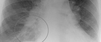
Dark spot on x-ray
In fact, the presence of dark spots in the lungs, visually visible on x-rays, does not in 100% of cases indicate the development of various ailments. Other organs are also localized at the level of the lungs, so that the radiation beam, while passing through the body, will project on film an image of all organs, as well as tissues that fall within its range.
Only a doctor can determine exactly what a spot on the lung found on an x-ray is. Sometimes additional tests are required to make a valid diagnosis.
If, during a routine annual fluorography, doctors discover a spot on a patient’s X-ray, this does not become a reason for immediate hospitalization in a tuberculosis clinic or for compulsory treatment. A patient with such a disorder is shown a detailed X-ray examination, when photographs of the chest are taken in various projections. This allows you to determine an accurate diagnosis.
What does darkening mean and how is it detected?
The pathology of the chest organs detected during fluorography is formulated by doctors as “darkening in the lungs.” Moreover, this formulation hides any reason that is not necessarily of a malignant nature. A large number of diseases can manifest as a shadow in the lung, ranging from banal pulmonary fibrosis to cancer. To exclude false data when pathological changes are detected, it is recommended to perform an X-ray examination of the chest organs.
However, it is not fundamental when making a diagnosis. But it allows you to eliminate errors in the interpretation of fluorographic data, excluding the presence of various artifacts, defects in the film itself and the examination technique.
The most detailed examination of the chest organs is computed tomography. It allows you to make a diagnosis with a high degree of probability and decide on further treatment tactics.
It is worth knowing that any lung disease is manifested by a change in the lung tissue, characterized by its compaction and subsequently a violation of airiness. It is these areas that form the pathological focus. In some cases, these changes may mask more serious pathology and cause poor outcomes. Moreover, the radiological term “darkening” is actually manifested by the appearance of light areas on the radiograph.
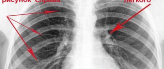
What does a classic x-ray look like?
The classic photograph is a reverse (negative) image. The difference between a negative and a positive radiograph is that there is an inverse shadow correspondence. That is, the light parts in the picture show organs with maximum density, which retain and absorb X-rays, and the darker areas, respectively, show less dense tissues and voids that allow radiation to pass through unhindered.
Healthy lungs in the picture look like this: the right one is short and wide, the left one is long and narrow, which corresponds to the norm. These areas should be transparent, as they contain a large volume of air and practically do not interfere with the passage of X-rays.
Classification of blackouts
Each problem has its own form of darkening in the image. This circumstance gives doctors the opportunity to make preliminary diagnoses before the upcoming examination, as well as most accurately give referrals to specialized specialists.
This saves time, which is very important when identifying and treating certain diseases, especially inflammation and tumors.
- Multiple opacities in the area of the apex of the lung. This location often indicates tuberculosis.
- Blurred spot boundaries. This indicates pneumonia. Additional signs include high fever and general weakness.
- Multiple blackouts. The spectrum is very wide - tuberculosis, inflammatory processes, tumors in other organs and systems. If such spots are detected, the examination will be lengthy and quite complex.
- Single clear spot. This is the most unpleasant option, since it speaks of a tumor. But in some cases, similar results are obtained with advanced pneumonia, foreign objects in the bronchi and heart problems, such as a heart attack.
When deciphering the results of fluorography, the doctor pays special attention to the geometric shape of the spots. It is she who speaks about suspected problems and allows us to adjust diagnostic measures.
Important! The most harmless cause of darkening is a defective film or a foreign object between the chest and the emitter. The subtlety is that a repeat shot to correct the result should not be taken earlier than six months later. You will still have to undergo the examination.
Types of darkening for different types of disease
When there is close collaboration between the doctor who ordered the chest x-ray and the radiologist, the examination makes it easier to make the correct diagnosis and further treatment.
The formation of morphological elements on the surface of the lungs during radiography may indicate the presence of certain pathologies.
Tuberculosis
Extensive damage to the upper parts of the lung occurs. The image clearly shows multiple dark spots up to 2 mm in size. Some of them merge, forming extensive foci.
Pneumonia
The disease is characterized by the appearance of dark circles in the lower parts of the lung tissue. As the pathology develops, the lesions become distinct, the pulmonary pattern changes, and shadows of different diameters appear.
Compaction syndrome. Infiltrate. Cancer.
Malignant formation
It is almost impossible to determine tumor formation in the initial stages of development using x-rays. Tumors smaller than 2 mm are extremely difficult to differentiate. A medical opinion will be made when the tumor reaches 3 mm.
In the picture, the cancerous tumor appears as a shadow without a clear outline, against which there is a white spot, indicating the disintegration of the formation.
Pleurisy (inflammation of the pleural layers)
The disease is characterized by darkening that does not have clear geometric contours. In addition, there is an accumulation of secretions in the pleural cavity.
Smoker's shot
Darkening on an x-ray does not always indicate a disease. Stains often occur as a result of prolonged smoking. The appearance of darkening is associated with an increase in pulmonary vessels during the development of respiratory failure.
Encapsulated exudative pleurisy
Foreign object
The reason for the appearance of light elements is often the presence of a foreign object. This is often diagnosed in children who swallow small objects and parts from toys. A foreign object is visualized as a spot with a vague outline, however, a qualified radiologist will be able to tell about this.
In addition, the artifact turns out to be acute helminthiasis, where the foreign bodies are helminths.
What should a patient do if elements in the form of darkening are detected on an x-ray? The right decision would be not to panic and not try to self-medicate. Shadows or clearings should not be treated until a full examination has been completed. The pathogenesis of the spots is extensive and may require detailed study. For this reason, it is not recommended to diagnose yourself; you should contact a specialized specialist with the diagnostic results.
Reasons for the appearance of spots in the photo
There can be many reasons for the appearance of spots. Only a qualified radiologist can give a complete transcript. Nevertheless, we can identify the main reasons why darkening occurs in a fluorography image:
- foci of active tuberculosis and post-tuberculosis changes;
- the occurrence of a benign or malignant tumor;
- consequences of smoking;
- post-traumatic lesions;
- accumulation of intrapleural fluid;
- bronchitis types of diseases;
- the presence of abscesses.
The location of the spot plays a big role. Depending on its location - on the right or left lung, in the lower or upper part - the value of this darkening may also change. To find out the true cause of the shadow in the picture, you need to consult a specialized specialist.
X-ray of the lungs
An X-ray of the lungs and bronchi shows the complications that arose as a result. If the patient has a direct inflammatory process in the bronchi, the following features will be noticeable in the image:
- decreased structure of the lung root - it is not easy to see it in the image. The right root is better visualized, but the left one is covered by the shadow of the heart. The root itself is initially heterogeneous; externally it will be formed by a plexus of blood vessels and bronchi. With bronchitis, the normal structure of the root is disrupted, since the bronchi “smear” the picture due to the existing pathology;
- vagueness and indistinctness of the contours of the root of the lung is also a consequence of inflamed bronchi, which is easily diagnosed in the image;
- strengthening of the pulmonary pattern - the pulmonary pattern is a shadow from the blood vessels. An increase in the pulmonary pattern provokes pathological changes in the bronchi, which are normally practically invisible;
- Thickening of the walls of the bronchi is the most specific sign that can be seen on an image of the lungs. It is decisive, together with other research data, when making a diagnosis.
Other signs
In addition to typical signs, such as decreased root structure, increased pulmonary pattern and thickening of the bronchi, other image descriptions are possible. For example, the image will show the curvature of the bronchi along their course, which is associated with the development of the inflammatory process and swelling.
Bronchitis on x-ray is characterized by the proliferation of connective tissue formations on the walls of the bronchi, as well as on the outside. The picture of complications may also be typical:
- bronchial obstruction (the presence of bronchial blockage, which is visualized as light “peas”);
- emphysema - transparent lungs due to accumulated air in them, which allows x-rays to pass through.
Additionally, the diagnosis is established by the abnormal condition of the diaphragm.
What is a lung x-ray and how does it differ from fluorography?
X-ray diagnostics, as an independent technique, appeared in 1895. Wilhelm Roentgen patented the rights to the invention of a tube of the same name, which emits microparticles that can penetrate soft tissue. A feature of the described phenomenon remains the different degrees of absorption of the corresponding rays. When a special film is fixed opposite the tube, structures through which the microparticles have passed will be displayed on its surface.
At the end of the twentieth century, the radiation diagnostic method for the first time allowed doctors to see the internal structures of the patient during his lifetime. Over the course of several years, the procedure spread throughout the world, where it was used to determine the causes of coughs, joint pain, bowel movements, and the like. The technique was improved, as were the devices.
In the twenty-first century, radiography is mainly used in traumatology (detection of fractures) and pulmonology (identification of the causes of cough and shortness of breath).
In practice, two main methods are used:
- traditional radiography;
- fluorography of the lungs.
The mechanism for obtaining the image in both cases is the same - the X-ray tube emits particles that pass through the chest organs, and the radiation with different intensities is deposited on the film located on the opposite side of the patient.
The difference between lung X-ray and fluorography lies in the purpose of the procedure and the size of the resulting image. The traditional procedure is used routinely to assess the condition of patients with cough, fever, and other symptoms of respiratory pathology.
Fluorography is a screening method used to detect tuberculosis and large tumors in the chest. The procedure is carried out regularly - once every 2 years for ordinary people. In the presence of harmful working conditions (miners, plant employees), the frequency of examinations increases.
Traditional X-rays of the lungs use films measuring 35 by 35 or 30 by 40 centimeters. In the case of fluorography - 2.4 by 2.4 or 7.0 by 7.0 centimeters.
In addition, during radiography, the patient receives a lower dose of radiation, which is due to the operating features of the devices. Fluorography only helps to detect the presence of pathology. Clarifying the causes of cough and choosing the appropriate treatment method requires the use of traditional radiation diagnostics.
Diagnosis of infectious lung diseases using x-rays
The X-ray method is used to diagnose the following inflammatory lung diseases:
- tuberculosis;
- pneumonia;
- bronchitis and bronchopneumonia;
- lung abscess;
- pleurisy, etc.
Pulmonary tuberculosis on x-ray
fluorography infectionimmunity The following radiological forms of tuberculosis are distinguished:
- Primary tuberculosis focus.
This picture is observed when mycobacteria first enter the lung tissue. The primary lesion is a rounded shadow measuring up to 12 mm with unclear contours. The root of the lung expands due to enlarged lymph nodes. From the shadow to the root of the lung there are small linear shadows from dilated lymphatic vessels. - Focal pulmonary tuberculosis.
Characterized by small shadows (
up to 6 mm
) in the amount of 2 to 5 pieces. Shadows are located in the upper segments of the lungs. - Infiltrative tuberculosis.
Represents limited shading of the pulmonary field, corresponding to a segment or lobe of the lung. The infiltrate may contain decay cavities or areas of mineralization, so the shadow is heterogeneous and large in size. - Disseminated tuberculosis.
With this form of tuberculosis, small shadows are found throughout the entire area of the lung fields. The pulmonary pattern is enhanced due to fibrosis of the connective tissue septa. - Cavernous tuberculosis.
The formation of a cavity (
cavity
) occurs as a result of the destruction of lung tissue during prolonged inflammation. Radiologically, the cavity is described as a round focus of clearing with a dense wall 1 - 2 mm thick. - Tuberculoma.
It appears as a single shadow on an x-ray image and is large in size. Tuberculoma looks dense on x-rays because it contains mucus, lymphatic fluid, and areas of calcification.
Inflammation of the lungs (pneumonia) on x-ray
bacteria staphylococci, streptococci, etc. X-ray examination for pneumonia reveals:
- foci of infiltration in the form of shadows of various sizes;
- the extent of the lesion ( segment, lobe, one or both lungs
); - inflammation of the pleura;
- inflammation of the bronchial tree;
- reaction from the lymphatic system ( expansion of the lung root
); - strengthening of the pulmonary pattern.
up to 1.5 cm
X-ray picture of a lung abscess
The following radiological signs of lung abscess are distinguished:
- at the beginning of the disease, intense shading of a round shape is detected;
- subsequently, the intensity of the shadow decreases, it takes the shape of a ring, in which the horizontal level of the liquid is determined;
- A chronic abscess is distinguished by a dense wall ( 3–4 mm in thickness
), in the center of it there is a clearing zone and there may be no fluid level.
Pleurisy on chest x-ray
injuries The following radiological signs of pleurisy are distinguished:
- uniform shading of part of the pulmonary field depending on the amount of exudate;
- shading shift when taking an x-ray in a different body position;
- with inflammation of the pleura in the interlobar fissure, shading in the form of a biconvex lens is determined.
from one third or more
puncture
of inflammation, injury or tumor
Whooping cough. X-ray signs
Pertussis vaccination Whooping cough on an x-ray is characterized by the following signs:
- extensive clearing of the lung fields;
- small multiple nodular shadows ( miliary whooping cough pattern
); - the pulmonary pattern is strengthened, branched ( takes on the appearance of a bush
); - expansion of the lung root.
Types of blackouts
Pulmonary diseases are generally accompanied by compactions in the lung tissues. This occurs due to a decrease or absence of air permeability in certain areas of the organ. X-rays show darkened spots. This symptom may indicate not only pathological processes in the lung itself.
Blackouts, the causes of which are associated with pulmonary pathologies, vary in their intensity, clarity, quantity and size. Darkening may indicate:
- on inflammatory processes and tissue compaction;
- on nodes of tumor formations;
- to an area impassable for air - collapse of the lung;
- on the development of tuberculosis;
- for the presence of fluid in the pleural area of the lungs (pleura is the membrane covering the lungs and chest cavity);
- for inflammation in the pleural area, possibly purulent (abscesses).
Pulmonary opacities, which appear due to problems with other organs, may also be visible on the images. These may include:
- swollen lymph nodes;
- formations on the ribs or spine;
- problems with the esophagus, for example, its enlargement.
X-ray and fluorography for bronchitis
Fluorography, like x-ray examination, shows the condition of lung tissue, helps to distinguish fibrosis or the presence of foreign bodies, and see signs of pathological changes.
These diagnostic methods are similar, but if signs of certain pathologies are detected, the doctor will prescribe an x-ray, since this method will give a more accurate result, make it possible to make the correct diagnosis and prescribe adequate treatment.
Bronchitis on fluorography can be suspected by the following changes:
- the pattern of the lungs is enhanced - this is a sign of acute inflammation of both the lungs and bronchi;
- the roots of the bronchial tree are deformed - evidence of probable tissue destruction and inflammation of the bronchi;
- stringy pattern of roots - usually observed in long-term smokers who are at risk of developing chronic bronchitis.
However, the doctor cannot say whether bronchitis is visible on fluorography. A fluorographic image alone cannot make an accurate diagnosis; this study does not provide a complete picture of the disease.
X-ray is a more informative and effective examination in terms of making a correct diagnosis. It will show in detail and accurately what cannot be determined with fluorography. On an x-ray with bronchitis you can see:
- small foci of infiltration, representing an accumulation of fluid in the lung tissues;
- increased thickness of bronchial tissue;
- due to inflammation, small vessels are not visible in the image, the contours of the lungs themselves are blurred in the image;
- focal and lamellar atelectasis - deformations in the form of collapse of part of the lung;
- the roots of the lungs spread and enlarge.
When the disease is advanced, signs of emphysema appear: the pattern of the lungs is significantly distorted due to the accumulation of a large volume of air.
What is the seriousness of darkening or brightening?
It is difficult to answer unequivocally only based on an x-ray image whether such enlightenment is dangerous. To assess the situation, you will need to take additional tests and analyzes and consider the patient’s condition.
X-ray is an auxiliary diagnostic method designed to supplement information about the condition of the patient’s tissues and organs. If you look closely, there is no “diagnosis” in the X-ray description report; instead, there is a “conclusion.” Only a specialized specialist can make a diagnosis based on the diagnostic results, as well as answer the question about the danger of such darkening during fluorography.
Interpretation of X-ray of the lungs. X-ray syndromes in various lung diseases
The following pathological symptoms and syndromes are identified on an x-ray of the lungs:
- shading of the lungs;
- clearing of the lungs;
- changes in pulmonary pattern;
- changes in the roots of the lung.
limited or extensive
Shading on an x-ray of the lungs
The following types of shading on a lung x-ray are distinguished:
- Extensive shading of the lung.
In this condition, the shadow occupies more than 80% of the lung field area.
This condition can occur when the lung collapses ( atelectasis
), edema or cirrhosis of the lung. Inflammation or neoplasms rarely occupy the entire lung, however, in the past, when medicine was not as well developed as it is today, such cases were observed. - Limited shading.
In this case, the shadow has the size of a lobe or segment of the lung.
Most often, this type of shadow occurs in inflammatory diseases ( segmental or lobar pneumonia
). - Round shadow syndrome.
It is a rounded single shadow with a diameter of more than 12 mm. This formation is stable and is found in all projections. This X-ray picture is characteristic of tuberculosis, tumors, cysts, pulmonary aneurysms and some other formations. - Focal shadows.
Such shadows have a size of less than 12 mm and can occur in quantities of several pieces (
from 2 to 5
). Focal shadows appear in the lungs with tuberculosis, cancer metastases, and pneumonia. - Extensive dissemination.
With this type of lesion, the pattern of the lungs is significantly changed; shadows from 2 mm to 12 mm are found in large quantities. Dissemination is considered extensive if it has spread to more than 2 lung segments. This X-ray picture is typical for alveolar pulmonary edema, tuberculosis, sarcoidosis and some other diseases.
Changes in the pattern of the lungs on x-ray. Strengthening, depletion or deformation of the lung pattern
There are three types of changes in the pulmonary pattern on x-ray:
- Gain.
In this case, the number of structural elements of the pulmonary pattern is increased. This is observed in congenital or acquired heart diseases, due to which the speed of blood circulation in the pulmonary circulation decreases. The pulmonary pattern can be enhanced by the uniform growth of connective tissue in the lung. - Weakening.
With this type of change in the pulmonary pattern, the caliber and the number of its elements are reduced. This can be observed with emphysema, pulmonary valve stenosis, and some heart defects. - Deformation.
In this case, the pattern of the lungs changes unevenly, and its contours are irregular. This picture is observed in infectious and inflammatory diseases, bronchitis, pneumonia, tuberculosis.
Changes in the roots of the lung on x-ray
expanded Lung root change syndrome occurs in the following diseases:
- tuberculosis;
- pneumonia;
- pneumosclerosis;
- pulmonary arterial hypertension;
- pulmonary edema;
- heart defects associated with pulmonary circulation disorders;
- malignant tumors ( lung cancer
); - cancer metastases to the lungs.
Main reasons
Darkening in the lungs on an x-ray - this can be inflammatory changes, neoplasms, infectious diseases . A similar picture is given by pathologies of the cardiovascular system, mediastinal organs, and metastases from other parts. Let's look at the main reasons that can cause spots on the lungs on X-rays.
Pneumonia
It is the most common pathology of the respiratory system. For this reason, when characteristic symptoms appear, the doctor sends the patient for testing. The main and main manifestation of inflammation in the lungs is darkening. They differ according to the type of infiltration, process activity, prevalence and localization. Pneumonia can occur for the following reasons:
- bacteria;
- viruses;
- fungal flora;
- protozoa;
- mycoplasma.
This is not a complete list of possible reasons for the development of this pathology. The darkening in the image depends on the stage of development of the disease:
- Tidal stage: decreased transparency of an uneven nature, without clear boundaries.
- Hepatization stage: distinct darkening with pronounced boundaries.
- Resolution stage: reducing the severity of infiltration in the image.
The outcome of this disease is complete normalization of the X-ray picture. But usually some changes in the pattern remain, deformation of the roots of the lungs, etc.
The X-ray picture of pneumonia does not have a pronounced specific character. Therefore, when diagnosing, they rely on general complaints, anamnesis and the effectiveness of the treatment.
Lung abscess
It is a formation that is filled with pus. More often it is the outcome of inflammatory lung disease. The infection can also enter through the blood and lymph. Dark spots in the image depend on the stage.
- intense uniform shadow;
- fuzzy outlines.
As the abscess develops:
- formation of a cavity with smooth edges;
- slight clearing in the middle;
- area of inflammation around.
Often, a horizontal liquid level is considered in the cavity. Focal shadows in the lungs due to an abscess can become chronic. In this case, the image will reveal an irregularly shaped cavity with a zone of inflammation (sclerosis) around.
This group includes lung diseases that are accompanied by diffuse changes. A common pathology is chronic obstructive bronchitis in smokers (black lung) and workers in hazardous enterprises. Detection of darkening is possible against the background of noticeable thickening of the walls of the bronchi. In this case, an intensification of the pattern and its mesh deformation will be formed in the image.
Bronchiectasis often develops with bronchitis. Main signs on x-ray:
- strengthening of the pulmonary pattern;
- cellular or looped seals with an area of inflammation.
Symptoms indicating the development of COPD are a persistent cough, sputum, chest pain, and difficulty breathing.
Emphysema
Accompanied by increased airiness of the lungs. It is an outcome of COPD, a pathology of the cardiovascular system . The image of emphysema shows increased pneumatization, depletion of the pulmonary pattern or its complete disappearance along the periphery.
Bronchial asthma
This disease also produces some changes when taking x-rays. Characteristic features include:
- swelling of the lung tissue;
- low diaphragm dome;
- increase in length of the respiratory organs.
Also, X-rays can reveal complications of asthma in the form of atelectasis. They represent areas where tissue collapse occurs.
If emphysema, bronchial asthma or COPD is suspected, CT is preferable. This method provides more information regarding these pathologies.
Lung cancer
When performing an X-ray, it is not possible to determine the histology (that is, the cellular composition) of the tumor. A dark spot on the lung due to cancer can be central, peripheral or bronchoalveolar.
Read also: Trigan d how to get stubborn
Signs that there may be cancer of central origin:
- the presence of a tumor originating from the large bronchi;
- characteristic expansion and deformation of the root;
- obstruction of the bronchial tree.
With this type, growth can be observed in the lumen of the bronchus, beyond it, or of a mixed type.
Peripheral cancer:
- more often in the form of a single education;
- the contours are lumpy and irregular in shape;
- the presence of a “path” to the root of the lung.
Pancoast tumor may occur in the upper lobes of the lungs. It is a type of peripheral species. It quickly spreads to the pleura, collarbone, and plexus of the shoulder, which leads to severe pain.
Symptoms of cancer may not appear for a long time. They are often preceded by exposure to adverse environmental factors (ecology), long-term smoking, and the presence of a genetic predisposition. Also, suspicion of a tumor may appear due to poor fluorography.
Metastases
Most neoplasms metastasize to the lungs. It is not difficult to identify them with x-rays. They can appear in different lobes, in the lower right or left lung. In this case, the lesions usually have a round shape. Spread from the primary tumor occurs through the blood or lymph. Therefore, neoplasms of the brain, liver, and mammary gland often metastasize to the respiratory organs.
Diffuse diseases
This group includes diseases that are accompanied by fibrosis and sclerosis. These concepts mean the replacement of healthy lung tissue with connective tissue or its reduction in size. They act as an outcome of other pathologies. Signs include:
- strengthening and mesh deformation of the pattern;
- high diaphragm position.
The honeycomb lung phenomenon is also revealed. Using X-rays, late stages of diseases are detected.
Pneumoconiosis
Includes a group of occupational pathologies. These include asbestosis, silicosis, and talcosis. Occurs when exposed to dust of various origins. Fibrosis gradually develops in the lungs. Signs:
- small outlined nodes;
- thickening of the pleura.
There is also an increase in peripheral nodes with the deposition of calcium salts.
Tuberculosis
Darkening of the lungs on fluorography is a suspicion of tuberculosis. This type of study is carried out to screen the population. Each type has its own characteristics on an x-ray. In clinical medicine there is a classification. Tuberculosis intoxication is accompanied by changes in the general functions of the body, a sharp increase in the size of the papule, and is practically not reflected in the image.
Primary has its own characteristics:
- swollen lymph nodes;
- enlargement of mediastinal organs;
- the lung tissue remains unchanged.
Tuberculosis of the intrathoracic lymph nodes is also distinguished. According to its classification, it belongs to primary. Accompanied by compaction and pockets of calcifications. Additionally, a complex is isolated, which includes all the above-described signs, plus the appearance of an inflammatory reaction track.
- multiple lesions in the lungs;
- round shape;
- size 1-2 mm;
- arrangement in the form of a chain.
Such lesions can merge over a long period of time. The classification distinguishes focal tuberculosis, which is characterized by:
- single or multiple lesions;
- size up to 1 cm;
- fuzzy outlines.
Then there is a form of infiltrative tuberculosis. It is characterized by the presence of shadows larger than 1 cm. When fused, such lesions can occupy several lobes of the lung at once. Caseous pneumonia is characterized by many confluent foci.
Tuberculoma is distinguished separately in the classification. Signs:
- lesion larger than 1 cm;
- clear contours;
- pronounced capsule.
The worst variants of tuberculosis are cavernous and fibrous . They are characterized by darkening in the form of a cavity with dense walls, without areas/with areas of fibrosis around.
Symptoms: fever, prolonged cough, positive sputum test, unsatisfactory fluorine result.
Pleurisy
Effusion into the pleural cavity occurs in the presence of various types of respiratory diseases. Normally, this area contains a small amount of fluid, which allows the pleura to move freely. X-rays can detect this disease. The image shows the liquid level as a concave shape. As a rule, it is bilateral in nature.
Changes on an x-ray can have a different shape and character, but in any case, this display is a reason to contact a specialist to correctly interpret the image. The identified pathology in this case will undergo immediate treatment.
Some patients panic when, after another fluorography done as part of a routine examination, a darkening is detected.
There are no particular reasons for concern yet - darkening in the lungs on fluorography does not necessarily imply some kind of “death sentence”. It is likely that this is simply a film defect or traces of constant smoking.
But in some cases there really is cause for concern. Final conclusions can be drawn only after additional examination has been completed. The doctor will definitely prescribe it in such a situation. So, what does darkening in the lungs hide under fluorography?
Bronchitis and its diagnosis
Bronchitis is a process of inflammation that affects the mucous membranes of the bronchial tree. Inflammation provokes swelling and hardening of tissues and, as a result, narrowing of the bronchial lumens.
Often this disease appears against the background of the patient’s colds, and can also be a consequence of exposure to toxic substances on the respiratory system.
There are three forms of the disease:
- Spicy. A sharp deterioration in the condition of a respiratory disease. First, a dry, barking cough appears; after a few days, sputum that is difficult to clear appears. Body temperature rises to 39 degrees.
- Chronic. Erased symptoms that do not go away for a long time - more than three weeks. Wet cough with sputum, body temperature fluctuates in the range of 37-37.5 degrees.
- Obstructive. It is characterized by attacks of debilitating cough, blockage of the bronchi leading to suffocation, and a severe course of the disease. When breathing, a characteristic whistling sound appears.
The diagnosis is made based on the patient's complaints and the results of a clinical examination. The main symptom of the disease is a cough, dry at first, and then, as the disease develops, turning into a wet one, with the discharge of sputum.
In addition, symptoms such as fever, difficulty breathing, and weakness appear.
Doctor for diagnosis:
- finds out when the first symptoms appeared and in what form they appear;
- listens to the patient's chest using a stethoscope;
- conducts blood, urine, and sputum tests to determine which infection caused the disease - viral or bacterial;
- prescribes an X-ray examination: in case of bronchitis, the image of the lungs will show a clear basal pattern of the bronchi and increased contrast of the large bronchi.
There is an opinion that x-ray examination is more dangerous to health than fluorography. In fact, the radiation dose during fluorographic examination is higher. Although in this case it does not go beyond the acceptable health norm.
X-ray as an examination method is more informative, since on a fluorographic image shadows of 5 mm can be distinguished, and on an x-ray - from 2 mm.
Thus, an x-ray will better help in making the most accurate diagnosis. Therefore, fluorography for bronchitis is usually not prescribed, but is used for preventive examinations once a year.
Learn more about shading types
The features of the geometric shape of lung darkening on fluorography were discussed above, now let’s talk about this in more detail.
Medical types of film stains:
- Focus . Round spots, the size of which exceeds 1 cm. They speak of pneumonia, asthma, abscess, fluid in the lungs. These are the most common pathologies accompanied by this type of darkening. A very thorough examination is required.
- Focal . They are nodular spots, their size does not exceed 1 cm. They talk about inflammation, tumors and problems with blood circulation. If the spots are accompanied by elevated temperature, then most likely we are talking about pneumonia. In any case, blood, urine and sputum tests should be taken.
- Segmental . Such darkening can be either single or multiple, affecting entire sectors of the lungs. In the first case, we are talking about injury or a foreign object in the bronchi, as well as a tumor at the initial stage. If the lesion is multiple, the doctor will suggest central cancer, fluid in the pleura, acute pneumonia, chronic tuberculosis. Also, such darkening may indicate cancer metastases from other organs.
- Equity . These spots are clear, clearly visible and have a regular geometric shape. The main causes are purulent processes and damage to the bronchi. If the spots are located in the middle and lower lobes of the lung, then we can talk about a neoplasm.
If none of the diagnoses listed above are confirmed after a thorough examination following fluorography, then the picture will have to be taken again. Moreover, you need to use a more modern device.
Important! Sometimes errors when deciphering images occur due to the inexperience of the doctor. In this case, you should contact a specialized clinic with experienced specialists for a repeat image. The harm from radiation is not comparable to the consequences of such mistakes, so there is no need to be afraid.
In most cases, darkening in the lungs on fluorography indicates a pathology, often very serious. If spots are found on the picture, you should immediately undergo a full examination.
There are frequent errors during decoding, which arise due to poor-quality film and inexperience of the doctor. In such a situation, fluorography needs to be done again. You should not be afraid of unnecessary radiation, because the consequences of incorrect diagnosis will ultimately turn out to be much more terrible!