X-ray examination: positive and negative aspects of the procedure
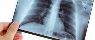
X-ray of healthy lungs
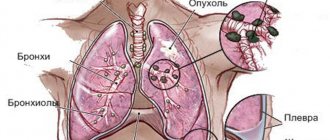
X-ray examination is based on the ability of X-rays to penetrate the human body. As the rays pass through fabrics of different densities, they will be absorbed differently, leaving silhouettes of shades of light and dark colors on the film.
Hard structures are light in color, while soft structures are dark. This property of the procedure allowed it to be widely used in the diagnosis of lung diseases, and low-dose x-rays (fluorography) began to be done en masse to primarily identify pulmonary tuberculosis.
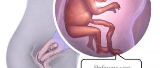
The disadvantages of the study are the possible risks to which patients are exposed. The examination is extremely undesirable for pregnant women.
Changes in the right cardiophrenic angle. Pericardial cyst.
Despite the negative aspects of X-ray irradiation, the procedure also has advantages. For example, the low cost of fluorography made it possible to make the study widespread and prevent the development of outbreaks of tuberculosis.
In particular, in such vulnerable social groups as children and the elderly. In addition, X-ray examination is non-invasive - it visualizes the condition of internal organs without opening the chest.
The diagnostic value of X-ray irradiation is also great - with the help of rays, severe pathological processes, abscesses, pneumonia, and tumor formations are visible. You can get results in the shortest possible time and begin treatment of the disease in a timely manner.
Preparation for fluorography and the mechanism of its implementation
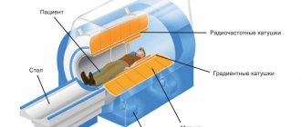
Digital scanning fluorograph (the safest and most modern diagnostic method)
Fluorographic examination does not require special preparation from the patient. However, before carrying out it is necessary to take into account some features. The result is not affected by:
- medications the patient is taking;
- smoking before the study;
- products that lead to gas formation in the stomach;
- physical exercise.
Before taking a picture, it is necessary to remove objects that can appear on the film and lead to difficulties in diagnosis. Doctors warn:
- women need to remove their bra or lower it at the waist so that the fasteners are not visible;
- patients need to remove body jewelry - chains, pendants, crosses;
- Patients with long hair are advised to pin it up so that it does not fall on the shoulders and create interference in the picture.
If fluorography is necessary, it must be taken into account that it does not affect the operation of pacemakers. Therefore, patients should not worry or refuse examination if they have such a device installed.
The x-ray is monitored by a medical professional who instructs the patient and helps him find the right position. The following key points are highlighted:
- after preparation, the person is asked to stand in the booth in front of the CCD matrix and press his chest as tightly as possible; he needs to stand slightly hunched over so that his shoulders are pressed against the surface of the screen;
- at the time of the procedure, the doors of the booth are closed, and the patient is given the command to inhale and not breathe - the breath is held for several seconds, after which the command to breathe is given and the doors open. The moment of photographing is carried out while holding your breath;
- After opening the doors, you can leave the booth and get dressed.
Fluorography codes
If it is necessary to shoot in several projections, the patient will be given appropriate recommendations from the medical staff. This concludes the research.
The transcript of the fluorography is sent to the doctor who gave the referral or is pasted into the patient’s card. If the study is preventive, the patient is given a receipt indicating personal data, the time of the study, and the result of fluorography.
How is the examination carried out?
In order to undergo a fluorographic examination, the patient must undress to the waist. Girls and women should remove their bras and expose their breasts. If you have jewelry on your neck, you will need to remove it or hold it in your teeth.
In order to take a photo correctly, your hands should be placed on your belt and your shoulders should be forward. In this position, you need to press against the scanning plate. In this case, the selection is installed on a special pad. Before the procedure, the x-ray technician will ask you to take a breath and hold your breath while the equipment operates.
To prevent X-ray radiation from damaging organs, the hospital provides a special apron with zinc plates to cover the lower part of the body.
How long will it take for the results to be ready?
You can get an answer immediately if you take digital fluorography. If the analysis is carried out using film equipment, you will have to come back for an answer in a few days.
If the doctor does not find any pathologies, in the fluorography room he gives you a certificate certified by a seal.
Decoding
Changes in the aorta.
Aortic aneurysm Decoding the procedure requires special knowledge, so it is carried out by radiologists. In the photo of the lungs there are pathological shadows, indicating the following abnormalities:
- the presence of free fluid in the lung cavity;
- lack of a clear contour of the respiratory organs;
- focal spots in various places;
- focused spots;
- fractional images on film;
- segment displays.
If spots are detected on an X-ray, doctors prescribe an additional study - it will give a detailed picture, since the chest organs are photographed in several projections.
Compaction syndrome.
Infiltrate. Cancer. If, when interpreting fluorography of the lungs, focal spots up to one centimeter in diameter are found, this may indicate abnormalities of a vascular nature, or the initial stage of the oncological process.
And this may also indicate a pathology of the respiratory system. To make it more specific, the patient will additionally undergo a computed tomography scan, and sputum will be taken for analysis.
In addition to these assumptions, focal shadows also indicate a possible myocardial infarction - it is this that is confused with the initial stage of the development of tuberculosis.
Another variant of deviation is segmental spots. They have clear boundaries and a triangular shape. The contour of the spots is clear, without blur. If a single segment is detected, the doctor suspects injury to the lung tissue. But it could be a foreign body or an endobronchial tumor.
In the presence of multiple segmental spots, the options for possible pathologies are wide. It can be:
- oncological pathologies;
- pulmonary tuberculosis;
- pneumonia in acute or chronic form;
- the presence of fluid in the pleural cavity;
- possible metastases.
Compaction syndrome.
Low to medium density lesion. Tuberculosis. If a lobar spot is detected, the doctor suspects bronchiectasis disease, purulent lung damage and other pathologies. But clearly focused spots are a sign of:
- pneumonia at various stages of pathology development;
- presence of fluid in the pleural cavity;
- bronchial asthma;
- purulent tissue damage as a result of the inflammatory process;
- helminthic infestation.
The spot may be the result of a callus in the bone tissue if the patient has previously suffered a bone fracture. For shadows of an indeterminate type (fuzzy, with “ragged” edges), doctors diagnose one of the pathologies:
- staphylococcal lung damage and inflammation that develops against this background;
- accumulation of fluid in the pleura;
- exudative pleurisy;
- heart attack condition.
Since deviations from the norm in the image can be interpreted ambiguously, a diagnosis is not made based on fluorography data.
For diagnosis, a set of signs is collected and additional tests are performed, including computed tomography. And only on the basis of a whole complex of symptoms is it possible to talk about the exact disease.
Encapsulated exudative pleurisy
When interpreting fluorography, not only the presence of spots and shadows is taken into account. Doctors also pay close attention to the structures being examined - the roots of the lungs, bronchi, and diaphragm. Their condition and display in the image allows us to illustrate a particular pathology.
For example, the following characteristics are noted:
Dextraposition of mediastinal and gastric organs
Compaction of the roots of the lungs is a characteristic not of the root itself, but of all structures that are visualized in a given area (main bronchus, vessels, lymph nodes).
If the roots are expanded and dense in structure, then this indicates a chronic pathological process, as well as recently suffered bronchitis or pneumonia.
If an x-ray shows that the roots are stringy, then this indicates chronic bronchitis in the patient. This sign can also occur with oncological pathology, with bronchiectasis disease, or with occupational hazards.
Expansion of the heart in diameter
Strengthening the pulmonary vascular pattern is the result of active blood supply, which occurs during an acute inflammatory process in the lungs, congenital heart defects, or cancerous damage to the organ. This symptom is also diagnosed in mild pathologies - bronchitis, ARVI.
If there is fibrosis in the image or fibrous tissue is detected, then this indicates a previous illness, for example, pneumonia, an infectious process, surgery, tuberculosis.
Foreign bodies in soft tissues
Calcifications are inclusions that resemble bone tissue in density and are therefore visualized in a light shade. Formed in a place where there was previously a suppurative process, helminthic infestation or a foreign body.
Calcifications are evidence of contact with a patient with tuberculosis, but in the patient himself the disease has not entered the active phase.
Adhesions and pleuroapical layers are signs of a previously suffered inflammatory process. Adhesions do not require intervention.
Pleuroapical layers are thickenings of the pleura, which also indicate a previous inflammatory process. The sinuses of the pleura are also reminiscent of inflammation. If the sinus is free or sealed, then this indicates normality or pathology, respectively.
Air in the dilated esophagus
Changes in the diaphragm are often not so much a sign of pulmonary pathology as evidence of a pathology of the digestive organs.
The shadow of the mediastinum is considered the distance between the lungs. The trachea, esophagus, glands, lymph nodes and blood vessels are located here. The mediastinal shadow is expanded and displaced with increasing heart size and hypertension.
Peculiarities of interpretation of the result in a smoker
Modern science has proven more than once that even one smoked cigarette can cause a number of certain pathological changes in a person’s respiratory system. That is why all passive and active smokers who regularly inhale air containing tobacco smoke need to have their lungs examined at least once a year.
Very often, patients who abuse smoking are quite skeptical about such a diagnostic method as fluorography. But timely detection of such a dangerous disease as pneumonia will make it possible to avoid the occurrence of many serious complications in the future.
Smokers are observed to have a thickening of the lung tissue structure along with the accumulation of fluid in their cavity or the formation of tumor-like neoplasms. In this case, a course of treatment measures aimed at removing the patient from the risk zone is urgently carried out.
How to find out the result of fluorography?
Contraindications for X-ray examination
The study cannot be carried out if the patient is not feeling well (high temperature, weakness, chills, fever), or if there is shortness of breath.
Fluorography is also not done for those patients who are unable to hold the body in the desired position. The study is not carried out on children and pregnant women.
Fluorography of the lungs is an effective diagnosis of the chest organs. The study determines not only the presence of tuberculosis, but also pathologies such as cancer, emphysema, and abnormalities in the structure of the heart and lungs.
Fluorography is carried out quickly and does not cause much harm. Nevertheless, doctors recommend it solely for indications, so as not to expose the patient to unnecessary radiation.