Malignant formation in the lungs is one of the most common cancers among men and ranks first, and fourth among women. It is impossible to detect lung carcinoma in the first stages, since the disease is practically asymptomatic, but lung cancer can be seen on fluorography.
Oncologists say that it is impossible to completely cure this tumor, but if you detect the tumor in the early stages, you can prevent the spread of metastases, thereby extending the patient’s painless life for several years.
About the disease
It is not for nothing that so many people are concerned about the question of whether lung cancer is visible on fluorography. This common cancer has a poor prognosis. There is a high risk of death. Therefore, it is so important to identify this oncopathology at the initial stage. It is dangerous because it is practically asymptomatic. Signs of the disease often begin to manifest themselves only at the stage of tumor metastasis.
The disease occurs in both sexes. However, men are still more likely to suffer from it. There are a number of factors that provoke lung cancer:
- Smoking.
- Alcohol abuse.
- Drug addiction.
- Unfavorable environmental conditions.
- Working in an environment with potential carcinogens.
- Genetic predisposition.
- Consumption of foods containing carcinogens.
- Radiation exposure.
Based on location, lung cancer is divided into central, peripheral and massive. The latter is quite rare. And the most common is the central one.
Is lung cancer visible on fluorography? This procedure is not included in oncological diagnostics. The latter is as follows:
- Examination of the patient.
- Collection of medical history and complaints.
- Laboratory tests of the patient's blood.
- Instrumental diagnostics. In particular, X-rays of the lungs.
Staging
Each stage of the oncological process is characterized by its own degree of cancer prevalence in organic structures. Staging of lung cancer is based on the assessment of tumor parameters, the presence of malignant cells in the lymph nodes, the spread of cancer to internal organs, etc.
Staging is one of the most important diagnostic moments that helps the oncologist choose the most adequate and effective method of therapy. Staging also helps in determining prognostic data regarding the success of the treatment process and overall survival.
Lung cancer is one of the most severe cancer conditions that takes the lives of many people every year. But it can be dealt with if you undergo a diagnostic examination in a timely manner.
Early diagnosis maximizes the success of therapy. Therefore, at the first symptoms you need to go to a specialist.
Video about bronchoscopic examination and endobronchial ultrasonography in the diagnosis of lung cancer:
Danger signs
Will fluorography show lung cancer at an early stage? As we will see later, not always. But sometimes the patient’s life depends on the slightest delay.
The insidiousness of lung cancer is that even their first symptoms do not cause serious concern in a person. That’s why so many people turn to specialists for help only when there are obvious signs of cancer.
The first alarming symptoms in this case are the following:
- Chronic causeless weakness, loss of strength.
- Cough of unknown origin.
- Dyspnea.
Is lung cancer visible on fluorography? Doctors advise that for the preventive diagnosis of this disease you should still use an X-ray of the lungs.
Hope for treatment
At an early stage, it is possible to cure lung cancer if a course of chemotherapy is carried out in a timely manner, and if necessary, the tumor is removed surgically.
These two methods are combined with each other to achieve good results. If cancer is detected at stages 1-2, then the prognosis is more favorable than detection of cancer in the last two stages, when patients are difficult to help.
Should you trust an X-ray showing a tumor? For most patients with suspected cancer, this is the first question that comes to their mind.
Of course, there is no need to panic, because diagnostics cannot determine whether it is cancer or not. But it’s definitely worth further examination, consulting with other specialists and undergoing a digital examination or MRI.
Fluorography - what is it?
It is possible to detect lung cancer using fluorography. But, unfortunately, not in all cases. And especially in the early stages. So why is this technique so common and included as part of a standard medical examination?
Fluorography is a fast and inexpensive way to examine the lungs. You can already get results in just a day. Using this technique, specialists can identify tumors and dysfunctions not only in the lungs, but also in other organs and systems: the heart, diaphragm, bone tissue, adjacent vessels, etc.
The method is also valued for being painless and harmless. The radiation dose that a person receives during the procedure is negligible. Therefore, there is nothing harmful in undergoing this preventive diagnosis every year.
Advantages and disadvantages of FLG
The main advantages of fluorography are the low cost of this method and its accessibility. The examination does not take much time and can be carried out in any clinic equipped with X-ray equipment. Disadvantages include the low degree of accuracy of diagnosis and the need to supplement this examination with accompanying diagnostic methods. In lung cancer, fluorography is the initial tool for prescribing further observation and, if necessary, adequate treatment. For this reason, it is not recommended to ignore FLG.
What can be seen on a fluorogram?
If the fluorogram is taken correctly, then from this image an experienced specialist can detect the following:
- Pleural inflammation.
- Calcium deposition in the heart muscle.
- Expansion of the roots of the bronchi and lungs, as well as fibrous tissues.
- Enhanced vascular pattern.
- Focal shadows of oncological tumors.
Fluorography is valued because with its help you can notice pathological changes not only in the structure of the lungs, but also in all organs that are somehow involved in the respiratory process: the heart, veins, arteries, etc.
Why does cancer appear?
Lung cancer is most often caused by smoking. Cigarettes contain harmful substances that, when burned, adversely affect the human respiratory system.
Among such substances are acids, gases, oils. They are deposited in the bronchi and lungs, and the carcinogenic effect on humans is truly colossal.
Under the influence of carcinogenic substances, pathological processes begin in cells, and they acquire the ability to undergo abnormal division.
Sometimes patients die from the tumor within a few months, while others undergo long-term treatment and do not always overcome the disease.
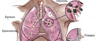
In case of a cancerous tumor, metastasis is of great importance, when pathological cells spread throughout the body through the bloodstream or lymphogenous route, affecting other organs in which tumors also grow.
You can observe not only organs, but also glands with metastases. For example, in lung cancer the thyroid gland is affected.
"Electric Smog"
When is lung cancer not visible on a fluorogram?
Does fluorography detect lung cancer? Yes, but, unfortunately, not in all cases. This is due to a serious drawback of the technique - the procedure is performed only in a direct projection. Therefore, there are a number of factors when fluorography fails to recognize a tumor in the lungs:
- The neoplasm is located in the basal segments of the lower right lobe of the organ. Since they are blocked by the liver, it is not possible for a specialist to examine the tumor.
- Too small oncological foci.
- The tumor is located too deep in the tissues of the organ.
Does fluorography show lung cancer in the early stages? Yes, but, as you have seen, not in all cases.
X-ray
Such a study is highly informative in 8 out of 10 cases of pulmonary oncology. Only in a few percent of cases of such oncology, the examination shows the normal condition of the organs.
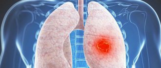
In the central cancer form, radiography reveals an expanded vascular network and opacified areas in the lungs.
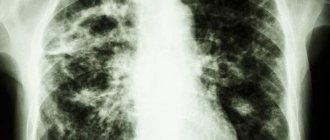
The photo shows well what central cancer of the right lung looks like on x-rays
If pulmonary oncology is peripheral in nature, then the picture on the x-ray will show the presence of a clear, uneven shadow, from which ribbon processes extend to the pulmonary root.
What does a neoplasm look like on a fluorogram?
What does lung cancer look like on fluorography? Of course, a tumor in an image is difficult to recognize for a non-specialist. A qualified doctor may suspect it based on the following signs:
- Availability of seal. Most often it is one-sided and casts a shadow. Strands may be adjacent to it. It is noticeable that the roots of the lung will be somewhat expanded.
- The shadow cast by the compaction varies in shape. But most often it is spherical. As a rule, it has fuzzy edges, and there may be some “radiance” around it.
What signs indicate the disease?
With lung cancer, symptoms such as:
- wheezing;
- Cough that does not go away for a long time and cannot be treated;
- Symptoms of shortness of breath;
- Pain syndrome that occurs with each coughing attack;
- Discharge of sputum with streaks and sometimes blood clots;
- Lack of performance;
- Fatigue, constant feeling of weakness, lethargy;
- Frequent causeless temperature fluctuations;
- Refusal of food.
The presence of such symptoms does not always indicate the development of cancer, but it is worth paying attention to, because it may indicate other pathological processes.
Gradually, the clinical picture begins to acquire more vivid symptoms. The patient is constantly bothered by intense shortness of breath, tachycardic manifestations, and a depressing cough with bloody discharge.
Even normal breathing causes intense pain, and the patient begins to rapidly lose weight. The patient's voice becomes hoarse, swallowing becomes difficult, cyanosis of the upper half of the body may occur, etc.
Indirect signs
The tumor is not always visible on the image when the cancer is already beginning to develop. A qualified doctor can suspect this disease based on a number of indirect signs:
- Hypoventilation (insufficient ventilation) of the lungs.
- Atelectasis of pulmonary areas.
- Compensatory increase in the airiness of the adjacent sections.
- Distal approximation of blood vessels (this can occur due to compression by the tumor).
- Thickening of the walls of the bronchi.
Fluorography and X-ray - is there a difference?
As we have seen, fluorography shows lung cancer, but not in all cases. A more accurate method of instrumental diagnosis of this pathology is x-ray.
An x-ray is taken faster than a fluorography. Moreover, if you undergo diagnostics in a private medical clinic, the cost of an x-ray is lower.
Another advantage of radiography: the radiation dose to the patient can be lower than with fluorography. By the way, the highest dose of radiation is during computed tomography. But the advantage of fluorography is its high throughput. Due to the speed with which results are obtained, the procedure is included in annual medical examinations.
The principle of obtaining an X-ray image is simple: a beam of rays emanates from the beam tube of the device. Accordingly, they will pass through the human body to varying degrees. The result is displayed on film. Because our organs transmit X-rays differently, the result is a photo-like image: the soft tissue is gray, the air spaces are black, and the bones are white.
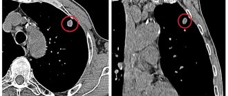
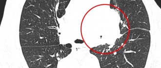
Two more alternative methods to fluorography are magnetic resonance and computed tomography. CT is a more accurate method, since the patient’s body is scanned with X-rays from several angles. But it is much more expensive than fluorography and radiography. The radiation dose from CT is much higher.
MRI is harmless because it is exposed to magnetic fields. But the procedure, again, is characterized by high cost. In addition, there are a large number of contraindications to MRI.
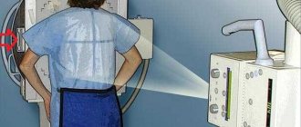
How is it carried out?
Fluorography is performed in a special X-ray room, isolated from the external environment by thick walls. The procedure takes less than a minute. The examinee must remove all clothing up to the waist and jewelry from the neck. Women always tidy up their hair so that it is not on their back or chest. The subject enters the apparatus and stands in the designated place.
The nurse lowers the special chin recess to the required level according to the person’s height. Next, the door closes and you need to take a deep breath and hold your breath for a few seconds while the picture is taken. Fluorography is performed digitally or film, when the image is transferred to a screen and then to a special film. This makes it possible to save the result for a long period, to review it by other specialists and compare it with the results of other studies.
Digital fluorography
Some Russian medical clinics today already have modern units installed that allow digital fluorography. Using the latest technology, you can get not only a standard image, but also study a digital image of the lungs, which is displayed on the monitor.
The advantage of the new method is obvious. If with standard fluorography only a frontal image of the organs was available, then with digital it is possible to view the lungs from all sides and angles. This greatly improves the quality of diagnosis. In addition, individual areas of the image that bothered the doctor can be enlarged and brought closer.
Does fluorography show lung cancer when performed using such equipment? The answer will, of course, be positive. It is important to note that the digital technique uses extremely sensitive sensors. This is good for the patient because the level of radiation exposure to his body decreases tenfold.
As for specialists, they additionally appreciate the new technique for the fact that they can transfer an image of the lungs to a stand-alone medium or print out its paper versions in the required quantity.
Fluorography: trust or check?
People mistakenly believe that during flug it is impossible to see changes that can be observed inside the respiratory organs. In fact, this opinion is considered fundamentally wrong.
In the image taken during an X-ray examination, a professional radiologist can accurately determine the “pattern” and then give an unambiguous answer to the question of whether changes are occurring in the respiratory organs or not. Is there a spot, darkening or compaction visible in the image in the area of the respiratory systems and what they may be associated with.
Fluorography can indicate the presence of possible problems with the respiratory system; as a result, the patient will be prescribed additional examinations that confirm or refute the disease.
Do not neglect conducting a study such as radiography; with its help you can determine the presence of most pathological processes that can be observed in the body. You can conduct research for different purposes, the main thing is to do everything on time, avoiding illness.
Alternative diagnostic methods
In addition to fluorography, there are a number of diagnostic procedures that can detect lung cancer in the initial stages:
- Cytology of sputum. Masses either coughed up or removed during bronchoscopy are examined. Cytological analysis allows us to identify atypical squamous cell fractions, which are characteristic of oncological pathologies.
- Pleural puncture. Allows you to refute or confirm the presence of cancer cells.
- Thoracotomy. A surgical procedure, the purpose of which is to “pinch off” a tumor particle for further analysis to determine its benignity.
- Mediastinoscopy. Laboratory examination of a sample of either tumor or lymph tissue.
- Needle biopsy. The material required for research is collected using a syringe and a very thin needle. The latter is injected into the tumor site. The procedure is performed under local anesthesia. It is valued for its 100% accuracy of research results.
- Positron emission tomography. During the procedure, tissue activity and metabolic productivity in the suspected area of cancer are assessed. However, it must be taken into account that the patient’s radiation dose here is approximately twice as large as that received during fluorography.
Let's summarize. Conventional fluorography is an outdated research method. But widespread because of its cheapness. The photo is unclear, from one angle. Why is it not always possible to recognize lung cancer at the first stage. An alternative is digital fluorography, x-ray, CT, MRI.
Could the diagnosis be inaccurate? Additional examinations to detect the disease
Making a preliminary diagnosis based on a fluorogram and the clinical picture of the disease can always be incorrect. Therefore, the doctor always prescribes additional examinations and tests.
To confirm the diagnosis, a specialist can send the patient for procedures such as chest X-ray, bronchoscopy , CT and MRI of the chest, analysis for Koch’s bacillus, and biopsy of the formation.