Such a severe pathology as epilepsy is traditionally difficult to diagnose. Its most characteristic symptom is an epileptic seizure, which cannot always be observed in a clinical setting, but if epiactivity on the child’s EEG can be recorded during examination, the diagnosis can be considered confirmed.
An extreme manifestation of the disease, or an epileptic seizure, is a short-term unprovoked stereotypical disorder in behavior, consciousness, emotional activity, sensory or motor functions. A similar, epileptiform seizure often occurs, which is not considered a basis for a diagnosis of epilepsy. In such cases, an accurate diagnosis is made if epiactivity manifests itself on the EEG.
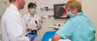
Video-EEG monitoring is important in the diagnosis and differential diagnosis of epilepsy.
How is the diagnosis of epilepsy confirmed?
The only reliable signs of the disease are epileptiform activity on the EEG and seizure patterns. As a rule, high-amplitude bursts of brain waves are recorded at the same time, but they cannot accurately indicate pathology. An EEG is necessarily prescribed to confirm the diagnosis, since epileptic discharges can be recorded outside of a seizure. In addition, the study allows you to accurately determine the form of the disease, which helps prescribe adequate treatment.
Epiactivity on the EEG in a child without seizures can be determined in various ways. Most often, rhythmic light provocation is used to stimulate epiactivity, but hyperventilation or other additional techniques can be used. Sometimes cases of epiactivity can be detected only during prolonged EEG recording, often performed during night deprivation sleep. Only a small proportion of patients cannot confirm the diagnosis with such a study.
epiactivity of the brain
about biliary dyskinesia
https://www.7gy.ru/rebenok/detskie-bolezni-simptomy-lechenie/125-diskinezija-zhelchevyvodjawih-putej-i-zhelchnogo-puzyrja-simptomy-lechenie.html
about leaky gut
https://mornaturalforums.ru/viewtopic.php?f=13&t=277&hilit;=leaky+intestines
I give trienza plant enzymes. I bought it on iherb.com.
tests for fatty acids in urine and other acids https://www.chromolab.ru/issledovaniya.html
https://www.chromolab.ru/zhirnye-i-organicheskie-kisloty-v-organizme.html
https://www.chromolab.ru/filtr-prays.htm
Physicians are increasingly recognizing the importance of the gastrointestinal tract in the development of allergic and autoimmune diseases. Leaky gut syndrome not only helps us understand why allergies and autoimmune diseases develop, but also provides an opportunity to bring the body into balance in safe and effective ways. Leaky gut syndrome usually accompanies autoimmune and allergic diseases. The main defect is the effect of increased permeability of the intestinal mucosa. This effect can be detected by microscopic examination of a fragment of the intestinal wall or by observing the bloodstream with phase contrast or dark-field microscopy. We put forward the following hypothesis. Changes in the structure of the intestinal wall lead to the absorption of large protein molecules, which are usually broken down into smaller fragments by the intestinal flora. The immune system begins to produce antibodies against large molecules. Since a healthy intestine would not allow such large proteins to appear in the bloodstream, the immune system begins to fight them. Antibodies are produced against proteins and safe foods. These antibodies can travel to various organs and cause an inflammatory response. This occurs because body tissues have antigenic sites, just like food, bacteria, parasites or fungi. Thus, autoantibodies are produced and inflammation becomes chronic. If inflammation occurs in the joints, autoimmune arthritis develops. If in the blood vessels, vasculitis occurs (inflammation of the blood vessels). If the inflammation occurs in the lungs, an asthma attack occurs every time a person consumes a food that triggers the production of autoantibodies. It is easy to see that food allergies due to leaky gut syndrome can affect any organ. Signs similar to those seen in chronic fatigue syndrome can be multiple and lead to severe exhaustion. The inflammation that causes leaky gut syndrome also damages the protective IgA coating of the intestinal wall that is present in healthy intestines. Since IgA helps us fight infections, the body becomes less resistant to viruses, bacteria, parasites and fungi. These microorganisms become capable of invading the bloodstream and colonizing almost any organ. In addition to food allergies, the body may be colonized by fungi and parasites that normally cannot penetrate the intestinal barrier. These microorganisms and their toxins, if present in large enough quantities, can impair the detoxification capacity of the liver. Leaky gut syndrome also creates a long list of mineral deficiencies because various transport proteins in the gastrointestinal tract needed to carry minerals from the intestines into the blood are damaged by the process of inflammation. For example, magnesium deficiency is common in fibromyalgia despite high dietary magnesium supplementation. If the magnesium transport protein is damaged, magnesium deficiency develops as a result of malabsorption. As a result, muscle pain and cramps may occur. Similarly, zinc deficiency due to malabsorption can lead to hair loss. Copper deficiency can lead to high blood cholesterol levels. Next, bone problems develop as a result of malabsorption of calcium, boron, silicon and manganese. Inflammation and the presence of harmful chemicals can block the absorption of vitamins and amino acids. A damaged intestine does not absorb nutrients properly. Ultimately, complaints arise - fatigue, headaches, weakening of memory and concentration, irritability.
Valeria, I would suspect parasitology, and in this case there could be problems with bile, pancreatitis, abdominal pain, colitis. You don't write anything about your son's diet. It’s interesting how palpation can show the malfunction of the gallbladder)) Present: bruxism, tossing about in bed when sleeping, circles under the eyes, drooling at night, anemia, elastase in the stool is increased, eosinophils in the blood are high or at the high limit of normal, decreased sharp appetite, or, on the contrary, gluttony, nausea, especially in the morning and after meals, itching of the anus, a capricious child? In addition to chronic dermatitis, urticaria, eczema. Take a full blood test, stool test for elastase, dysbacteriosis, coprogram, liver biochemistry: AST, ALT, GGT, alkaline phosphatase, bilirubin and its fractions, blood sugar, Igs for parasites, unfortunately, they can be false negative, but you can do the same. Mucus in the stool, most often candidiasis, candida is also a parasite, by the way. While, high-quality vitamin/min complex, digestive enzymes, lecithin, magnesium, artichoke extract -1 month, probiotics 6 months intensively, prebiotics, probiotics. Exclusion of dairy, wheat, simple carbohydrates AT ALL, strictly for 3 months. “Norm” tests don’t sound convincing to me; they still need to be interpreted professionally.
Valeria, what I write below is at the forum level; on the forum we (doctors, forum participants, independent researchers, etc.) cannot advise and recommend, as you understand. I can only give you directions, and then in conditions of a lack of complete information about the child and even about the mother. “Increase in bile”, i.e. the outflow of bile is affected, “reactive changes in the pancreas”, i.e. due to problems in the outflow of bile, inflammation of the pancreas occurs, “biliary system of the liver” - bile inflows in the liver. Lecithin, ursodioxycholic acid preparations, choleretic herbs such as artichoke, vitamin C for the pancreas, turmoric as an anti-inflammatory, digestive enzymes - plant-based, probiotics. Moreover, if you understand that the child’s diet is such that he does not get a lot of things from food, the same zinc and group B will help with appetite. And of course, check or treat, just treat with herbal antiparasitic drugs without tests, I can just understand, but pharmaceutical drugs. With medications we need to know what we are treating specifically. Mucus in the stool is candidiasis, most often. Herbal preparations for Candidiasis: Candida Complex - Complementary Prescriptions For the biofilm - a membrane made of metals, the cat protects the community of unfriendly flora. Enzymes - Interfase Plus - Klaire Lumbrikinase is good for this, excellent, but an expensive drug. A diet is required, the cat alkalizes the body (we have a whole topic on the forum) and taking into account food intolerances/sensitivities. You are talking about yogurt, what is it? Store-bought with sweets in it? ESR - high - inflammatory process in general.
CYSTIC FICIDOSIS
https://rmj.ru/articles_7705.htm
succinic acid, etc. for rejuvenation, improvement of condition, prolongation of life, etc.
https://bessmertie.ru/vk.lek11.shtml
https://mamadeti.ru/doctors/irina-titova/
https://www.medkrug.ru/question/display?syspopup_id=question_ask_premoderate
https://guest.policlinica.ru/answer.php?id=140612&success
https://doct.ru/index.php?m=mform
https://medharmony.ru/index.php?do=faq&action=addquestion&subactio=addquest
https://www.03.ru
https://www.consmed.ru/qpanel/?qid=824198 question 8241 98 (and 89)
Hello, my name is Valentina, the child is 3 years old, a couple of weeks ago I was diagnosed with MMD and ZRR, did we conduct an EEG, according to the EEG results, there are no epi signs, some waves are slow, as the doctor explained, we have very poor interaction between the right and left hemispheres, hence the ZRR and in the future there may be problems with training, i.e. He seems to understand everything, but he gets it. as they say, like a duck, very slowly... and so - a normal child, climbs everywhere, pokes his curious nose, loves children, animals, draws, cuts, with fine motor skills everything is ok. whatever you ask, he will bring it and do it (according to his mood), he understands everything, but speaks poorly, sometimes 2-3 simple words, mom to eat, mom to pee, give me moko, Misha has gone, etc.... but does not distinguish between colors and shapes , there was porridge in his head, the doctor immediately told us that there were probably problems with colors and shapes - straight from the door..... he was born at 32 weeks, weight 2260 g, breathed on his own, ate on his own, but very poorly, there were choroid plexus cysts , but we did ultrasounds 5-8 times up to 1.5 years old - they are not growing, and the doctor said that we can no longer control them. There was also retinopathy of prematurity, i.e. again problems with blood vessels in the head area, more precisely in the eyes, but everything is interconnected there.
.. The neurologist said that her son was given the same diagnosis, but she did not treat the child, because... Grandmothers took over, they didn’t take him to school the first time - he was not prepared, they said, now he seems to be studying normally, with practically no C grades, but in many subjects he has to hire a tutor..
The doctor prescribed Cortexin and Kindinorm for us for now, I read here on the net that it can provoke seizures (although there seem to be no signs on the EEG), I scared myself (and my husband) that a simple EEG is not enough to determine the possibility of epiactivity, that I should probably do it at night , but then I see that they do the nightly with Epi in order to determine the degree or type of attacks, i.e. a detailed examination, and it seems like we don’t need this...
I think I’ve already decided to inject, but my husband now says that there are no injections for now, we’ll wait for the operation... We are going to have a small operation with mask anesthesia - perhaps we should avoid injections, but I’m thinking, what if, on the contrary, it’s better for us to inject? so to speak, to “protect” the brain before anesthesia?
questions
1. Do you think a simple short EEG is enough to exclude epiactivity or signs?
2. Is Cortexin really indicated in this case? It seems that they are prescribed to almost everyone, but no particular research has been carried out, they are prescribed as guinea pigs.
3. Is it better to pierce it before or after anesthesia? (although my husband probably won’t agree
4 In our case, is it also possible to use Cogitum to improve blood circulation?
What can be seen on the EEG with different forms of epilepsy?
Each type of this pathology is characterized by its own clinical symptoms. In addition, epilepsy looks different on the EEG.
Benign rolandic epilepsy:
- During an attack (exacerbation), a focal epileptic discharge is noted in the middle temporal and central leads. It looks like high-amplitude spikes, in addition to which a combination of sharp and slow waves is observed. Exit beyond the boundaries of the original location is noted.
- In the absence of an attack, focal spikes are often recorded, combining waves that arise simultaneously in several leads. Often, epileptic activity on the EEG does not manifest itself in any way during the daytime while the person is awake. In this case, it necessarily appears as soon as the person falls asleep.
Rolandic epilepsy is the most common form of childhood epilepsy
Epilepsy form Panagiotopoulos:
- During an exacerbation, an epi-discharge is recorded - high-amplitude spikes, combined with slow and sharp waves that rarely remain within the initial localization.
- At rest, multifocal low- or high-amplitude complexes can most often be seen. It is characteristic that the complexes arise serially - at the moment of closing the eyes, and at the moment of opening they are blocked. An attack can be triggered by photostimulation.
Generalized idiopathic epilepsies
EEG patterns can be more often observed in a child and in juvenile epilepsy with absence seizures:
- During an exacerbation, the equipment can show an extensive discharge in the form of rhythmic activity in excess of 10 Hz with a steadily increasing character, as well as sharp waves and high-amplitude delta and theta waves. They are unstable and asymmetrical.
- Outside of an exacerbation, the EEG picture may remain standard, with uncharacteristic activity occasionally present.
Typical absence seizures are short, generalized epileptic seizures with sudden onset and termination.
Early infantile epileptic encephalopathy:
- An exacerbation causes an increase in the number and amplitudes of the spike in combination with sharp waves.
- Outside the exacerbation, there is extensive activity, where the outbreak is followed by its disappearance. Possible hypsarrhythmia.
Lennox-Gastaut syndrome:
- An exacerbation is characterized by the occurrence of numerous spikes and sharp waves; a “spike-slow wave” combination can be recorded. Desynchronization develops.
- Outside of exacerbation - hypersynchronous activity accompanied by sharp waves, spike-slow wave complexes, focal disorders.
Lennox-Gastaut syndrome is a severe form of epilepsy that begins in childhood
Seizure patterns can also be recorded in phenotypically healthy patients. In this case, a diagnosis of epilepsy is not made, but it is believed that such people have a genetic predisposition to this pathology. As a rule, periodic examination is recommended.
Preparation and carrying out the procedure
Preparation is carried out as follows:
- Before the EEG, you need to inform the specialist about the medications you are taking. Anticonvulsants and tranquilizers are canceled 2-3 days before the procedure, as they can affect its result.
- The day before and on the day of electroencephalography, it is prohibited to drink alcohol, drink coffee, tea, cocoa, or eat chocolate products.
Before performing the manipulation, you will need to remove metal jewelry (earrings, hair clips, piercings).- Hair must be washed, skin must be clean and intact. It is advisable to loosen dreadlocks and braids. It is better not to use hairspray and styling foam.
- You need to eat a hearty meal before the procedure.
- If electroencephalography is performed in the sleep phase, then it is advisable for the patient to remain awake for 1-1.5 days before the procedure.
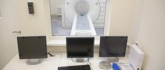
Options for conducting an EEG: during wakefulness, sleep, and also with deprivation of sleep at night. Diagnostics are usually performed during the day. The examination lasts about an hour. A person enters a dark, soundproof room. He is put on a cap with attached sensors or electrodes (about 21) are fixed on the scalp.
The patient sits on a chair or sits down on the couch. The examinee can remain alone in the room, and communication with the doctor will be carried out through a microphone and camera. The electrodes are filled with an electrically conductive substance and connected to an electroencephalograph. The received signal is reflected as wavy lines.
Interference during EEG recording
Several times at the beginning of the manipulation, the patient is asked to blink in order to monitor the nature of the changes in the electroencephalogram. The rest of the time the eyes are closed. The recorded vibrations are brain waves. They come in different frequencies and amplitudes, and differ in shape. Doctors distinguish several types of waves. Each of them is associated with the state in which a person is:
- beta waves are recorded in an awake patient;
- during rest, alpha waves are taken into account;
- delta waves are recorded in sleep,
- Theta waves occur during sleep.
To detect hidden pathologies, hyperventilation and stroboscopic stimulation are prescribed. If there is a need to leave for natural needs or to change body position, the person reports this and the diagnosis is suspended.
What is epileptiform activity based on?
In epilepsy, a periodic “explosion” of the cell’s action potential is observed as a result of a paroxysmal shift of its membrane. After this, a fairly long period of hyperpolarization begins. This mechanism is relevant for any form of pathology, regardless of what kind of epileptiform activity is recognized by sensitive equipment.
To generate epileptiform activity, a huge number of neurons must be activated. Two types of nerve cells are always involved in this process. The first are “epileptic” neurons that generate autonomous bursts. The second - their surrounding nerve cells, as a rule, are under afferent control, but occasionally enter into an active process.
EEG and sleep
Patients often ask what an EEG is and whether the procedure is performed during sleep.
Carrying out the procedure during this particular time period makes it possible to determine brain activity directly at night. Often, symptomatic manifestations of the disease in children occur during sleep. An increased value of nerve cell activity is easily determined. For comparison, this is almost impossible to do during the day.
Often a symptom of deprivation, in other words, sleep deprivation of the patient is a provocateur. The technique is often used to identify the hidden form of the disease. The procedure is carried out exclusively under the supervision of a doctor.