The essence of skiascopy
Skiascopy is a medical procedure aimed at determining the ability of the pupil to refract light.
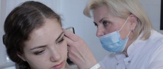
The technology dates back to the 19th century - it was then that the doctor-scientist Cunier proposed this method of refraction. The name comes from an instrument called the skiascope. It has a concave shape on one side and flat on the other. There is a hole in the middle through which the doctor observes the patient's eye. An ophthalmologist directs a beam of light into the pupil. Then, he observes the reflex - the reaction that occurs in the fundus of the eye. This method gives the most complete picture of the refractive ability of the pupil. If necessary, the patient is prescribed the necessary medications.
Contraindications
Skiascopy involves cycloplegia, or artificial paralysis of the ciliary muscle of the eye using special eye drops. They can cause temporary blurred vision, pupil dilation, and increased intraocular pressure. This imposes certain restrictions on the use of this technique to determine the type and magnitude of refraction:
- glaucoma and suspicions of it;
- intolerance to drugs from the cycloplegic group (Cyclomed, Atropine, Cyclopentolate);
- the patient is drunk, under the influence of narcotic and psychotic substances;
- inappropriate behavior and aggressiveness of the patient, mental disorders;
- fear of bright light, or photophobia, which is characterized by increased tearing.
If there are contraindications or concerns, it is possible to perform skiascopy without the use of drops that lead to paralysis of the eye muscle.
Indications for use
Often this technique is used to check refraction when others cannot be used for some reason. Basically, the following cases are indicated:
- Intellectual impairments have become noticeable, but the patient cannot explain what they are associated with;
- Due to age-related changes, a person cannot specifically describe his feelings of deteriorating visual acuity;
- The patient independently simulates refractive errors;
- Myopia, or farsightedness, as well as astigmatism appears.
Myopia means a clear distinction between objects only nearby, and with farsightedness, on the contrary, visual acuity increases with the distance between the eye and the object being examined. Astigmatism is expressed in a decrease in visual acuity at any distance from an object. Most often, this is due to age or illness.
| Skiascopy is aimed at establishing a specific diagnosis and treatment method. |
Sometimes it is carried out for preventive purposes to identify emerging eye diseases, or for progressive existing diseases.
Method Definition
Skiascopy is a method whose purpose is to check the functional state of the organs of vision in terms of their ability to refract light (refraction). Two structures are responsible for the refraction of the eye - the lens and the cornea. Using this procedure, the degree of impairment is determined even when a person is simulating a disease. This method will also be useful for studying refraction in a child or mentally retarded patient. In these cases, it is impossible to perform conventional visometry or refractometry. Synonymous names for the method are retinoscopy and keratoscopy.
Skiascopy is a common technique that has such advantages as:
- Accuracy of results. And this is very important for identifying the cause of an eye disease and making a diagnosis by a doctor.
- Painless. The examinee does not experience any painful sensations during the examination, which is very important in cases of examination of a child.
- Simplicity. This procedure does not require complex medical equipment. Everything is quite simple, and the accuracy of the results depends only on the professionalism of the ophthalmologist. The doctor observes the patient’s reactions and draws certain conclusions. The patient simply sits on a chair and follows the doctor’s instructions.
- Economic benefit for patients. This procedure is inexpensive even in private clinics, since it does not require the use of expensive equipment.
Skiascopy is the most accurate and objective way to study the refraction of the eye, the results of which are especially important in professional selection or determining a person’s ability to work.
Who should not undergo skiascopy?
Who should not undergo skiascopy:
- The procedure is prohibited for people who already have a list of ophthalmological or other ailments. Among them:
- Glaucoma – existing or suspected of its development;
- Prohibited for photophobia;
- The examination cannot be carried out while under the influence of drugs or alcohol;
- Children under 7 years of age;
- If you have diseases associated with mental disorders, in which an aggressive reaction to light exposure may occur.
In the absence of contraindications, skiascopy is performed at any age.
| It is the most gentle compared to other types of examinations. |
Most contraindications are related to the body's reaction to light radiation. If you have doubts about the presence of certain contraindications, it is better to consult a specialist in advance.
Return to contents
What is a shadow test and why is it done?
Skiascopy allows you to check the condition of a person’s eyes and determine the most distant point of clear vision. The essence of the method lies in determining the clinical refraction of the eye through the directional illumination of the pupil. Refraction is the ability to refract light rays by the optical structures of the organ of vision.
Synonyms for skiascopy are retinoscopy and keratoscopy.
The optical system includes the cornea, the fluid-filled anterior chamber, the lens, and the jelly-like contents of the vitreous. Having passed through all these areas, the light hits the retina, which is capable of converting light particles into impulses that enter the brain, where the image is formed. The units for measuring visual acuity are diopters.
Clinical refraction is the location of the main focus, that is, the point at which light rays intersect, in relation to the retina. If this back focus is located on the retina, then the vision is one hundred percent, that is, absolutely normal - emmetropia. If the focus position changes, visual acuity is impaired. So, with farsightedness, the location of the intersection point is behind the retina of the eye, and with myopia, in front of it.
Skiascopy determines clinical refraction, which is the location of the intersection point of refracted light rays in relation to the retina
Skiascopy allows you to objectively assess the degree of refractive error in almost any person, including the smallest children. This is especially important if it is not possible to determine vision through visometry (using tables) or perform refractometry (assess visual acuity using special equipment).
Skiascopy can be performed under conditions of cycloplegia (artificial shutdown of the muscle responsible for accommodation with the help of medications) or active accommodation (the adaptive ability of the eye to focus in order to see objects located far or close equally clearly).
The study is indicated for various visual acuity disorders:
- farsightedness, when a person has trouble seeing nearby objects,
- myopia, in which the patient sees well near, but distant objects are blurred for him,
- astigmatism is a pathology in which several foci are present at once, while different types of refraction (+ or -) can be combined in one eye.
The shadow test is a valuable diagnostic method for examining children in whom it is not yet possible to perform refractometry using a device and carry out diagnostics using ophthalmological tables. The method is used to make a diagnosis, to assess the effectiveness of therapy and at the stage of clinical observation.
Hardware refractometry is carried out using devices that cannot be used on very young children.
Contraindications to the procedure are:
- intolerance to cycloplegics - drugs used for temporary paralysis of the ciliary (ciliary) muscle responsible for accommodation,
- glaucoma is a progressive disease that occurs with increased intraocular pressure and leads to blindness,
- photophobia - fear of bright light, manifested by increased tearfulness,
- mental disorders with inappropriate behavior of the patient,
- state of intoxication (alcohol or drugs).
Currently, refraction studies are carried out not only by means of a shadow test, but also with the help of computer devices - refractometers. Both of these methods are objective, reliable and easily accessible for assessing the refractive power of the optical system of the eyes.
The advantage of skiascopy is that it can be performed on the smallest patients who cannot be seated in front of the device, and the advantage of automatic refractometry is a more accurate determination of the degree of astigmatism in a person. The advantages of refractometry include its faster implementation compared to skiascopy, as well as the possibility of performing visometry immediately after the procedure due to the absence of the blinding effect that skiascope has on the eyes when performing retinoscopy.
Carrying out a shadow test requires certain professional skills from the ophthalmologist, and the data obtained during this manipulation may have minimal errors, as with an examination using a device.
Research methodology
Before the test begins, the patient receives a dose of atropine or cyclodol. This drug reduces the movement ability of the eye. This is necessary in order to eliminate errors in the procedure. Gradually, the effect of this drug decreases and the eyeball can actively move, as before.
Another contraindication is the presence of an allergy to this drug. Therefore, a check is being carried out. When arriving for the procedure, the patient is placed in a dark room and a special device is placed at eye level.
The ophthalmologist is located about a meter from the subject of examination, from his position he directs the light, controlling the beam. The doctor ensures that it reaches the fundus of the eye through the pupil. There is a mirror near the person being examined. The doctor turns it, getting shadows on the fundus from the light. They are the ones who provide the examination results and diagnosis.
Next, the results obtained are deciphered. It is possible to prescribe the use of atropine or cyclodol 3-4 days before visiting an ophthalmologist. Sometimes special rulers or lenses are used. Skiascopy with a “wide pupil” is used for the most accurate determination of refraction.
How the degree of hypermetropia or myopia is determined based on the results
To establish an accurate result in refractive errors, the specialist uses the method of neutralizing the fallen shadow. Skiascopic rulers (or lenses used to select corrective glasses) help him with this.
Results taking into account the use of skiascopic rulers:
- The absence of a shadow when a ray of light hits the pupil indicates myopia, which is equal to no more than 1.0 diopter.
- In case of myopia with a large deviation, the shadow moves. Then apply a ruler with negative lenses. First, the weakest lens is applied to the eye organ, gradually increasing it. The procedure stops at the lens on which the shadow disappears. Next, 1 diopter is added to its optical power and the degree of deviation for myopia is obtained.
- To determine the degree of hypermetropia, an identical procedure is performed. The difference lies in the type of lenses used (positive lenses are used for this diagnosis), as well as in the calculation of refraction. In this case, you need to subtract 1 diopter from the readings of the optical lens.
The results of skiascopy will indicate the exact degree of refractive error of the eye at any stage of development of the pathology. Skiascopy is painless and takes up to 10 minutes. This is an excellent option for those who care about their health and want to know about the slightest changes in the structure of the eye organ.
Sources used:
- Angelova, Diana Ultrasound in ophthalmology / Diana Angelova. - M.: Palmarium Academic Publishing, 2012.
- Arkhangelsky, V.N. Morphological foundations of ophthalmoscopic diagnostics / V.N. Arkhangelsk. — M.: State Publishing House of Medical Literature, 2020.
- Baltin, M. M. X-ray diagnostics and radiotherapy in ophthalmology: monograph. / MM. Baltin. - M.: State Publishing House of Medical Literature, 1987.
- Ophthalmological Clinic of the State Budgetary Educational Institution of Higher Professional Education of the North-Western State Medical University named after. I.I. Mechnikov of the Ministry of Health of Russia
Advantages of the technique
| This technique does not take much time and has few contraindications. In one procedure, you can get the most accurate examination result and referral for treatment in connection with the diagnosis. |
Another advantage is the proven method and its reliability. The practice of skiascopy began a long time ago; now the instruments used have been brought to perfection. It is also ideal for children, even babies. Skiascopy is also ideal for children, even infants.
Who is the procedure indicated for?
Skiascopy is an eye examination procedure, so it is intended for those people who have vision problems. Skiascopy is indicated for the following ophthalmological diseases:
- farsightedness;
- myopia;
- astigmatism.
Skiascopy is also indicated in cases where it is necessary to determine the effectiveness of treatment, as well as to determine the rate of progression of eye diseases. This method has many advantages, but despite this, there are also a number of contraindications.
Decoding the research results
Determining the type of refraction
Emmetropia occurs when a dark spot on the bottom of the eyeball moves in the same direction where the doctor moves the mirror. This indicates good normal vision, or unnoticeable mild impairments due to the diagnosis of myopia or farsightedness. If the shadow moves to the other side or there is no movement at all, this indicates myopia above 1 diopter.
| Erratic movement indicates a severe degree of astigmatism, that is, poor vision, unclear vision of objects. This phenomenon is otherwise called scissor syndrome. |
Determination of the degree of myopia and hypermetropia
This is the second stage of skiascopy. Here, 1 diopter is subtracted from the lens for farsightedness and vice versa, 1 is added for myopia. That is, lenses are individually selected depending on the tendency to myopia or hypermetropia. They should be a little more than a centimeter away from the eye - 12 mm. The result obtained usually lies in the range from 0.25 to 0.5 diopters. A complete picture is created after turning off the devices and completing the procedure.
Determination of refraction for astigmatism
Astigmatism is fundamentally different from myopia and hypermyopia. It is characterized by very poor visual acuity. If, for example, with myopia or farsightedness a person sees clearly at a certain distance, then with astigmatism any objects at any distance are poorly distinguishable and “smeared out”. Therefore, for this category of disease, a slightly different procedure is performed - cylindroskiascopy, that is, cylindrical lenses.
Also, this process is accompanied by other techniques and tools. Sometimes there is a need to study peripheral vision, in which case computer eye perimetry is used.
Return to contents
How is retinoscopy performed?
Preparation for the procedure involves performing cycloplegia. In order to temporarily disable the ciliary muscle, a solution of atropine is instilled into both eyes in a certain age-specific dosage twice over three days and in the morning of the fourth day. The shadow test can be started an hour after the last instillation. If the results are controversial, atropinization is extended to 7 or 10 days. Standard three-day cycloplegia is performed before the first skiascopy in children, as well as in adults in complex cases. The use of atropine has a certain disadvantage - after instillation, the patient experiences difficulties for a long time in visual work at a short distance, for example, reading.
Before skiascopy, cycloplegia is performed - drugs are instilled into the eyes, causing temporary paralysis of the ciliary muscle responsible for accommodation.
Recently, to relax accommodation, ophthalmologists have been using mild and short-acting drugs - solutions of scopolamine, homatropine, cycloborine, amizil or ready-made drugs - Tropicam, Midriacil, Cyclozhel. They are instilled 1 drop at an interval of 10 minutes and a shadow test is carried out after 45 minutes. Ophthalmologists use such drugs for repeated retinoscopy procedures in children and when it is necessary to disable accommodation in adults. For patients over 40 years of age, drugs for cycloplegia are used after mandatory measurement of eye pressure and only in situations where it is impossible to do without them. This is due to the fact that such medications can trigger an attack in people predisposed to glaucoma.
Classic cycloplegia involves instilling an atropine solution into the eyes.
Cycloplegia is necessary for a full examination of the patient - the pupil dilates, and the doctor has the opportunity to see not only the central area of the fundus, but also peripheral areas.
The shadow test is carried out in a darkened room. The subject is seated on a chair, on the side of which a light source is placed - at the level of the patient's ear. Most often this is a regular incandescent lamp. The light should not fall on the face of the person undergoing skiascopy. The ophthalmologist sits opposite, maintaining a distance of 67 cm or 1 meter. To carry out the procedure, you need a skiascope - a device that is a round mirror, concave on one side and flat on the other, with a hole in the middle and a handle. The doctor takes the device in his hand and directs a beam of light reflected from the lamp into the eye of the person being examined so that it hits the fundus of the eye through the pupil.
Skiascopy is performed using a skiascope - a mirror with a hole in the middle
If cycloplegia has been previously performed, the patient is instructed to look into the center of the skiascope, and, with accommodation preserved, past the ophthalmologist’s ear on the side of the eye being examined.
Then the doctor begins to slowly move the device around the vertical and horizontal axis of the handle, while the area of illumination of the fundus of the eye shifts, forming a shadow (dark spot). Usually, the flat mirror side of the skiascope is used for examination, since in this case the spot is clearer and more pronounced, and its movement is easier to assess. Based on the direction in which the darkening area moves, the ophthalmologist draws a conclusion about the nature of the patient’s refraction.
When performing skiascopy, the doctor can be at a distance of 1 meter or 67 cm from the patient
After determining the type of visual impairment, the doctor makes more accurate measurements of the refractive power of the optical structure of the eyes, for which he uses a device - skiascopic rulers. They are frames between which lenses of different optical powers are fixed; on each instrument there are only negative or only positive glasses.
A method is used to neutralize the movement of the dark spot. A ruler with the required lenses is given to the examinee’s hand, and it should be positioned vertically no closer than 12 mm from the cornea of the eye. The doctor directs the beam into the pupil through the lenses, starting with the smallest diopter (0.5) and gradually, moving towards the strongest glasses, determines the one at which the dark spot disappears. Neutralization of the shadow occurs when the eye is in the very center of the focus of the rays reflected from the fundus of the eye.
After determining the type of refraction, the ophthalmologist measures the degree of myopia or hypermetropia using skiascopic rulers
Instead of skiascopic rulers, lenses with different optical powers are sometimes used, which are inserted into a special frame. This technique requires time, but it has advantages - greater accuracy compared to rulers and the ability to diagnose astigmatism using cylindrical lenses (cylindroskiascopy). Before this study, the doctor may use strip or bar-skiascopy. In this case, special attachments are used for the skiascope, which do not have a hole, but a strip-shaped slot.
Video: carrying out the procedure
Features of skiascopy in children
The first ophthalmological examination is carried out no later than 3 months from the date of birth, then at the age of 6 and 12 months. The examination process is complicated by the fact that in infants it is very difficult to reduce the mobility of the eyeball, even with the help of drugs. Often, instead of a skiascope, special rulers are used. The accuracy of this procedure is necessary in order to identify the occurrence of diseases as early as possible and prevent them as soon as possible. At an early age, there are correction methods that help bring children’s vision back to normal.
| By the way, most babies are farsighted. |
In the first months of life, their vision is better at distinguishing distant objects than those that are close. The ability to recognize and recognize objects/people at a distance of less than 1 meter is formed after 3 months. Gradually the refraction is normalized. At the age of more than 5 years, the ophthalmologist checks visual acuity using a familiar table.
Light sensitivity develops the functioning of the visual system, which means that there can be no contraindications for skiascopy in children. They have not yet developed myopia and hypermetropia, astigmatism and mental illness, as well as light intolerance.
In what cases is skiascopy indicated and are there any contraindications to the procedure?
There are a number of indications for which skiascopy may be prescribed:
- Myopia (myopia) - when the patient distinguishes objects close to him relatively well, but at the same time does not clearly see the outlines of objects located in the distance;
- Farsightedness - when a person sees poorly near, but distinguishes objects well at a distance;
- Astigmatism is a special pathology that combines the presence of several foci at once (sometimes different types of refractive error are combined in the eye).
However, despite all the harmlessness and simplicity of the “shadow test”, this method has contraindications, such as:
- Glaucoma - in this case, the procedure can provoke an attack;
- Photophobia, or special sensitivity to light;
- State of alcohol or drug intoxication;
- Acute mental illnesses in which the patient can harm others or himself.
