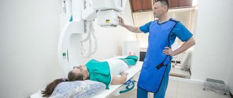
Magnetic resonance imaging (MRI), a method of studying the condition of the internal organs of the human body using strong magnetic fields and radio waves. The popularity of the procedure is explained by its safety for the patient and the high informative value of the results obtained. As diagnostic technology develops, so does the equipment used in it. For this reason, it is not easy for an untrained person to immediately understand which MRI device is better, the result of which type of study will be more informative.
Choosing the right equipment
In order not to make a mistake and choose a high-quality model, it is important to take into account the main technical parameters:
- Power. This indicator is measured not in the usual Watts, but in Teslas. There are four popular varieties: low-field, mid-field, high-field, and the most expensive and advanced devices - ultra-high-field. The latter are present only in expensive clinics, since their cost starts from 30,000,000 rubles.
- The scanning time completely depends on the power of the device; accordingly, the higher it is, the faster the examination of internal organs takes place.
- The last thing to consider is the type of MRI. Today, only two popular varieties are sold - open and closed models.
Types of MRI by area of interest
Open type MRI machine.
Types of MRI studies differ depending on what area of the body they are aimed at studying. Modern diagnostics uses a whole range of studies:
- Head and brain.
- The spine and all its sections.
- Joints (knee, shoulder, hip and others).
- Abdomen.
- Paranasal sinuses.
- Kidneys.
- Liver.
- Small pelvis.
- Mammary gland.
- Lungs.
- Heart and other organs.
The images obtained as a result of the scan show tumors, inflammation, developmental anomalies, injuries, cysts and other pathologies of the organs being examined.
More about power
Before purchasing the most powerful or low-floor option, you should learn more about the advantages and disadvantages of each type. This will help you find a suitable tomograph and not overpay.
Low-field and mid-field devices
This technique has become more widespread in Russia and some CIS countries. The power of such equipment rarely exceeds 1 T; models with an indicator of 0.35-0.5 T are more common. The main advantage of the models is the cost-effectiveness of consumable materials. But many users also highlight the user-friendly interface and affordable cost.
But the shortcomings here are significant, since the quality of the survey results obtained is low. Therefore, it is impossible to talk about 100% reliability.
Most often, low-profile devices are used to detect cardiac pathologies, and also thanks to them, disorders of the brain are detected. There is no special functionality in such options, which is why it will become useless if complex diseases develop.
High-floor models
This equipment is much better and more expensive than previous copies. Here the power is 1.5 Tesla. There is high-quality cooling, which is made in the form of a cryogenic gel composition. This invention is distinguished by its quality and reliability. Why is it used not only in Russia, but also in many European countries.
Using a high-field device, the doctor will be able to conduct a reliable and complete diagnosis of the human body. Every internal organ and tissue is exposed to a magnetic field.
High-field MRI is used to identify benign or malignant tumors and is also used to identify aneurysms, which has a positive effect on functionality. In addition, with the help of this equipment, comprehensive diagnostics are carried out.
The limit of perfection – ultra-high-field MRI
This category includes all devices that have a power rating exceeding 1.5 Tesla, but not more than 7 Tesla. The cost of such a unit is very high and it is used only for research purposes. The main advantage of this product is high accuracy.
Ultra-high-field MRI is rarely used to identify brain problems, only in extreme cases. The equipment most often used is spectroscopy, MRA of the cerebral vasculature, etc. In addition, the device is excellent for studying the microstructures of the human brain.
What types of MRI are there?
Magnetic resonance imaging is classified based on the area being studied, the use of additional imaging tools (a contrast agent based on the rare earth metal gadolinium), and the type of device.
MRI of the head
The scan is not very informative regarding bone structures and well reflects the condition of soft tissue elements. The high level of detail in the photo allows you to study any part of the anatomical zone down to the smallest detail. Magnetic resonance imaging can be used to examine organs surrounded by bones.
When performing an MRI of the head, information is obtained about:
- brain structures (pituitary gland, hypothalamus, etc.);
- cerebral vessels;
- paranasal sinuses;
- elements of visual, hearing aids, etc.
MRI of the head
Magnetic resonance scanning of the head is used in neurology, neurosurgery, otolaryngology, rheumatology, dentistry and other branches of medicine. Using the method, congenital anomalies, benign and malignant neoplasms, inflammatory processes, purulent changes, consequences of hemorrhages, and demyelinating diseases are identified.
Vascular MRI
In an MRI study, a magnetic field changes the direction of movement of hydrogen atoms in water molecules. Under the influence of radio frequencies, the element returns to its normal state with the release of energy. The tomograph captures the impulse and converts it into a picture. Due to the high fluid content, the method is most informative regarding soft tissues. Magnetic resonance imaging is used to study blood vessels in any part of the body.
To obtain more detailed images, a drug based on chelated gadolinium salts is injected into the patient’s body. The substance increases the vibration level of hydrogen atoms and the outgoing momentum, which increases the contrast of images.
MRI angiography allows you to detect:
- violation of the integrity of the vascular walls;
- pathological expansion or narrowing of veins and arteries;
- blood clots;
- wall delamination;
- vasculitis (inflammation of blood vessels);
- occlusion (obstruction), etc.
The diagnostic procedure helps to detect circulatory disorders that cause pathological changes in tissues.
MRI of joints
Using magnetic resonance imaging, the structure of the joints is studied. The method is more informative than ultrasound and x-rays. The study shows the details of the structure of soft tissue articular elements and helps to establish the causes of impaired motor activity.
Reasons for scheduling a scan:
- stiffness, hypermobility of joints;
- pain;
- tissue swelling in the joint area;
- crepitus (crunching, clicking), etc.
The study is carried out if damage to joint elements (bursa, cartilage, articulating surfaces of bones) is suspected.
MRI of the spine
Magnetic resonance imaging involves examining the entire structure or specific sections (cervical, thoracic, lumbosacral). The spine is a complex organ consisting of bones, cartilage (discs), spinal cord, blood vessels, and nerve trunks. Visualization of the listed elements allows us to identify:
- congenital developmental anomalies;
- consequences of injuries;
- malignant and benign tumors and metastases;
- vascular diseases;
- damage to nerve fibers.
MRI of the cervical spine
Changes in the structures of the spine are accompanied by disturbances in motor activity, dysfunction of internal organs, and neurological symptoms. Based on the results of magnetic resonance imaging, the doctor will determine the cause of the deviations and select an effective way to eliminate the problem.
MRI of the abdominal cavity
MRI examination provides information about the size, location, and structure of internal structures. An abdominal scan provides visualization of:
- liver;
- gallbladder;
- stomach;
- lymph nodes;
- pancreas;
- kidney;
- adrenal glands, etc.
The method is actively used in oncology. In cross-sectional images, small pathological changes can be detected, which is important for the early diagnosis of cancer.
Soft tissue MRI
MRI scanning of soft tissue elements of any anatomical zone is prescribed after injuries for a detailed examination of the condition of ligaments, tendons, and muscles. Based on the scan results, foreign objects, purulent and necrotic changes, and space-occupying formations can be detected.
Optimal power for brain examination
To get a high-quality and reliable photo, there must be high tension. Almost all high-field models meet this requirement, so thanks to them, getting a high-quality photo will not be difficult. The doctor will be able to distinguish even the smallest structures.
For powerful equipment, the cut thickness is greater than or equal to 1 mm. This allows for more accurate diagnosis at an early stage of organ damage.
When using a low-floor device, you cannot expect an accurate result on the first try. Often it is necessary to conduct the examination again. Only then will it be possible to talk about the accuracy of the results obtained. This is due to the fact that the device displays a picture of low quality.
What will a tomography of the musculoskeletal system show?
MRI of the musculoskeletal system helps diagnose:
- thoracic spine;
- lumbosacral region;
- cervical spine;
- coccyx.
This type of magnetic resonance imaging will reveal changes in the musculoskeletal system, establish spinal canal stenosis, areas of disc displacement, determine infectious and inflammatory processes, tumors, neoplasms, metastasis and spread to neighboring organs.
Most often, such studies are prescribed for those who have suffered a serious spinal injury, with constant cramping back pain, if other forms of diagnosis do not show specific causes.
Features of open and closed type tomographs
Another important criterion for choosing equipment is the type of MRI. Today there are two popular options, one closed and the other open. The products differ not only in their external design, but also in their technical characteristics and additional capabilities.
When using a closed-type MRI scanner, the magnetic field induction does not exceed 3 Tesla, while the minimum value is 1.5 Tesla. In open models, the parameter is 0.5-1 T.
Another feature of the closed version is the presence of a retractable reel, which easily extends and enters the tunnel. This is not provided for in open models, since all the magnetic rays are collected around the working surface: above the couch, under which there are special magnets.
When using closed-type equipment, it is easier to obtain a reliable result, since the magnetic field will be everywhere. And in open products, where the working area is located above and below, the resulting field is not very powerful, which as a result affects the image quality.
Types of tomographs
The power of MRI devices is indicated in T (tesla). This affects the duration of the scan. On more powerful devices, the examination is carried out faster. Tomographs are used to diagnose the entire body or its individual parts (for example, head, pelvic area, etc.).
The devices can be closed, in the center of which there is a large elongated ring into which the patient is placed. The tunnel only has openings at both ends. Open tomographs do not have side walls, only a couch and a hinged part with built-in instruments. MRI machines can be:
- Permanent with stable magnets. The field is formed between the poles. This technique does not require a cooling system and consumes little electricity. However, the magnetic field is inhomogeneous and its power does not exceed 0.3 Tesla.
- Resistive when current is passed through the coil. As a result, a field of about 0.6 Tesla is formed. This technique consumes a lot of electricity and is equipped with a strong cooling system.
- Superconducting, in which the field is generated by current in a wire made of a special material. At the same time, the power of the tomograph is over 0.5 Tesla.
- Hybrid, where the field is formed using special coils and magnetized material.
All MRI devices are divided according to the magnetic field power into low- and high-field. In the first case, these are mainly open-type devices, in the second - closed ones. Depending on the power (in T), tomographs are divided into:
- ultra-low (less than 0.1);
- low-floor (0.1-0.4);
- mid-field (approximately 0.5);
- high-field (1-2);
- ultra-high-field (more than 2).
Basically, manufacturing companies produce devices with medium power. Ultra-high ones are used mainly in special laboratories. Tomographs can withstand human weight in the range of 80-200 kg. Some closed devices cannot accommodate people over 150 kg. In this case, open tomographs are used. The best manufacturing companies include Philips, Siemens and Samsung.
Advantages and disadvantages of MRI
Before making a purchase, you should learn in more detail the key features and disadvantages of closed and open type devices. This will help you quickly find a profitable and reliable product.
The main advantages of a closed type device include:
- High power, which makes it possible to carry out any research, even the most complex;
- The quality of the images allows you to establish an accurate diagnosis and choose the right course of treatment.
Disadvantages of closed type models
- High noise level, which cannot be avoided, as it is caused by the operation of the device itself. To prevent the patient from experiencing discomfort, the doctor must give him special headphones.
- Medical personnel cannot fully control the position and condition of a person, since the doctor will be present in the next room. If there are any difficulties, there is a chance that he will not hear the patient.
- In closed devices it is impossible to conduct a full diagnosis for people who are afraid of closed spaces. But if there is no other option, some doctors prescribe special sedatives.
- If a person has an injury to the head, neck, arm, leg, etc., it will be difficult to conduct an examination.
- The closed-type design is not intended for small children; it is suitable only for adults.
- It is impossible to conduct a good and complete examination of people who are obese.
In contrast to this model, the open-type design is simpler and is suitable for most simple diagnostic purposes. If complex diseases or cancer are detected at an early stage, there is a chance that MRI will not be effective.
Types of tomograph
In modern clinics, two types of tomographs are used. We are talking about an open and closed device. The first is necessary if there is a need to examine patients with large weight, height, and small children. The closed one has a tunnel design. The patient must lie on the couch and the tomograph will move inward with him. However, during the examination, many patients may experience an attack of claustrophobia, which is why clinics also install an open apparatus.
All described types of MRI are an excellent method for diagnosing various pathologies. Tomography is used in orthopedic practice, oncology, gynecology, and so on. What type of diagnostics to prescribe for a patient will be decided only by a specialist, of course, coordinating all issues with the patient.
Rating of the best budget models
MAGNETOM SYMPHONY 1.5T SIEMENS
One of the best devices in terms of cost and clinical capabilities. With this device, more than 2,700 scans can be performed. The device is focused on in-depth cardiological studies. MRI has a wide range of clinical software packages.
Sufficient patient comfort is ensured thanks to the anterior-frontal approach; this solution helps reduce claustrophobia. The controls are simple and understandable for every user, there are no complicated actions. This simplifies many routine operations and increases throughput.
The average cost is 10,500,000 rubles.
MAGNETOM SYMPHONY 1.5T SIEMENS
Advantages:
- Convenient table for positioning the patient;
- Good console with keyboard;
- More than 2700 scanners;
- Suitable for neuroresearch;
- Functionality;
- Price.
Flaws:
- Not found.
Philips Brilliance CT Big Bore
A high-quality tomograph that is suitable for examining various patients. There is CT motion control in 4D mode. Stability is ensured during scanning, the field of view is maintained at 60 cm. There is a wide aperture, which reduces the time it takes to prepare the patient for the procedure.
The device also features noise reduction, so any implants will not affect image quality. Three modes of information collection make it possible to more accurately assess the movement of organs and targets. The platform is made with high quality and provides confidence, as well as constant operation even with active use.
The average cost is 19,000,000 rubles.
Philips Brilliance CT Big Bore
Advantages:
- Scanning stability;
- Clear interface;
- Supports O-MAR technology, which reduces the level of artifacts;
- Three modes of information collection;
- Patient marking from the console.
Flaws:
- Not found.
HITACHI OASIS 1.2T
An open type device that is suitable for some clinics. The maximum magnet voltage is 1.2 T, which is the optimal solution for high SNR. High-quality design ensures patient comfort and reduces anxiety for people with claustrophobia.
The device can be used to solve basic clinical problems, which makes it universal. Good image quality affects the accuracy of disease detection. Full panoramic visibility is made possible by the two-post asymmetrical design. The tabletop can withstand loads of up to 300 kg.
Sold at a price: from 11,540,000 rubles.
HITACHI OASIS 1.2T
Advantages:
- Comfort for the patient;
- Image quality;
- Maximum tension;
- Multi-coil connector;
- Functionality;
- Price.
Flaws:
- Not detected.
Top effective models
EVIDENCE 0.35 DIXION
A high-quality device, which is equipped with 6 radio frequency coils, ensuring good information transmission. The design includes an air conditioning system, which allows you to automatically maintain a normal temperature. The software package quickly processes and analyzes the image.
The open type reduces noise levels and makes it possible to examine a variety of patients. There is a "Do Not Enter" signal, as well as a hand-held metal detector. The rate of increase of the gradients is 55 mT per second. The slice thickness in 3D mode varies from 0.5 to 5 mm. The image acquisition matrix is 1024x512.
The cost is specified when ordering.
EVIDENCE 0.35 DIXION
Advantages:
- Equipment;
- Maximum field of view – 40 cm;
- Convenient console;
- 6 RF coils;
- Premises protection;
- Good software.
Flaws:
- Not detected.
Toshiba Excelart Vantage Atlas-Z 1.5T
Reliable modern tomograph with a power of 1.5 Tesla. The manufacturer has added its own unique technologies to the MRI, making it easier to use the device. The rate of change in intensity is 130 mT/m/m. This makes it possible to examine the patient in a short time, while obtaining a high-quality and reliable image.
The parallel information collection technology allows for accurate diagnosis in a short period of time. Maximum patient comfort is achieved by a short open gantry. In addition, there is an additional option that reduces the level of extraneous noise.
Sold at a price: from 31,050,000 rubles.
Toshiba Excelart Vantage Atlas-Z 1.5T
Advantages:
- Emergency call function;
- Over a dozen applications;
- Convenient patient monitoring system;
- High-quality oxygen monitor;
- SAR calculation;
- Safety.
Flaws:
- Not detected.
BRIVO MR355 1.5T GE
Powerful device with 1.5 Tesla magnet. The equipment is equipped with the necessary functions and capabilities, a safety unit and a cabinet for storing phantoms are also provided. The product is highly accurate. The cooling system helps create a favorable climate.
No electrical interference will cause any problems as there is a high quality UPS present. A 14-element coil is used to examine the head and neck, providing accurate and reliable results.
The average cost is 29,500,000 rubles.
BRIVO MR355 1.5T GE
Advantages:
- Convenient use;
- High quality construction;
- Good built-in UPS;
- Set of phantoms;
- Efficient cooling system.
Flaws:
- Not detected.
Which machine is better for doing MRI?
Conducting MRI of the heart, spine and other organs requires high-tech equipment, as well as new tomograph software and extensive employee experience. Therefore, before carrying out all procedures, it is necessary to find out which device is best for diagnostics.
The most important factor to consider when choosing the most suitable MRI machine is its power. After all, the quality and reliability of the resulting images depends precisely on the power of the magnet. Image clarity is the most important point for the doctor when making a diagnosis. Thus, a high-voltage closed-type magnetic resonance device is better than a low-field open device.
Modern MRI machines are equipped with high-power magnets and user-friendly software. Thanks to this, the doctor has more opportunities to make an accurate diagnosis. The devices installed in the diagnostic center are much quieter than their predecessors. Thanks to this, comfort during the research process increases significantly.
High-field MRI machine
The diagnostic center has a 1.5 Tesla high-field tomograph. One and a half tesla devices provide a wider study area with a minimal difference in reliability (from three tesla systems). Therefore, specialists can diagnose various organs. A tomograph operating with a magnetic field strength of 1.5 Tesla is quite informative equipment that is used to obtain highly accurate data for making a correct diagnosis. The latest model equipment allows you to obtain all the necessary information in a short time.
Mid-field and low-field devices
The main difference between tomographs lies in the technical part, namely, in the power of the electromagnetic field, which is created artificially. Thus, the following types of MRI scanners can be distinguished:
- mid-field;
- low-floor.
The magnetic field power of mid-field devices is 1-1.2 Tesla. Low-floor devices have a lower power rating (it is 0.2-0.35 Tesla). Such devices are capable of demonstrating the general condition of the brain without detailed studies. This is very convenient if the doctor does not suspect the presence of serious pathologies.
Get a free consultation Consultation on the service does not oblige you to anything
Channel architecture of tomograph spectrometer
The 32-channel tomograph has higher power. Such equipment is equipped with particularly sensitive coils. 32-channel architecture (expandable to 128) enhances diagnostic capabilities. An 8-channel tomograph has less power. To obtain more accurate and reliable results, the diagnostic center uses a 32-channel tomograph. The highest image quality opens up wide opportunities for studying various body functions, as well as identifying pathology in a short period of time. Rich in technological and functional characteristics, the device provides the best image quality. You can undergo an MRI in Moscow at any convenient time.
No special preparation is required before conducting this study. The only requirement may be to limit the intake of certain foods. For example, your doctor may recommend eliminating foods that cause gas from your diet. Before the scanning procedure, the patient must remove jewelry, watches, as well as removable dentures and hearing aids. Phone and other electronic devices are also left with metal objects. During the procedure, the patient must lie still. In this case, he will not experience any discomfort.
You can undergo a high-quality MRI in our diagnostic center. Specialists will advise the patient on all questions that arise. The procedure will be carried out at any time convenient for the client. The extensive experience of our staff, the availability of modern equipment, as well as powerful software allows doctors to obtain reliable, most accurate research results.
Make an appointment Make an appointment and get a quality examination in our center
Popular MRIs with wide functionality
MPF 3000 AMICO
The main advantage of the device is its efficiency, low costs and dimensions. MRI is an open-type device, which allows examination of various groups. A distinctive feature of the tomograph is the open two-post magnet design. This solution makes it possible to gain access to the patient’s body from different sides.
The examination table is convenient and easy to control. In addition, it is equipped with a music center and a microphone, which makes it possible to talk with the patient during the examination. The projection can be displayed in two modes: 2D and 3D.
The cost is specified when ordering.
MPF 3000 AMICO
Advantages:
- Good visualization;
- High quality RF system;
- Convenient examination table;
- Power;
- Design.
Flaws:
- Not detected.
Canon (Toshiba) Vantage Galan 3T
Convenient and safe equipment, which is designed to obtain high-quality images, while the noise level is minimal, so there is no need to issue special headphones. All photographs are high quality and reliable. In addition, this MRI can be used not only with adults, but also with children.
To reduce the level of artifacts, a special Quick Star technology is used. The product is made in Japan. The brain examination is carried out in 5 minutes. After registering the patient, the equipment automatically selects the scanning protocol.
Price on request.
Canon (Toshiba) Vantage Galan 3T
Advantages:
- High-quality image;
- Reliability and safety;
- High performance;
- Research in 5 minutes;
- Automation of routine actions.
Flaws:
- Not detected.
What will vascular tomography show?
Perhaps the most scrupulous study is the circulatory system and blood vessels. Here they highlight:
- general vascular MRI;
- vascular system of the brain;
- vessels of the neck.
These studies are very important when the patient’s condition worsens, against the background of general health, after injuries, falls, or operations. MRI is required when planning neurosurgical interventions and during monitoring after operations. Tomography is an improved version of Dopplerography, which has a wider range of capabilities.
