During the development of osteomyelitis, an X-ray examination is performed in order to establish a final diagnosis, as well as clarify the extent of the pathological process and monitor the dynamics of the disease.
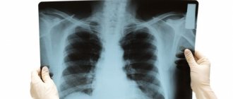
Osteomyelitis on x-ray
In an image taken in the initial stages of the disease, it is impossible to localize intermuscular and fascial septa, which are normally visible in healthy people. During the pathological process, the line between the muscle structure and subcutaneous tissue disappears, and the saturation and volume of soft tissue structures also increases.
X-ray examination in the initial stages
The first signs of pathology in the bone structure in hematogenous osteomyelitis can be detected on x-rays already on the sixth day of the disease.
In order to diagnose osteomyelitis, multi-projection x-ray is usually used. If the osteoporosis at the site of osteomyelitis is massive and has reached the second or third stage, x-rays with hard radiation are used.
The choice of X-ray rigidity is determined by the course, phase and rate of development of osteomyelitis. For example, acute hematogenous osteomyelitis is visible on x-rays already in the initial stages of the disease. It is characterized by compaction and loss of sharp boundaries of the deep layers of the muscle structure that envelop the bone at the level of the primary focus of osteomyelitis, this indicates the presence of foci of the inflammatory process.
Features of the examination
X-ray diagnosis of osteomyelitis is a key method, but if infiltration inflammatory processes have affected soft tissues, namely muscles, tendons or fatty structures, x-rays are not informative. However, the medical literature describes cases where the inflammatory process in soft tissues was diagnosed using x-rays with reduced beam rigidity.
Chronic recurrent osteomyelitis
It is also necessary to remember that the diagnosis of chronic osteomyelitis should include not only x-rays, but also tomography to create a more complete clinical picture. Tomography is especially recommended in cases of damage to large bones and joints in the pelvis or spine.
When a specialist draws up a clinical picture, the presence of key signs of osteomyelitis in the image is of great importance, namely:
- osteonecrosis;
- sequesters.
In order to determine the signs of their presence using x-rays, you need to have enormous experience. This often involves repeated x-rays or the use of a tomograph.
Diagnosis of osteomyelitis using x-rays. What does osteomyelitis look like on an x-ray?
Get a description of your medical examination
Definition
Characterized by thickened, uneven, sclerotic bone with areas of clearing and widespread periosteal new bone formation.
Epidemiology
It occurs primarily in children and young adults, but can occur at any age.
Clinical picture
General condition is satisfactory. There is moderate local pain, there may be swelling of the soft tissues and slight hyperemia of the skin. In some cases, moderate leukocytosis and accelerated ESR are noted.
Morphology
Usually one bone is affected, most often a long tubular one. The favorite localization is its metaphyseal section.
Localized in a typical location, most often in the metaphysis of a long tubular bone, one or several foci of bone tissue destruction are identified with fairly smooth and clear contours, limited by a strip of sclerosis. With subcortical or intracortical localization of foci of destruction, a periosteal reaction may be observed in a limited area.
Signs of remaining activity or reactivation:
- change compared to the previous photo,
- poorly demarcated areas of osteolysis,
- layered periosteal reaction with thinning,
- poorly demarcated bone outgrowths and sequestra,
- fistulas or abscesses in soft tissues.
Forms of chronic osteomyelitis
- Garre's sclerosing osteomyelitis is a mild infection without purulent discharge, represented by focal or round thickening of the cortical layer and sclerosis of the mandible (most often) or the diaphysis of long tubular bones.
- Ollier's albuminous osteomyelitis.
- Brody's bone abscess.
A. Chronic osteomyelitis. Thickened, sclerotic distal portion of the humerus diaphysis, containing several lucencies.
B. Chronic recurrent multifocal osteomyelitis. Thickened and sclerotic distal tibia with several medullary and cortical osteolytic lesions.
C. Garre's sclerosing osteomyelitis. Circular thickening of the cortical layer and sclerosis of the tibial diaphysis.
- Antibiotics for bone osteomyelitis: drugs, use, contraindications
Chronic recurrent multifocal osteomyelitis, chronic symmetrical plasma cell osteomyelitis most often occurs in children, is characterized by a long clinical course and often symmetrical involvement of the medial ends of the clavicles and metaphyses of long bones in combination with osteolysis and osteosclerosis and widespread periosteal reaction and the formation of new bone tissue. SLOP syndrome (synovitis, pustulosis, acne, hyperostosis, osteitis) may be a concomitant condition.
Differential diagnosis
- osteoid osteoma,
- "fatigue" fracture,
- Ribbing's disease (hereditary multiple diaphyseal sclerosis with a typical asymmetric distribution),
- eosinophilic granuloma - a single focus of bone tissue destruction is detected, which can be localized in any part of a long tubular bone, most often sub- and intracortically, the focus of destruction has an irregularly rounded or elongated shape along the length of the bone, sometimes bone bridges can be traced against the background of the focus of destruction.
- fibrous dysplasia - the process can be polyostotic in nature, deformation of a long tubular bone, its expansion in diameter, in the meta-diaphysis or diaphysis, more often sub- or intracortically, several foci of destruction of bone tissue of an elongated shape with unclear contours, with a “ground glass” symptom, combined with irregularly located, ill-defined areas of osteosclerosis. There is unevenness of the inner surface of the cortical layer, and there is no periosteal reaction.
Complications
Epidermoid carcinoma occurs in 1% of cases of osteomyelitis at the site of a chronically draining fistula and is clearly visible as an enlarged tumor-like soft tissue mass eroding the bone affected by osteomyelitis.
Sources
- “Differential diagnosis of diseases of bones and joints” Textbook. St. Petersburg, 1985 The textbook was prepared by the departments of radiology (head of the department - Prof. M.K. Mikhailov) and radiology-radiology (head of the department - Prof. G.I. Volodina) of the Kazan State Institute for Advanced Training of Doctors named after V.I. Lenin.
X-ray technique
If osteomyelitis is suspected, it is necessary to select the optimal dose of X-ray radiation. The higher the hardness of the rays, the more contrast the image and the better the inflammatory areas are visible.
The chronic course of the pathology requires mandatory research. This is necessary to clarify the degree of damage and control the dynamics of the development of the process, since the study expands the field of view and helps the doctor fully imagine the volume of the lesion.
In the chronic course of hematogenous osteomyelitis, the technique is not very informative, since dense foci of sclerosis make visualization difficult. In this case, a fistula tomographic examination is required.
How is it carried out?
Using X-rays, you can obtain the most accurate information about the course of the disease. This type of diagnosis is the main method for determining bone tissue pathology.
However, it is not suitable for detecting inflammatory processes in soft tissues. To diagnose osteomyelitis, polyprojection x-rays are used. The more severe the stage of the disease, the higher the radiation severity with which the images are taken.
During the manipulation, the doctor, laboratory assistant and patient are protected from dangerous rays using lead-lined items.
Manifestations of the disease in photographs
Brody's Abscess
In the initial stages, osteomyelitis on an x-ray image has the following signs:
- The bone is clearly thickened in places where the inflammatory process develops.
- The x-ray shows sequestra - foci in which necrosis of the bone or muscle structure is observed. They appear on an x-ray as dark circles on the bone, or light circles on soft tissues that have an irregular shape.
- On an x-ray with osteomyelitis, it is impossible to detect the bone marrow canal.
Modern X-rays allow the use of direct magnification of the X-ray image. This method allows you to analyze small foci of necrosis and cavities in which chronic inflammation is localized. However, when the inflammatory process has gone too far and pronounced bone sclerosis has formed, and intraosseous regenerates have also appeared, x-rays do not provide a spatial representation of the bone structure and intraosseous canals. In such cases, the patient is prescribed fistulotomography.
Diagnosis of osteomyelitis using x-rays
Osteomyelitis is an infectious pathology that provokes inflammation of the bone marrow, bone, and periosteum. The disease has different forms of progression. Timely diagnosis helps to identify changes at the initial stages of development. One of the diagnostic methods to detect osteomyelitis is x-ray. The images visualize signs characteristic of pathology.
Features of the procedure
X-rays are used to detect all types of disease. The examination allows you to localize areas of bone tissue damage and determine the stage of necrosis.
In most cases, polyprojection radiography is used for the procedure. In the chronic form of the disease, the use of hard radiation with greater penetrating power is required. This degree of irradiation allows for better visualization of areas of inflammation.
X-rays can be performed using a contrast agent, which helps to obtain a more detailed description of the ongoing pathological processes. However, the use of contrast has a number of contraindications.
Indications for diagnosis
The study is prescribed if pathological changes are suspected, to monitor the dynamics of the disease.
The etiological factor of the disease is bacteria. A common type of pathology is hematogenous osteomyelitis, which occurs due to the entry of infectious agents through the circulatory system from other pathological foci in the body. This type of disease can be localized in any part of the skeleton, but in most cases the infection affects long tubular bones.
The development of the disease occurs within 2 days. Symptoms of osteomyelitis may not appear much at this time. Possible muscle pain and malaise. As the infection progresses, many toxins are released and general intoxication of the body begins. Symptoms may indicate the presence of pathological processes:
- elevated temperature;
- excessive sweating;
- swelling, redness of the skin in the affected area;
- discomfort, aching joints;
- pain in the affected area that increases with movement;
- the occurrence of abscesses;
- severe weakness;
- nausea, vomiting.
Toxic osteomyelitis is characterized by severe intoxication, accompanied by fainting, convulsions, vomiting, and a critical decrease in blood pressure. For this form, the probability of death is high.
The traumatic type of pathology is characterized by the presence of acute symptoms. A person develops a high temperature, there is severe pain in the wound area, and purulent discharge appears.
Lack of diagnosis and medical care leads to complications, transition from the acute form to the chronic stage. If alarming symptoms occur, it is important to consult a doctor immediately.
How much does the examination cost?
The cost of radiography depends on the region, medical institution, and type of equipment. In Moscow, prices range from 380 to 1300 rubles. In the regions, the cost of the study averages 350-500 rubles.
X-ray techniques with contrast are more expensive.
Is osteomyelitis visible on x-ray and how exactly?
The X-ray method is used to diagnose any affected area of the skeletal system. Depending on the form, osteomyelitis looks different on x-rays. Each stage has characteristic features.
The procedure is not suitable for a detailed examination of pathological lesions in soft tissues. Magnetic resonance imaging is used for this purpose.
X-ray signs of osteomyelitis at different stages
When the disease is present, inflammation affects the soft tissue structures around the affected bone. Swelling of muscle tissue and subcutaneous fat occurs. The x-ray shows blurred contours between these structures. Fiber density increases.
Lightened areas are visualized in the outer layer of the bone, which indicates a decrease in tissue density. In many cases, they are located in the metaphyses and diaphyses of tubular bones. Sequestra are observed - necrotic areas surrounded by living tissue, which on x-rays look like dark circles of irregular shape.
To describe an x-ray, careful examination is important. Identification of sequestration, which is a decisive sign of the presence of pathology, requires the experience of a specialist. In some cases, it is necessary to re-examine X-ray images and perform tomography.
In the early stages of the acute course
Radiological signs in the early stage of the acute form appear approximately 6 days after the onset of the disease. The images show thickening of the bones at the location of the pathological focus. Necrotic areas are present. The medullary canal is not visible or is poorly visible.
With timely diagnosis and appropriate therapy, it is possible to stop destructive processes. In many cases, the disease becomes chronic.
Chronic
This form of pathology is divided into primary chronic and secondary chronic types.
There are 3 forms of primary chronic osteomyelitis:
- Ollier's albuminous osteomyelitis. On the X-ray image, changes in most cases are present in the metadiaphyseal region of the femur. The cortical layer thickens, areas of tissue destruction are noted.
- Brody's bone abscess. In this type, the main X-ray sign of osteomyelitis is the presence in the metaphyseal section of the bone of a single area of destruction, which has a round shape and smooth outline. Sclerotic changes in bone tissue are observed.
- Garre's sclerosing osteomyelitis. One bone is affected. On an x-ray, it appears thickened and has clear outlines. Thickening and sclerotic changes in the cortical layer are noted. A narrowing or filling of the medullary canal with sclerotic mass is visible. Necrotic areas and fistulas occur in rare cases. Areas of destruction can only be accurately determined using MRI and CT.
The secondary chronic type of pathology occurs as a consequence of the acute stage of the disease.
There is a compaction of the cortical layer, thickening of the bone tissue, and significant thinning of the periosteum. The images are dominated by osteosclerotic changes. There are sequestra that have a denser shadow than the surrounding structures. However, they are difficult to detect on an x-ray.
To obtain reliable information, an important factor is the correct choice of projection. Otherwise, it will be impossible to see the changes in the image.
It is important to note that standard radiography may not be sufficient to make an accurate diagnosis of chronic osteomyelitis.
These research methods make it possible to identify complications and distinguish osteomyelitis from other diseases.
In the presence of fistulas, the necessary procedure is fistulography. The examination involves filling the fistula tracts with a contrast agent and taking an x-ray. Such diagnostics helps to identify the prevalence of fistulas in soft tissue and bone structures and to clarify the scope of surgical intervention.
Source: https://iDiagnost.ru/issledovaniya/rentgen/diagnostika-osteomielita-pri-pomoshhi-rentgena