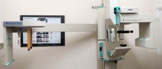
The essence of MRI, computed tomography and x-rays: distinctive features
The operating principle of a computed tomograph (CT) scanner is based on X-ray radiation. The rays pass through tissues and are absorbed by the latter with varying intensities. The information processed by the computer program is translated into a layer-by-layer 3D image.
MRI uses electromagnetic radiation that passes through organs, stimulating the reaction of hydrogen compounds. The observed changes are recorded, and on their basis a three-dimensional image of the anatomical structure is formed.
Radiography (x-ray) and fluorography are based on the action of x-rays. Ionizing radiation passes through the human body until it reaches the receiving matrix.
Due to the different throughput of tissues, the output is an image (not three-dimensional) with darkened and lightened areas.
MRI
X-ray Fluorography
The difference between radiography and MRI and X-ray and CT lies in the principle of operation of the equipment and other diagnostic features:
- a few seconds are enough to carry out radiography; magnetic and computed tomography will require 10-60 minutes (depending on the examination area and type of diagnosis);
- Each of the methods is applicable for certain diagnostic purposes. X-ray radiation helps in identifying pathologies of the musculoskeletal system and bone injuries. Magnetic tomography is powerful in diagnosing infectious zones, diseases of the spinal cord, digestive system, etc.;
- X-rays and computed tomography have an adverse effect on the patient’s body, MRI does not.
Is it possible to do an MRI after an x-ray: limitations and contraindications
Both of these studies have limitations; their list does not change if both methods are used together. MRI is contraindicated if the body contains:
- metal structures;
- grafted implants;
- electromagnetic devices (especially pacemakers).
Relative contraindications are pregnancy in the first trimester, claustrophobia and the general serious condition of the patient requiring emergency intervention.
There are no absolute contraindications to X-ray examination. Relative contraindications include pregnancy (especially the first trimester), childhood and the patient’s serious condition.
How much time should pass between procedures
The procedures, if they did not involve contrast enhancement, can be performed almost immediately one after another. Traditionally, MRI is performed after X-rays. But if a patient is prescribed an MRI of one area and an x-ray of another, then an x-ray after the MRI can also be done almost immediately.
CT (computed tomography) and MSCT (multispiral computed tomography) are also performed using X-rays. These methods, like MRI, are high-resolution imaging techniques. To some extent, their areas of application overlap with magnetic resonance imaging. Therefore, although the combined use of MRI and CT (MSCT) is not contraindicated, the advisability of this step should be discussed with your doctor.
Combination of X-ray and CT: is there a danger?
It is undesirable to do a CT scan after radiography, since both methods are based on X-ray radiation, which is harmful to the patient’s health.
It is necessary to delay diagnosis for 1-2 months. If it is impossible to organize a break, resort to CT immediately after an x-ray only if the diagnostic benefit outweighs the harm.
If it is necessary to perform repeated diagnostic procedures based on X-ray radiation, they rely on the maximum permissible annual exposure for the population - 5 mSV. Use the “Dosimeter” to determine the examination dose.
In general, X-ray scans are performed once a year. If there are grounds for an additional procedure, the diagnosis is carried out as many times as is permissible in a particular case.
Important! The radiation dose received depends on the equipment used, the organ being examined and the number of images taken.
Are x-rays and fluorography done on the same day?
X-ray is considered one of the most accurate and informative methods for studying the human skeleton, soft tissues and internal organs. However, the examination involves the patient receiving some dose of radiation. Typically, the exposure is small, but it accumulates.
Radiation of 50 mSv (millisievert) per year is considered hazardous to health, while with fluorography the body receives from 0.05 to 0.5 mSv. A person receives a comparable amount of radiation per month from natural sources. With an x-ray, the patient can receive radiation from 0.015 to 8 mSv, depending on the examination technique and the area being examined.
Therefore, specialists try to maintain certain deadlines for how many days or weeks after the x-ray the patient can undergo fluorography. In special cases, both examinations may be scheduled on the same day.
What is more harmful: MRI, CT or X-ray
In the case of MRI, there is no talk of harm at all. When undergoing radiography and CT scanning, the human body receives a certain dose of x-ray radiation.
The x-ray lasts for several minutes, the x-ray is ready in a split second. Computed tomography is accompanied by the reproduction of a series of images; the more slices, the longer the scan and the higher the load - this is the price for the high quality of the final images.
To reduce possible harm from radiation examination, special protective equipment is used (lead aprons, plates, collars).
It is possible to do an X-ray on the same day as an MRI, this is explained by the harmlessness of the second procedure. The situation is different with a combination of radiography and CT.
Since we are talking about studies that involve radiation exposure, diagnostics in a short period of time are not recommended.
Under such circumstances, it is necessary to take a break of at least 1-2 months. Conducting research in a short period of time is justified only if the benefits of diagnostics exceed the possible harm.
Diseases for which MRI and X-rays are prescribed
Both methods are externally similar, but the content of Article 1 MRI recommends using the procedure only for examining soft tissues and organs. This helps to avoid errors during decoding and reduces the risk of misdiagnosis. In this case, the picture is obtained in three dimensions, taking into account several planes. If there is no need to examine the skeleton, the MRI procedure will help diagnose the following diseases:
- pathologies of the cardiovascular system;
- severe dysfunction of the kidneys, liver, pancreas;
- neoplasms and metastases in internal organs, lymph nodes;
- brain damage;
- hernias and injuries of intervertebral discs.
Radiography is an indispensable technique that is recommended for various pathologies:
- diseases of the lungs and bronchial tree;
- disruption of the tissue structure of the bones of the limbs and ribs;
- severe injuries, fractures, punctures;
- metastases in bone tissue;
- curvature of the spine, displacement.
Therefore, it is possible to do an MRI and X-ray on the same day in case of combined injuries or the patient’s serious condition after an accident.
Contraindications to computed tomography
For all its informativeness, the computed tomography method is not indicated for everyone and not always. First of all, it is harmful to fetuses and small children, due to the fact that they are in a state of rapid growth, and fast-growing tissues are especially susceptible to the effects of X-rays. Pregnancy is also an absolute contraindication, with the exception of brain tomography to protect the abdomen from radiation and only in exceptional cases.
CT scanning is incompatible with drinking alcohol, and it is also contraindicated for people with mental disorders. The obstacle is a lot of weight, which one depends on the design of the particular tomograph. Braces can distort the results of tomography of the jaws, everything else will be illuminated without interference.
MSCT with contrast is not performed for patients with renal failure, heart failure, multiple myeloma, thyroid pathologies, or allergies to iodine.
Is it possible to do a CT scan during pregnancy?
CT scanning during early pregnancy can cause miscarriage. If this does not happen, abortion is still recommended, since the teratogenic effects of radiation (i.e., capable of causing underdevelopment or functional immaturity of organs in the fetus) used in CT scans are known.
An exception (and only in very rare situations when it comes to a woman’s life) can be made if a pregnant woman needs a CT scan of the head. Then the rest of the body must be covered with a special lead apron that does not transmit x-rays. In this case, even if a woman had a CT scan during pregnancy, the manifestation of pathologies due to this in the unborn child is unlikely.
At what age can a CT scan be done?
Considering the possible harm from radiation, and for a child it is many times higher than for an adult, computed tomography is prescribed for children only in cases where it is a vital indication. For example, to identify and examine tumors. Of course, if it is possible to obtain the necessary information in other ways - for example, MRI or ultrasound, then they are carried out.
Is it possible to do a CT scan during menstruation?
Menstruation itself does not interfere with CT scanning, including when examining the abdominal cavity and generally all parts of the body except the uterus: in this case, the diagnostic results may be distorted and it is better to wait until the end of menstruation.
Is it possible to do a CT scan after an x-ray?
A CT scanner, which uses the same radiation as conventional X-ray machines, produces very clear images. This especially applies to bone tissue and hollow internal organs. Often what is seen on X-rays requires the detail that tomography can provide. And it is not only possible, but must be done if there are serious reasons for this. The same can be said about MSCT after fluorography. But if the case is not an emergency, it is better to leave a gap of several weeks between examinations.
Similar questions arise regarding CT scanning after chemotherapy. Will the harm from it increase against the background of the toxic drugs received? Studies are required to evaluate the success of treatment; they cannot be avoided. But you need to maintain a certain time interval between the use of chemotherapy and CT.
Complications after CT
The most terrible complications of diagnostics based on X-rays include the development of oncology. We are talking only about an unlikely possibility, but the fact itself must be taken into account. When prescribing procedures, the doctor takes into account the total number of procedures, so as not to exceed permissible radiation exposure standards. If a patient feels unwell after a CT scan, it is unlikely that it is related to the procedure itself. Poor health and fever after a CT scan can be explained by the general painful condition of the patient. In addition, if a contrast agent is used, an allergic reaction to it is possible. You should tell your doctor if you experience itching, swelling, nausea, or tightness in the airways during or after diagnosis.
When should you choose CT instead of X-ray?
X-rays in the form of flat images are simpler and safer for humans than a computer tomogram. However, the work of organs in real time cannot be seen on x-rays.
In the same way, x-rays are not able to show volumetric problems masked by other contrast inclusions, which often include tumors, problems with blood vessels, and complex injuries.
CT scans are also performed after operations, especially when metal prostheses, shunts or pacemakers are introduced
Therefore, CT scans are prescribed if an urgent decision is needed when serious problems of the heart and brain are suspected, as well as other emergency conditions, which include:
- head injuries accompanied by fainting,
- headache if there is a suspicion of serious disturbances in the functioning of the body,
- convulsions that appeared for the first time without a reason, and in combination with suspected systemic diseases,
- mental state disorders with suspected brain damage.
Routine and non-urgent CT examinations are carried out strictly as prescribed by the doctor in cases where simpler methods do not produce results. Otherwise, the radiation exposure is unreasonably high.
Fluorography after magnetic resonance imaging
MRI examination is considered a highly accurate way to study a huge range of diseases. Medical practice and customer reviews confirm that almost any part of the body can be checked using MRI. There is absolutely no pain when performing an MR procedure, there is no harm from the operation of the equipment, and today it is widely used to find various tissue anomalies.
Despite its proven advantages, tomography has limitations consisting in insufficiently objective imaging of hollow organs (intestines) and lungs. Often, to confirm the information obtained after undergoing magnetic MRI diagnostics, doctors order fluorography.
Fluorographic research, compared to the NMR procedure, uses x-ray irradiation to form an image.
Why is fluorography dangerous?
Considering the harm of ionizing radiation, experts try not to prescribe x-ray examinations for diagnosing patients under 14 years of age and women carrying a child.
X-ray fluorography helps to detect diseases of the respiratory system (tuberculosis and malignant neoplasms). The procedure is of particular value because it provides the opportunity to detect pathologies that progress without symptoms.
Is it possible to do fluorography after an MRI?
The appointment of X-ray fluorography after the study is allowed, since the principle of nuclear magnetic resonance does not have a negative effect on the human body.
The radiation exposure after a fluorographic examination is insignificant, so examinations can be done before and after an MRI examination.
In what cases can a doctor prescribe both an ultrasound and an x-ray?
Both diagnostic procedures may be required for many reasons. You may encounter, for example, a situation where it is urgently necessary to establish an accurate diagnosis, but the conducted (or one or the other) study provides insufficient information. In another case, for example, a routine examination is required. In one day in a private clinic you will undergo an ultrasound, an X-ray room, an ECG, and other studies.
Another option for the need to undergo both diagnostic procedures in one day could be, for example, an injury. If a person had an ultrasound in the morning and later (on the same day) twisted his leg, then, of course, they will take an x-ray at the emergency room.
Some medical centers today even offer comprehensive diagnostic services (for preventive purposes or according to indications), in which both the ultrasound and X-ray procedures are performed at the same time by the same doctor. This approach is believed to be more effective.
Who should not have a CT scan and when?
Children undergo CT scans only for serious illnesses
Tomography is strictly prohibited for pregnant women and is allowed only if necessary for children.
With “contrast” - iodine-containing drugs - there are much more restrictions.
In addition to those diseases for which it is expressly prohibited to take iodine-containing drugs, this type of examination is contraindicated for diabetics, persons with a body weight exceeding that allowed for the device, and patients in severe conditions, if the risk of the examination outweighs the benefit.
For everyone else, it can be done without fear - but only taking into account the total annual radiation dose, which should include flights, X-ray examinations and all other cases of interaction with ionizing radiation.
There are few of them in our lives, but they still exist.
Which organs can be examined by MRI?
What organs are examined using MRI? Magnetic resonance scanning makes it possible to check almost all internal organs - the head, any part of the spinal column, hard joints, and the vascular system.
MRI diagnostics of soft tissues of the cervical area reveals abnormal abnormalities in the larynx, endocrine gland, and lymph nodes. MRI of the abdomen examines the liver, the organ that stores bile, and the spleen. When examining the retroperitoneum, the adrenal glands and nearby lymph nodes are checked.
MRI of the pelvis allows the doctor to determine a malignant tumor, inflammatory processes (acute and chronic), and vascular defects. The non-invasiveness of nuclear magnetic resonance allows private clinics to use the method without the consent of attending physicians in public clinics.
Before examining the pelvic organs, preparatory steps are carried out (you will need to drink more than 1 liter of water).
What diseases can be examined using MRI units?
The following types of internal organ diseases can be examined using MRI:
- Inflammatory processes (pathology of the urinary and reproductive systems);
- Diseases of the spinal cord, head (lesions of the pituitary gland, spinal column, nerve fibers);
- Neoplasms of various etiologies. Whether complex forms of cancer can be diagnosed using MRI depends on the specific situation. The equipment makes it possible to detect formations at the earliest stages;
- Diseases of the heart and its vascular channels;
- Traumatic injuries;
- Anomalies of the structural elements of the joints.


