Human internal organs, including the gallbladder and ducts, and pancreas, require x-ray examinations. Liver angiography, portography, splenoportography and others are the most accurate research methods. Indications are pathologies, inflammation, neoplasms in organs. The diagnostic method is determined by the doctor. Deciphering the results helps determine the disease.
Why do you need radiography?
The contrast agent used to visualize the gallbladder and ducts, bilitrast, is iodine-containing. The scanning machine concentrates and directs x-rays, which pass through the soft tissue and the injected substance in different ways. A contrast agent is administered intravenously immediately before the procedure after a tolerance test.
From the blood, the contrast agent enters the liver, gallbladder and its ducts. The results of cholecystocholangiography are based on photographs taken that record the passage of the contrast agent. With its help, it is possible to assess the degree of damage to the bladder and ducts, see tumors, stones, as well as other formations and anomalies that are not visible during other research methods. Images are taken after 20 minutes, 30 minutes and 40 minutes after the administration of contrast, for a step-by-step assessment of the function of the entire biliary system.
In what situations are x-rays of the liver and gallbladder prescribed?
Liver X-ray is prescribed in the following cases:
- Suspicion of the presence of multiple cysts;
- Disruptions in the circulatory system;
- Suspicion of liver infarction;
- Increased liver size for unknown reasons;
- Degeneration of liver cells into adipose tissue;
- Tumors of various types;
- Suspicion of cirrhosis;
- Tuberculosis;
- Purulent inflammation;
- Hepatitis;
- Formation of blood clots in blood vessels;
- Organ injuries.
An X-ray of the gallbladder is prescribed for cholecystitis, tumor formations in the organ, or if the presence of stones is suspected. It is also carried out when there is a violation of the flow of bile into the duodenum.
Contraindications to the procedure
The main contraindications to this contrast X-ray examination are intolerance to iodine-containing drugs, severe damage to the liver and kidneys.
The procedure is not performed on pregnant women in the first trimester unless absolutely necessary. For children, cholecystocholangiography is prescribed in cases where the benefits of the information obtained during the study outweigh the risks of radiation exposure. During the examination, additional protective aprons are used (protect the chest, thyroid gland and genitals).
Features of the event
During the study, additional pain or symptoms of malaise may be observed. Before prescribing the procedure, the attending physician assesses the risks and prescribes additional studies. Complications after cholecystocholangiography in rare cases may include:
- prolonged headaches;
- disturbances in the gastrointestinal tract;
- nausea and vomiting (symptoms of body intoxication).
Cholecystocholangiography is performed only in cases where standard research methods do not help establish an accurate diagnosis (biochemical blood tests, ultrasound). The dose of X-ray radiation received during the study should be summed up with the total dose received during the year.
Also, cholecystocholangiography is performed before liver surgery, when it is necessary to assess the condition of the excretory ducts. This research method is a mandatory part of preoperative preparation. The results (images) show neoplasms that may be a contraindication to surgery.
Carrying out the procedure
The contrast agent is administered in two ways - orally or intravenously. In the second method, the substance is administered immediately before the procedure. After installing the catheter, the patient is positioned on the table (images are taken standing and lying down). The medical worker takes several pictures by changing the direction of the main scanner of the device.
The procedure is carried out in two opposite positions. The patient must remain still while the scan and target image testing is performed. After taking the yolks (an hour later), pictures of the emptied organ are taken. The images show shadows of the bile ducts. If the liver clears the contrast agent, the gallbladder will be visible on images without interference. If the passages are obstructed, the contrast agent will begin to accumulate, which will appear on the resulting images.
After the procedure, the patient can consume food and water.
What can you see in the picture?
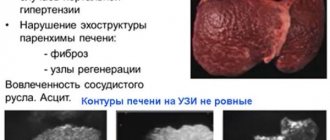
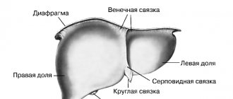
A healthy liver looks like a uniform triangle with smoothed corners. Its contours are clear and even. The norm is determined by the following criteria:
- Uniformity of structure;
- Density greater than that of the pancreas, gallbladder, spleen and kidneys;
- Less density in the area of liver vessels;
- Invisibility of the liver artery and bile ducts of the organ to the eye;
- The ability to identify the bile duct and common hepatic duct in the image.
What does a liver x-ray show? The image can reveal the presence of benign and malignant tumor formations, impaired functioning of blood ducts, and poor bile conductivity. A healthy liver is shown in the photo.
An x-ray of the gallbladder allows us to determine the presence of stones, inflammatory processes in the mucous membranes, the ability of the bladder to supply bile to the intestine in a timely manner, the presence of adhesions and scars. A healthy gallbladder in the picture looks pear-shaped, has clear edges and thin walls.
Although a plain abdominal radiograph may reveal hepatomegaly or ascites, physical examination is more helpful. On a plain X-ray, stones containing calcium can be detected in the bile ducts, but it must be taken into account that 15% of cholesterol or mixed stones and 50% of pigment stones are X-ray negative.
Oral cholecystography can be informative in identifying gallstones. The day before the study, the patient takes iopanoic acid (cholevid). It is absorbed in the intestine, secreted into the bile canaliculi, and concentrated in the gallbladder. On an x-ray, stones are revealed as defects in the filling of the gallbladder (Fig. 7-6). In this study, side effects in the patient may include nausea, vomiting, and diarrhea, which requires a second dose. The lack of visualization of the gallbladder after taking the second dose of the drug indicates its poor ability to concentrate bile, which can be the case with chronic cholecystitis.
Currently, ultrasound examination (ultrasound), as a simpler and more easily tolerated method, has almost completely replaced cholecystography (Fig. 7-7). Ultrasound of the abdominal cavity allows visualization of the bile ducts (for example, dilatation of the common bile duct in choledocholithiasis), liver, spleen, pancreas, and kidneys. Ultrasound partially helps in the differential diagnosis of cystic and space-occupying formations in the liver, and is more sensitive in the diagnosis of ascites (detects even 200 ml of fluid) than physical examination. The use of Doppler ultrasound allows one to assess the velocity of blood flow in the hepatic, portal and splenic veins and is used to diagnose hepatic portal or splenic thrombosis (Budd-Chiari syndrome).
Fig.7-6.
On a cholecystogram, gallstones appear as a filling defect
Radioisotope liver scanning is performed by injecting special isotopes that are selectively absorbed by the liver. When scanning, colloidal sulfur labeled with technetium (99mTe) is used, which is captured by Kupffer cells. Changes in the structure of the liver in the form of metastases or abscesses are perceived as areas of reduced uptake - “cold” spots (Fig. 7-8). In diffuse hepatocellular diseases (hepatitis, fatty hepatosis or cirrhosis), uneven uptake is noted, the so-called colloidal shift, in which the mesenchymal substance of the spleen and bone marrow absorbs the isotope-labeled substance more intensely than the liver. Technetium (99mTc) labeled red blood cells are used to identify liver hemangiomas. The property of gallium-67 (67Ga3+) to accumulate in tumor and inflammatory liver cells in greater quantities than in normal cells can be used to diagnose carcinomas and abscesses in the liver. For hepatobiliary scintigraphy of the liver, iminodiacetic acid labeled with 99mTc is also used, with which the rate of hepatic and biliary secretion is assessed. The absence of visualization of the gallbladder during slow-motion scanning can help in the diagnosis of acute calculous and non-calculous cholecystitis.
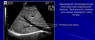
Rice. 7-7.
Ultrasonography of gallbladder stones located parietally and giving a “shadow” or “track” when scanning
Rice. 7-8.
In which an adenoma is detected in the right lobe of the liver in the form of a “cold focus”
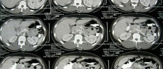
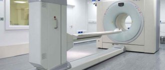
Computed tomography allows you to visualize the contours and structure of internal organs on serial slice images (Fig. 7-9).
Rice. 7-9.
. (A) - peripheral localization of the hemangioma shadow. (B) - central shadow enhancement in a later photograph
Direct injection of contrast agents into the biliary system can be carried out using so-called endoscopic retrograde cholangiopancreatography (ERCP) (Fig. 7-10) or using percutaneous transhepatic cholangiography. RCCP is a priority in diagnosing pathology of the distal part of the biliary tract, especially before performing a sphincterotomy operation, as well as in cases of the presence of signs of ascites or coagulopathy. Percutaneous transhepatic cholangiography can be used in the assessment of proximal localization of biliary system pathology or in cases of anatomical disorder in the gastroduodenal gastrointestinal tract. Both methods are important in the diagnosis of obstructive jaundice.
Which is better, a colonoscopy or an X-ray of the intestines? We will describe the article in this vein, since the question is relevant for patients. In practice, doctors are faced with the fact that people refuse probe examination in favor of X-ray contrast techniques (irrigoscopy).
X-ray examination of the intestine and probe examination have different purposes and purposes, so the methods complement each other. For example, colonoscopy can reveal superficial defects of the mucous membrane (ulcers, cancer, epithelial hyperplasia, polyps).
The introduction of contrast makes it possible to evaluate the external contours of the intestine and identify exophytic forms of neoplasms, fistulas and diverticula.
Modern cholecystocholangiography
Currently, percutaneous transhepatic cholangiography is used. A laparoscope is used to administer an iodine-containing substance. In this study, contrast is injected directly into the bile ducts during percutaneous liver puncture.
This method is suitable for examining the biliary tract for the presence of fistulas or neoplasms. The choice of contrast injection method depends on the area being examined. Cholecystocholangiography is performed in a special room protected from background radiation. Image recording is carried out by medical workers from a designated protected room, using protective aprons and plates.
Indications
An X-ray of the liver and biliary system helps to see the structure and size of the organs being examined. There are many computer diagnostic methods, such as MRI, ultrasound, CT, but X-ray studies using contrast are the most informative. The examination method is chosen by the doctor. Indications are the diagnosis of such diseases:
- cirrhosis;
- malignant and benign tumors;
- dysfunction of the liver and gallbladder;
- disruption of the biliary tract.
Return to contents
Results of the procedure
Best materials of the month
- Why you can't go on a diet on your own
- 21 tips on how to avoid buying stale food
- How to keep vegetables and fruits fresh: simple tricks
- How to curb your sweet cravings: 7 unexpected products
- Scientists say youth can be extended
Cholecystography is prescribed to study the anatomical structure and functional activity of the gallbladder and bile ducts. On the resulting image, you can evaluate the shape and position of the area under study, the displacement of its position, which is deviated from the norm. The size of tumors and stones is assessed from several photographs taken in different planes. The two-dimensional image allows you to evaluate large abnormal formations, tumors and polyps that interfere with the functioning of the gallbladder or ducts.
Cholecystocholangiography provides a clear image of the internal organ: the gallbladder is pear-shaped with smooth outlines and a thin contour. Any deviations from the norm are recorded by a radiologist and are the reason for prescribing additional research methods. The shape of the gallbladder may differ from the norm due to the design features of the body. In hypersthenics, the bladder is round in shape, while in asthenics it is elongated upward: the structural features and position of the organ are assessed by a doctor who writes a conclusion on the cholecystocholangiography performed.
CT scan
This method has significantly improved the capabilities of x-ray research.
J.D. Hounsfield (1973) created the first CT scanner and proposed that the image be enhanced during examination to improve its clarity. With the introduction of the method, it became possible to clearly diagnose the presence of hydatid cysts in the liver and even identify daughter blisters in the cyst cavity. CT scans record changes in the contours of the liver, displacement of the great vessels and ducts, which also helps in diagnosis. With secondary (metastatic) liver tumors, the organ contains round, low-density, often multiple formations. Primary liver cancer is characterized by a heterogeneous decrease in image density and blurred contours of the pathological focus.
Buy cheap medicines for hepatitis C
Hundreds of suppliers bring Sofosbuvir, Daclatasvir and Velpatasvir from India to Russia. But only a few can be trusted. Among them is an online pharmacy with an impeccable reputation, Main Health. Get rid of the hepatitis C virus forever in just 12 weeks. High-quality drugs, fast delivery, the cheapest prices.
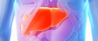
Human health depends on the functioning of the liver and gall bladder. If there are problems with the liver, metabolism suffers, digestion and blood circulation processes are disrupted. The gallbladder is responsible for storing bile, which is produced by the liver, and takes part in the digestive process. To monitor the functioning of these organs and to check their structure, an X-ray method with a contrast agent is used. What does an x-ray show? This can be seen in the photo.