Take an MRI of the male genital organs and find out the cause of concern within 60 minutes
Make an appointment by calling +7 (495) 120-55-67 or 24 hours a day using the registration form on the website
Sign up for an MRI
- Restrictions on the procedure: weight up to 120 kg, volume up to 140 cm;
- The procedure time is 40 minutes;
- Imprisonment time 20 minutes;
- Preparatory procedures
- The study is carried out strictly on an empty stomach.
- The day before the test, exclude gas-forming foods (legumes, cabbage, brown bread, sweets, raw vegetables and fruits).
- Within 2 days before the study, take 1 tablet. "Espumizan" (gas suppressants) 3 times a day.
- In the evening before the examination, we clean the intestines (with Duphalac or a classic enema - contraindications - “Colitis”).
- On the morning of the examination, drink 2 No-Spa tablets and 0.5 liters of water.
- Upon arrival at the center we do not go to the toilet.
- In men, it is recommended to conduct an examination with contrast.
- Application of construal Recommended;
Indications
- fracture of the penis, rupture or severe bruise of the testicle due to injury,
- changes in skin color,
- rash or sores of unknown nature,
- signs of inflammatory processes,
- congenital and acquired pathologies.
Contraindications
- uses a cardiac or neurostimulator,
- the body contains metal and metal-containing prostheses and implants,
- the man weighs more than 120 kg.
MRI of male genital organs in Moscow is prescribed in cases where making an accurate diagnosis by visual examination or palpation is impossible. Injuries to the penis and scrotum are in most cases accompanied by severe pain, making it impossible to use other diagnostic methods.
The structure of the organs does not allow one to obtain a clear picture of abnormalities with ultrasound, therefore. If the doctor sends you for a tomography, you should not refuse.
| Field of study | Prices in Kurkino (SZAO) | Price on Tsvetnoy Boulevard (TsAO) | MRI prices without discount | Prices for MRI ON PROMOTION in Kurkino | Prices for MRI + online consultation with a doctor after MRI | ||||
| Equipment power | 1.5 Tesla | 1.5 Tesla | |||||||
| MRI of the pelvic organs and external genitalia | 6000 | 7 000 | 5,500 rub. | — | |||||
| Bladder | 5500 | 5500 | 6 000 | 4,900 rub. | — | ||||
| External genitalia | 6800 | 6 000 | 4,900 rub. | — | |||||
| 500 | 2 000 | 1,300 rub. | ||
| Recording an MRI study on flash | 800 | 800 | 1 000 | 800 rub. |
| Photographs on film | 600 | 600 | 700 | 600 rub. |
Sign up for an MRI of the male genital organs right now!
Possibilities of MRI in the diagnosis and diagnosis of scrotal diseases
MRI shows a high percentage of specificity and sensitivity in identifying benign and malignant scrotal masses, and in assessing the involvement of regional lymph nodes in the process.
The method visualizes inflammatory diseases of the scrotum, torsion of the testicle and epididymis, traumatic damage to the testicle (rupture of the parenchyma, membranes), fracture of the penis.
In case of anomalies of testicular development, MRI is used to differentiate changes by assessing the structure, position and presence of an accessory organ.
Based on the tomography conclusion in conjunction with the clinical picture, the following diagnoses are made:
- orchiepididymitis;
- scrotal abscess;
- pneumatosis of the scrotum;
- cryptorchidism;
- polyorchidism;
- hydrocele;
- testicular cyst;
- tumor of the testicle and epididymis;
- tumors of the penis.
Indications and contraindications for MRI of the genital organs in men
Before you sign up for an MRI of the male genital organs, read the contraindications
Experts recommend MRI as the best way to detect diseases of the bladder, prostate, seminal vesicles, testicles and penis after inconclusive results of ultrasound and CT, and sometimes the procedure is considered as a first-line examination in younger patients with suspected neoplastic process. Magnetic resonance scanning is a highly informative diagnostic that provides a multiplanar assessment of suspected or known pathologies of the genital organs and related conditions affecting the perianal and perineal structures, for example, rectal-urinary fistula. Complaints that patients come with for MRI of the genital organs:
- an increase in the size of the scrotum, pain, hyperemia, palpable neoplasm in the testicle or its epididymis;
- curvature of the penis, swelling, pain;
- absence of testicle in the scrotum;
- difficulty urinating, nocturia, feeling of incomplete emptying of the bladder;
- hemospermia, macro- and microhematuria;
- sexual dysfunction: premature ejaculation, dyspareunia, loss of orgasmic sensations, weakened erection;
- pain in the lower abdomen, dysuric disorders accompanied by fever, lack of effect from treatment or a tendency to frequent relapses, etc.
All of the listed symptoms and complaints, with little success from other examinations and the appropriate clinic, can be considered as indications for MRI of the genital organs.
MRI of the genitals is performed for prostate cancer. The high specificity of magnetic resonance imaging of the prostate (about 88-90%) allows this method to be used in patients with moderate and high risk of extra-organ tumor spread to clarify staging. Based on the results, indications for surgical or conservative treatment are determined. MRI of the prostate is necessary when the level of the tumor marker PSA increases above 10 ng/ml. Magnetic scanning allows you to assess the condition of regional lymph nodes, which is not always possible with computed tomography, and also to identify metastases in the pelvis and lumbar spine. The final verification of the diagnosis occurs after a morphological examination, which will show the type of tumor, its prevalence, and invasiveness.
Contraindications to MRI of the genitals
Considering that MRI uses a magnetic field and impulses to construct three-dimensional images that do not have a harmful effect on the body, there are few contraindications to the study:
- the presence of metal components: cochlear implants, pacemakers, vascular clips, Ilizarov apparatus, foreign body in the bladder, etc.;
- the patient’s weight is too high (obesity 120 kg and above);
- claustrophobia;
- inability to remain motionless in a lying position.
There are patients who have undergone low-dose brachytherapy for prostate cancer. This type of treatment involves implanting radioactive seeds into the gland with gradual irradiation of the tissue to control the tumor. When exposed to a magnetic field, they create interference on the films. There is evidence that with a tomograph power of 3 Tesla or more, satisfactory visualization is possible, but it is better if the question of the advisability of MRI in this situation is decided by a doctor.
When performing MRI with contrast, it is necessary to inform the staff if a significant allergic reaction (such as generalized angioedema) has previously been noted to the administration of a paramagnetic substance, or if there is a history of kidney disease with severe impairment of functional capacity (end-stage chronic renal failure).
When an MRI of the scrotum is prescribed, restrictions
MRI is indicated for pain in the scrotum, increase in size, change in color (cyanosis) and local increase in temperature.
These disorders are characteristic of acute disease of the epididymis and testicular parenchyma due to an inflammatory process or traumatic injury.
If palpation reveals a change in the size of the testicles or their absence, tomography is used to verify the diagnosis.
The procedure is prescribed for patients with leukemia, lymphoma, and malignant tumors of the pelvis in order to identify metastatic lesions of the penis.
The choice in favor of MRI is made in cases of erectile dysfunction (impotence), diagnosing the consequences of priapism, and in case of trauma to the penis.
Contraindications to tomography:
- installed pacemaker;
- implants, fragments in tissues located near blood vessels;
- risk of heart attack;
- unconscious state;
- intoxication with alcohol and drugs;
- fear of confined spaces.
Contraindications to the procedure
It is important to always carefully study the contraindications for a particular study, and neglecting them can lead to extremely dangerous and unpredictable consequences. Here is a list of the main reasons why doctors refuse magnetic resonance imaging of the female organs:
- the presence of any metal devices and elements in the body, for example, implants, clips on blood vessels or anything else;
- tattoos, the dye of which contains metal;
- excess body weight - more than 120-130 kg (the device is not designed for people whose body weight exceeds these figures).
Note! The situation related to implants is not so clear-cut. The fact is that some of them are already made of inert alloys or, for example, titanium, which is also not affected by the magnetic field created by the tomograph. In this case, most likely, the procedure will be possible, but all this must also be discussed with the doctor individually.
There is another group of contraindications that are relative, that is, with them, magnetic resonance imaging can still be performed, but if absolutely necessary and if there is a threat to the patient’s life:
- an extremely serious condition in which a person cannot lie without moving for the required time;
- claustrophobia (fortunately, this problem can be solved by using an open tomograph, and even sedatives can help with mild forms of claustrophobia);
- pregnancy in the first trimester (there is an assumption that the magnetic field can still have a negative effect on the fetus when it is just beginning to develop, so MRI is practically not performed in the first trimester).
Let us mention that the contrast agent also has its contraindications, the main of which are:
- individual intolerance to any components of the contrast agent;
- renal or liver failure;
- diabetes.
Preparation procedure for MRI of the scrotum
Before the tomography, the patient provides the doctor with a medical history and the results of other diagnostic methods of the area being examined, if available. When repeating an MRI, they take the previous study protocol with them in order to track the process over time.
If a contrast agent is used for MRI of the scrotum, then 3-7 days before the study, blood is donated to determine the creatinine level. Before the procedure, patients do not adhere to a diet. Sexual contacts are not prohibited.
How to prepare for an MRI of the male genital organ
A light snack will help you endure the MRI procedure with contrast more comfortably
No special preparation is required; normal hygiene procedures and a change of linen are sufficient. Before administering a contrast agent, you should have a light snack 40 minutes before the procedure, this will avoid unpleasant vegetative symptoms: drooling, nausea, dizziness, etc. They are registered in approximately 40% of people and go away on their own within 3-5 minutes. The study is carried out with a full bladder; for this you need to not urinate for 1.5-2 hours before the start of the procedure.
You must take with you a referral from a doctor, which clearly indicates the type of diagnostic procedure, the area being examined and the proposed diagnosis, the results of extracts and previous MRIs, CT scans, ultrasounds (if any were performed). Clothing should be comfortable and without any metal parts. Before the examination, you will be asked to remove cell phones, magnetic cards, watches, and jewelry. If desired, you can change into a disposable robe.
Examination technique
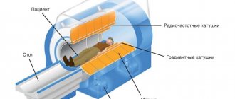
The patient is placed on a table, his ears are covered with headphones or earplugs, and a signal button is given to his hands. Then the table is immersed in the tunnel of the apparatus, where the body is exposed to a constant magnetic field.
The study takes 30-40 minutes to obtain images weighted in T1, T2 and proton density in three projections. A special surface coil is used to differentiate the scrotal organs.
At the end of the diagnosis, the doctor draws up a protocol and displays the resulting images on film, disk or flash card.
Diseases of the urinary system
An examination of the bladder is usually carried out in the general diagnosis of the pelvis. Because the symptoms of pathological changes in this organ are closely related to other diseases of the urinary system. A prescription for additional diagnostics may be the results of a visual examination or biopsy. A detected tumor becomes the reason for an MRI of bladder cancer. To enhance and provide high-quality images of cancerous tumor clots, the patient is often injected with a contrast agent. It is absolutely harmless.
Visualization of pathology of the scrotal organs on MRI
When tomography, an inflammatory process in the testicle or epididymis is indicated by an increased signal on T2 VI, an increase in the size of structures, and the formation of a hydrocele (fluid in the membranes of the testicle).
If, with orchitis and epididymitis, the patient’s condition worsens during therapy, tomography helps to exclude right-sided testicular abscess as a complication of orchiepididymitis.
Testicular torsion is diagnosed when a twisted spermatic cord, hemorrhage and edema in the testicular parenchyma and epididymis with a change in their size are detected.
Cryptorchidism is diagnosed when a reduced testicular volume is detected outside the scrotal cavity, with a reduced signal on T1 and T2 images.
An intratesticular epidermoid cyst on photographs appears as a delimited formation with a homogeneous liquid structure.
Lipoma of the spermatic cord and epididymis has a high signal intensity on T1 VI, T2 VI, characteristic of adipose tissue.
For inflammation of the corpus spongiosum of the penis (spongiositis), a characteristic sign in the photographs is an enlarged corpus spongiosum, surrounded by infiltrative changes. Thrombosis of the corpus cavernosum is manifested by changes in blood flow and uneven expansion of the cavernous bodies.
Testicular cancer
Fibrous changes in the corpora cavernosa are recorded at low signal intensity, due to reduced blood supply, and when fibrous foci are detected in normal tissues of the corpora cavernosa.
In Peyronie's disease, fibrous plaques are detected on the tunica albuginea in the form of its thickening on T2-weighted images.
Disadvantages of MRI
Like any medical procedure, MRI, in addition to its advantages, also has some disadvantages. These include a number of contraindications: it cannot be performed on pregnant women, patients who have metal structures in their bodies or an artificial heart pacemaker, or those suffering from claustrophobia. Also, the disadvantages of this type of diagnosis include the duration of the procedure and its high cost. Despite the fact that the examination result is very detailed, sometimes the use of MRI is not enough. This applies to stones and calcifications in the pelvic organs; here the reliability of magnetic resonance imaging leaves much to be desired. If a doctor during an examination is interested in the presence of adhesions, for example, in the fallopian tubes, then focusing on MRI data should be a stretch. The fact is that only the roughest adhesions will be visible in the pictures, and minor pathologies will be invisible.
MRI of the scrotum using contrast agent
A study using the paramagnetic gadolinium is used for suspected tumor diseases of the penis and testicles in men. The substance introduced into the vascular bed accumulates in tumor cells, limiting them from healthy tissues.
No contrast
With contrast
The pictures provide a clear image that helps determine the location of the pathological formation.
Benefits of pelvic MRI
When examining the pelvic organs, not only MRI is used, but also ultrasound and CT. Each of these methods has its own advantages and disadvantages, but as a result of MRI of the pelvis, information is obtained not only in the most detailed manner, but also this method is one of the safest, especially when comparing CT and MRI.
- As a result of the examination, images of organs are obtained in several planes (sections, angles).
- The volume of data is very large, the information is reliable and clear.
- There are no age restrictions, that is, MRI of the pelvic organs can be performed not only on adult women, but also on girls.
- There are no requirements for the level of bladder fullness.
- Allows you to monitor the dynamics of treatment, since MRI of the pelvis is possible with the required frequency.
MRI of the pelvis is very important when there is a suspicion of cancer. Such a study not only gives a clear and unambiguous picture of the disease, shows the boundaries of the tumor, the spread of metastasis, but also allows you to make an accurate prognosis regarding the course of the disease and evaluate the effectiveness of treatment.
Advantages of MRI over ultrasound in diagnosing scrotal diseases
MRI, unlike ultrasound, provides a detailed assessment of the condition of the parenchyma of the testicle and epididymis, indicating the extent and stage of the inflammatory process.
In tumor diseases, tomography establishes the nature of the tumor and stage, and ultrasound shows an uncertain result and is informative in identifying simple and epidermoid testicular cysts.
The accuracy of MRI diagnostics and the absence of radiation exposure makes this method a reliable and safe method for studying diseases of the scrotal organs, the timely detection of which improves the prognosis and prevents complications.
How is the examination carried out?
MRI is performed with the patient immobile. The duration depends on the number of shots and projections. It is possible to conduct a study in 30 minutes if all rules are followed. The procedure does not require pain relief, but if it is difficult for the patient to remain still, sedatives are used.
Contrast is used during the study to identify the condition of the testicular parenchyma, as well as to diagnose vascular pathologies. This method allows you to determine the presence of tumors and other neoplasms. Most often, during the study, medications based on gadolinium are used, which are quickly and without harm removed from the human body.
