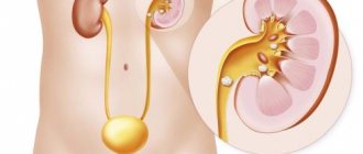
In urological practice, urinary tract infections are the most common bacterial diseases, the treatment and diagnosis of which have their own difficulties, most often associated with the atypical nature of the pathogen and its resistance to antibiotic therapy.
Therefore, according to modern medical recommendations, urine culture is mandatory for diagnosing and monitoring the course of urinary tract infections in high-risk individuals. In this material we will look at what urine culture is, what this analysis shows and how to properly prepare for it.
Bacteriological culture (bacteriological culture or bacteriological examination of urine) for the presence of pathological microflora is a highly accurate microbiological analysis that allows not only to isolate and identify the causative agent of urinary infection, but also to determine its exact concentration (degree of bacteriuria), as well as the degree of sensitivity to the most important groups of antibacterial agents.
The research allows:
- 1Identify the causative agent of infection;
- 2 Determine its diagnostic titer in 1 ml of urine;
- 3Detect the presence of antibiotic resistance (resistance or sensitivity to antibiotics);
- 4 Monitor the decrease in pathogen titer during therapy;
- 5Choose the most effective antibacterial drugs for treatment;
- 6Monitor the prevalence of antibiotic-resistant strains of microorganisms in the population.
Indications for bacteriological examination of urine
The main goals of the analysis pursued by the attending physician when prescribing urine culture are the diagnosis and monitoring of bacterial infections of the urinary tract, as well as determining the sensitivity of the pathogen to antibiotic therapy.
Sometimes bacteriological examination (bacteriological culture) of urine is used as a screening for the prevention of inflammatory diseases of the urinary tract in people at high risk (pregnant women, the elderly, patients with diabetes mellitus). The main indication for urine culture is the presence of symptoms of urinary infection.
However, according to Russian urological recommendations, if a patient has an uncomplicated urinary infection, as well as in the absence of concomitant diseases, antibacterial therapy can be prescribed empirically, without first identifying the pathogen. Empirical prescription of antibiotics achieves a positive effect in approximately 75-80% of cases.
Absolute indications for conducting a urine microflora test are:
- 1Urinary tract infections in pregnant women;
- 2Suspicion that the patient has pyelonephritis;
- 3All cases of urinary tract infection in men;
- 4Outbreaks of hospital-acquired urological infections;
- 5 Fever during prolonged catheterization of the bladder, as well as suspicion of a urological infection associated with medical procedures (cystoscopy, catheterization);
- 6 Fever above 38 C for no apparent reason in children under 3 years of age;
- 7 Recurrent urinary infections, ineffectiveness of previously conducted empirical antimicrobial therapy;
- 8Complicated urinary infections over the age of 65 years;
- 9The appearance of symptoms of urological infection in persons with impaired immune status, chronic kidney disease, diabetes mellitus, congenital anomalies of the structure of the kidneys and ureters, after kidney transplantation;
- 10The patient had taken antibacterial drugs during the previous three months, which theoretically could lead to the formation of resistant forms of pathogens.
According to the recommendations of the Ministry of Health of the Russian Federation, mandatory screening bacteriological urine culture is regulated, even in the absence of symptoms of urinary system pathology, in the following population groups:
- 1Women during pregnancy, after the 14th week, even in the absence of any symptoms of pathology, which reduces the risk of developing pyelonephritis in pregnant women;
- 2Patients with planned surgical intervention on the organs of the urinary system;
- 3Patients with a kidney transplant during the first two months after surgery, and then with any deterioration in the functioning of the transplanted organ.
Why do you donate a urine culture tank during pregnancy?
During the period of bearing a child, a woman's immunity is weakened, and she becomes susceptible to many infections. The most common are diseases of the urinary system (cystitis, pyelonephritis, etc.). For timely detection of infection, bacteriological culture of urine is performed. Based on the results of the study, the patient is selected an effective treatment regimen and antibacterial drugs that kill pathogenic microorganisms.
Rules for collecting material
We will discuss below how to properly take a urine culture test during pregnancy, in adults and children. To ensure the accuracy of laboratory diagnosis of urological infection, it is important to comply with all stages of diagnosis, including its preanalytical part - the collection of urine samples and their delivery.
It is the need to determine the exact amount of bacterial agents in the test sample that places special demands on sample collection.
When conducting research in a hospital setting, the nursing staff of the hospital is responsible for preparing the patient, collecting material and transporting it to the laboratory. For such cases, laboratories have written instructions regulating all diagnostic steps.
When examined on an outpatient basis, the patient collects urine independently, without the supervision of a medical professional. In connection with the above, it is necessary to carefully instruct the patient on the technique of independently collecting the test material in order to avoid false analysis results.
- 1Urine collection must be done in a sterile disposable container specially designed for this purpose, which can be purchased at the pharmacy chain. It is not allowed to collect urine in non-sterile, previously used containers, jars or bottles;
- 2If possible, urine collection should be carried out before starting to take antibacterial drugs or in the intervals between drug courses, to avoid distortion of the information received;
- 3The most reliable study is the average morning urine sample - after a night's sleep and before breakfast;
- 4 In the evening, on the eve of urine collection, it is recommended, if possible, to refrain from taking diuretics, since when they are taken, the urine is diluted and the total number of bacteria in one milliliter is reduced;
- 5 To collect urine from infants, you should use special children's urinals with a hypoallergenic adhesive; the collected sample is subsequently poured into a urine collection container, labeled and transported to the laboratory.
Figure 1 - Sterile containers for collecting urine for analysis (without a spatula)
What does it show
To find out what urine culture shows, you need to consult with your doctor who ordered the test. In simple terms, this study makes it possible to identify and identify microorganisms in urine, as well as to estimate their number.
If concentrations of microorganisms are detected at which infection may develop, then doctors consider the result of the culture to be positive. In this case, in order to combat the found pathogen, the patient is prescribed an antibacterial drug to which this microorganism is especially sensitive.
The main stages of self-collection of urine
The urine collection procedure described below is used for all adults, including pregnant women. The optimal volume of urine for microbiological analysis is approximately 10-20 ml, and the minimum volume should not be less than 1 ml.
The stages of independent collection of the research material can be presented as follows:
- 1Wash your hands thoroughly with soapy water and dry them with a clean (preferably disposable) towel;
- 2 Toilet the external genitalia using warm soapy water, without using skin antiseptics, dry the groin area with a clean disposable napkin;
- 3Open the previously prepared sterile container, avoiding touching its internal surfaces with your fingers;
- 4Release the first portion of urine, stop urinating;
- 5Collect the next (middle) portion into a prepared sterile container, without touching the skin in the groin area with the container;
- 6Finish urinating into the toilet;
- 7 Close the lid of the filled container tightly, sign it, attach the direction for analysis to the container with a thin rubber band, and deliver it to the laboratory.
It is unacceptable to use urine from a bedpan and urine from women during menstruation for microbiological research.
How to properly collect urine for bacterial culture from a child?
- 1Give the baby warm water or other liquid to drink (you can give the baby breastfeeding);
- 2Wash your hands with soapy water and dry with a clean towel;
- 3 Toilet the child’s external genitalia;
- 4If possible, collect a medium portion of urine by sitting the child on the assistant’s lap. If it is impossible to collect the middle portion, you can use a specially designed children's urinal, which should be checked at least every 10 minutes.
- 5Urine from a pot cannot be used for analysis.
Figure 2 - Collecting urine from an infant into a sterile urine bag
Rules for delivering material to the laboratory
The conditions under which a urine sample is delivered to the laboratory have a direct impact on the accuracy of the bacteriological examination, since they can both reduce the viability and stimulate the growth of bacteria in the submitted urine sample.
The sample should be delivered to the laboratory no later than two hours after collection. The optimal period for inoculating urine samples is less than 30 minutes after urination, however, due to the difficulty of implementing this condition, this period in many laboratories is regulated to 1-2 hours.
If it is impossible to deliver within the specified period, it is allowed to store the material at a temperature not exceeding +40C for 24 hours from the moment of collection. Freezing a urine sample is prohibited - this leads to the death of some types of microorganisms.
To improve the quality of the study, as well as to ensure longer storage, the use of special stabilizers, for example, 1% boric acid, is allowed. However, the addition of such preservatives is permissible only to a strictly permitted extent.
In this regard, when conducting bacteriological examination of urine, it is advisable to use commercial transport systems (special tubes), in which a certain concentration of a preservative is achieved by adding a certain amount of urine.
If special test systems are available, it is possible to carry out express bacterial culture without leaving the patient’s bedside (Diaslide, Deepstreak test strips), but this is not common in Russia due to the high cost of the equipment.
All urine samples received by the laboratory must be labeled by the laboratory assistant in any available way, allowing one to accurately identify one sample from another.
The following are not accepted for research:
- 1Urine samples without labeling or direction;
- 2Samples without indicating the date, time and method of collection;
- 3Stored for more than 24 hours from the date of delivery;
- 4If the integrity of the collection container is damaged;
- 5Spilled and opened samples.
Execution technique
Russian laboratories carry out urine microflora testing in accordance with the order of the Ministry of Health No. 535 of April 22, 1985 “ON THE UNIFICATION OF MICROBIOLOGICAL (BACTERIOLOGICAL) RESEARCH METHODS USED IN CLINICAL AND DIAGNOSTIC LABORATORIES OF TREATMENT AND PREVENTIVE INSTITUTIONS”
Over the past few years, the order has been supplemented and updated several times.
A bacteriologist and a laboratory assistant (paramedic laboratory assistant) are involved in carrying out bacteriological culture. The doctor’s responsibilities include monitoring the technology of the study and directly performing all the necessary stages of diagnosis.
The laboratory assistant is responsible for receiving incoming urine samples, preparing accompanying documents for them, preparing the necessary diagnostic media and reagents, cultivating cultures, quantitatively counting grown bacterial colonies, destroying consumables and sample residues.
Several types of nutrient media are used for sowing:
- 1Universal (blood agar, CLED) – supporting the growth of both gram-positive and gram-negative microorganisms;
- 2Differential diagnostic (chromogenic) – used for differentiation of uropathogens, changing color upon contact with the products of their vital activity;
- 3Selective (Columbia agar, Endo agar, MacConkey) - allow you to separately grow gram-positive and gram-negative pathogens.
The acquisition of nutrient media for the cultivation of pathogens occurs in ready-made form or in the form of specialized dry mixtures for self-preparation. For dry mixtures, instructions are required that precisely regulate all stages of preparation.
To obtain the result of bacterial culture of urine using a non-sector method, a certain volume of the test material is applied to a Petri dish filled with a non-selective medium (in this case, exactly 1 µl of urine). Then a special microbiological loop, tated to the appropriate volume, is immersed into the sample under study.
The material is distributed over the surface in the central area in the form of several straight lines, and then horizontal strokes perpendicular to them.
Figure 3 - Urine culture, application of material to the nutrient medium in a Petri dish
When using the sector sowing method, the Petri dish is pre-divided into 4 equal sectors, designated by letters of the alphabet. A sterile loop is used to inoculate urine in an amount of 0.005 ml, alternately in four sectors. When moving to the next sector, the loop is first burned through.
Culture of urine on non-selective media allows for quantitative culture, while selective media are used to obtain isolated colonies and primary determination of the microorganism, so urine is not applied to them evenly.
Petri dishes are kept in a thermostat at a temperature of 35-37 for 18-24 hours, after which the result is calculated. In some cases (weak growth of flora, discrepancy between the result and the clinical picture, suspicion of the presence of fungi, etc.), incubation can be extended to 2 days.
When bacterial growth is detected, using special tables, all types of grown colonies are taken into account, and their possible pathogenicity is also assessed.
When probable uropathogens are detected, they are further identified by studying cultural, biochemical, tinctorial and agglutinative properties, and the degree of their sensitivity to antimicrobial drugs is determined.
Determination of antibacterial sensitivity
Determination of the sensitivity of uropathogens to antibiotics is carried out using one of three methods recommended by the European Committee on Antibiotic Resistance:
- 1Disk method - a suspension of bacteria of a predetermined density is applied to the surface of the agar in a Petri dish, then disks containing a certain concentration of antibiotic are placed on top, which leads to the appearance of a zone of inhibition of bacterial growth, the diameter of which is used to judge the sensitivity of the microorganism and determine the minimum inhibitory concentration of the drug;
- 2Gradient-diffusion E-test - the method is similar to the disk test, but instead of antibacterial disks, E-test strips containing a concentration gradient of the antibiotic from minimum to maximum are used. In this case, the minimum inhibitory concentration of the drug is also determined;
- 3 Using the serial dilution method - the antibiotic is diluted in several, previously known concentrations, and then introduced into agar for cultivating bacteria. The degree of suppression of bacterial growth can also be used to judge the presence or absence of sensitivity to the antibiotic.
Efficiency of the method
To carry out laboratory analysis, appropriate algorithms and standard actions are used to determine the type of certain microorganisms. During the initial study, the appropriate environment is selected for a specific class of pathogens. Then the culture is grown in a nutrient solution and further research is carried out:
- primary microscopy of sediment;
- bacterial culture to isolate the infectious agent;
- accumulation of pure culture;
- study of the properties of the obtained microorganisms;
- precise identification;
- antibiotic vulnerability assessment.
If the class of the causative agent of the disease was not determined during the initial studies, it is possible to inoculate the biological sample in different media. Depending on the type of microorganisms, active growth will be observed in one of the tanks.
The method is highly sensitive and allows the detection of a pathogenic microorganism, even if its content in a biological sample is low. Also, bacterial culture has a high specificity and is used for accurate identification of bacteria.
Interpretation of results
When deciphering the result of urine culture, the severity of the clinical manifestations of urological infection, compliance with the method of collecting material, and the rules for transporting and storing samples must be taken into account.
All types of urological infections can be divided into mono- and mixed, occurring with the release of up to two types of pathogens.
If three or more pathogens are detected in the test material, the situation is considered as a sign of accidental contamination (contamination) of the urine sample. In this case, it is recommended to repeat the study in compliance with all stages of sampling and diagnosis.
Currently, all bacteria found in the urinary tract are usually divided into three groups, which affects their minimally significant diagnostic titer (when inoculating 1 μl):
- Primary pathogens (Escherichia coli, Salmonella, Mycobacteria, Leptospira). These microorganisms are obligate pathogens, their minimally significant diagnostic titer is more than 10x3 CFU/ml;
- Secondary pathogens (enterobacteriaceae, Klebsiella, Pseudomonas aeruginosa, Staphylococcus aureus, Haemophilus influenzae).
These pathogens are pathogenic only in certain conditions - when the immune system is weakened, after invasive medical procedures, with concomitant chronic conditions, their minimum diagnostic titer is 10x4 CFU/ml;
- Doubtful pathogens (coagulase-negative staphylococci, streptococcus agalactia, acinetobacter, Pseudomonas spp, their minimum diagnostic concentration is more than 10x5 CFU/ml.
Isolation of representatives of normal microflora, such as diphtheroids, gardnerella, alpha-hemolytic streptococci is not diagnostically significant.
Detected pathologies
In relation to urogenitalia, only streptococci of the viridans group, Corynebacterium spp., Lactobacillus spp., Neisseria spp., Staphylococcus spp. are considered friendly. Therefore, their presence in the titer is considered normal, in contrast to the presence of bacteria such as:
- Candida spp;
- Escherichia coli;
- Klebsiella spp.;
- Proteus spp.;
- Staphylococcus saprophyticus and Pseudomonas spp;
- Enterobacteriaceae (Klebsiella, Salmonella, Citrobacter).
This is just a short list of likely pathogens. In fact, microflora culture provides more extensive information, which makes it possible to accurately determine the infection, and therefore select the most effective treatment for numerous diseases of the urinary system. These include:
- cystitis. Infectious inflammation of the bladder, accompanied by discomfort and frequent urination;
- pyelonephritis. An inflammatory process that affects the kidneys and has a bacterial etiology. If treated incorrectly, it can develop into acute renal failure or urosepsis;
- urethitis. Common diseases caused by microorganisms and occurring in acute and chronic forms. Often accompanied by purulent discharge and negatively affect pregnancy.
Bacterial infections contribute to the development of prostatitis. They also cause acute urethral syndrome and other urological diseases.
Reasons for false results
One of the most significant reasons for the appearance of false results of bacteriological analysis of urine is non-compliance with standardized research technology, in particular:
- 1Increase the seed dose of the test material;
- 2 Failure to comply with the duration of the preanalytical period of urine storage;
- 3 Violation of urine collection standards;
- 4 Lack of hygiene of the external genitalia before collecting urine;
- 5Collecting urine in inappropriate containers;
- 6 Violation of temperature conditions for storage and transportation;
- 7Conducting research during antibiotic therapy or during menstruation in women;
- 8 Non-compliance with the holding time of the material in the thermostat.
Video Photo Tables
Decoding
If, as a result of the analysis, a value of up to 1000 CFU/ml is determined, it means that bacteria from the environment entered the urine during the delivery of the material. The indicator is considered normal and does not require treatment. If the value is over 100,000 CFU/ml, we can talk about the development of extensive inflammation associated with the identified microorganism. In such a case, a specialist should prescribe additional diagnostics and antibiotic therapy.
If the colony forming unit ranges from 1000-10,000 CFU/ml, the test result is considered equivocal. It is necessary to undergo diagnostics again. Additional data can be obtained through an antibiogram. The study helps determine the most effective means for treating the identified infectious agent.


