What pathologies of the elbow joint are detected on MRI?
In the elbow area there are three joints combined into one - it is formed by the ulna, radius and humerus. They are surrounded by powerful ligaments that form a capsule. Several muscles, tendons, branches of blood vessels and nerves are thrown through the joint.
Due to such a complex structure, radiography and CT do not always reflect pathological changes in the joint and surrounding structures.
MRI shows the condition of bones and soft tissues - the images are used to diagnose:
- developmental anomalies - the picture shows anatomical structures;
- injuries - damage to bones, joint capsule;
- arthritis - inflammation of the joint;
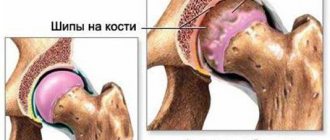
- arthrosis - dystrophic destruction of cartilage;
- ligamentitis, ligamentosis - similar lesions of the ligaments;
- bursitis - inflammation of the joint capsule;
- myositis - inflammation of a muscle (brachialis, forearm muscle);
- tendonitis - inflammation of a tendon (for example, the triceps muscle);
- damage to blood vessels and nerves;
- tumors - the use of a tomograph helps to identify tumors of any size.
The effectiveness of diagnostics depends on the power of the device used for scanning. High-quality images are obtained on devices from 1.5 Tesla and above.
What does an MRI show?
An MRI of the elbow allows you to see the condition of the internal tissues, cartilage, bones, and synovial membranes.
If certain symptoms are present, determine the severity and extent of the disease, which is of great importance in treatment and the future.
The study allows you to see:
- arthritis, arthrosis;
- nerve damage;
- tumors;
- injuries;
- presence of infections;
- fractures;
- bursitis;
- abscess;
- osteonecrosis;
- inflammation of the synovial membranes;
- ganglion cyst;
- various injuries to muscles, ligaments, tendons.
Advantages of MRI
Doctors are inclined to prescribe a magnetic tomography scan, arguing for the following advantages:
- Safety - during procedures, the device generates a magnetic field that does not harm the body. The list of restrictions is minimal, the examination is carried out repeatedly and the patient’s condition is monitored.
- High diagnostic value - the tomograph scans in a three-dimensional projection and makes layer-by-layer sections. The photographs reveal swelling of soft tissues and small pathologies.
- Identification of physiological abnormalities - the use of diffusion-weighted MRI helps to assess the characteristics of blood flow and lymph movement.
Unlike other research methods, MRI does not require preparation and is performed with caution on pregnant women and children.
How is the scan performed?
Magnetic resonance imaging is performed in open and closed type devices. The examination is carried out with or without contrast.
It is believed that open MRI is less accurate than closed MRI. An open tomograph scans patients who are overweight or who suffer from claustrophobia.
The open apparatus is a table above which there are recognition scanners. The patient lies down on the table and the elbow is scanned. The duration of the study varies from 15 to 45 minutes.
Scanning progress in a closed-type tomograph
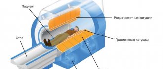
If the diagnosis is carried out in a closed apparatus, then the patient lies in the capsule, his head is fixed in a special device, his arms and legs are secured with belts.
To eliminate discomfort from the noise of the tomograph, earplugs are inserted into the ears. To signal the doctor if a person feels unwell, there is a special bulb that the person holds in his hand during the entire procedure.
The doctor is in another room and monitors the progress of the scan on a computer monitor. After the procedure is completed, the patient, without making sudden movements, carefully gets up and leaves the diagnostic room. A person receives the results of MRI interpretation on the same day or on the day prescribed by the doctor.
When to Apply Contrast
MRI with contrast is used if there is a suspicion of a tumor in the bone tissue. This is done to more accurately determine the location of the tumor and its size.
Gadolinium is used as a contrast agent, on the basis of which contrast agents are produced.
The technique involves injecting a contrast agent into the human body, which enhances the magnetization effect of tissues. The contrast solution accumulates in areas with good blood flow, which is typical for tumors due to their rapid growth.
Thanks to the contrast, these areas are clearly visible in the photographs. The procedure itself is quite painful and is supervised by an anesthesiologist.
How long does the examination take?
A standard magnetic resonance diagnostic procedure lasts 15-45 minutes. If an elbow scan with contrast is ordered, an MRI takes much longer.
To begin with, a standard scan is performed, then the patient returns to the doctor's office and is given an intravenous contrast agent. After this, a re-diagnosis is carried out. The duration of an MRI study with contrast is approximately 50 minutes, in each case individually.
Recommendations for MRI
A CT scan is recommended if there are symptoms indicating disease in the elbow area. Sometimes the test is ordered for clinical reasons. Like any diagnostic method, MRI has a list of limitations.
List of indications
MRI of the elbow is prescribed in the following cases:
- Previous injuries - after bruises there is pain that does not go away for a long time and limits movement.
- Lumps - the neoplasm is palpable through the skin and makes it difficult to flex or straighten the forearm.
- Constant pain in the elbow and below - this symptom indicates possible destruction of bone tissue, damage to blood vessels and nerves.
- Limitation of movements - even in the absence of pain, it is recommended to undergo a tomograph to find out the cause.
- To clarify the diagnosis - if after a CT scan it was not possible to detect changes.
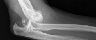
Epicondylitis
After an MRI, the doctor carefully examines the images, and if pathology is detected, treatment is prescribed.
Contraindications
Despite the safety of magnetic waves, there are limitations to research. Ignoring contraindications harms the patient’s health and spoils the quality of the images:
- Research is prohibited if there are metal implants in the body - they create interference on the monitor.
- You cannot carry out procedures if there are electronic devices or sensors - the tomograph will disable them.
- Research is limited in cases of claustrophobia and mental disorders - if desired, sedatives are used for medicated sleep.
- The device is harmful in the 1st and 3rd trimesters of pregnancy; scanning is not recommended for children under 5 years of age - in this case it is better to choose another method.
How does the procedure work?
Preparation for the scanning process comes down to the fact that the patient is asked to remove all metal and metal-containing objects. If you are intolerant of a confined space, you should notify your doctor. In such cases, sedative medications are taken.
When scanning with a contrast agent, inform the doctor about the possible occurrence of allergic reactions to iodine.
Scanning algorithm
If the examination is carried out using a closed tunnel scanner, the patient lies on his back or stomach. When obtaining images using an open-type tomograph, the patient undergoes the procedure in a chair, reclining.
- The patient is placed on a mobile table or chair, while his outstretched arm is secured to the bolster with special straps.
- The tomograph ring begins to move around the joint, transmitting a layer-by-layer image to the computer screen.
- The examination time is about 30 minutes; when using contrast, the time increases.
- After deciphering the examination results, the patient is given a photo.
Scanning a diseased joint using a contrast agent is considered a more complex procedure. The administration of a drug based on iodine is necessary if the doctor suspects various types of tumors. The contrast helps to determine the exact boundaries of the spread of the tumor and examine the network of blood vessels. The substance is carried throughout the body by the bloodstream and accumulates in those places where the inflammatory process occurs. After a day, the contrast agent is removed from the body.
Scanning procedure
A closed tomograph is used for the examination - it consists of a tunnel in which there is a ring-type magnet.
Most medical institutions are equipped with such devices; they are universal and are used to scan any area of the body.
You can undergo an MRI on a specialized tomograph for extremities - the arm is placed in it up to the elbow and a scan is performed. These devices are designed for people with claustrophobia.
Scanning in a closed tomograph
Let's consider the procedure on a closed tomograph for examining the extremities:
- before the examination, the patient removes metal jewelry, clothing containing steel and its alloys;
- the subject lies down on a special chair with an armrest, the arm is fixed with straps to limit mobility;
- the platform slides into the tunnel to the middle of the shoulder;
- A scan is performed, after which the limb is released.
For MRI of the extremities, various models of devices are used, and the methodology is sometimes different.
Study with contrast
A contrast agent is required to detect vascular changes and detect tumors.
This addition improves image quality and information content. It is not recommended to eat 4-5 hours before the procedure.
The introduction of contrast is prohibited:
- with individual intolerance;
- for diseases of the kidneys and liver.
To exclude allergies, tests are carried out in which contrast is introduced in a minimal amount.
If there are no side effects, the elbow scan begins - during the injection the patient feels discomfort.
Preparing for an MRI of the elbow joint
Lateral view of MR imaging of the elbow
No special preparation is needed before MRI. The patient is allowed to spend the day without any special features: take food and medications. It is recommended to arrive at the office 10-15 minutes in advance to remove all metal objects (jewelry, piercings, etc.) in a calm environment. These measures are necessary to prevent image distortion and maintain its quality.
When a person spends a long time in the tunnel of the device, fear may appear. If there is a fear of closed spaces, the possibility of using sedatives or sedatives should be discussed before the study. The doctor will prescribe the medicine and the patient will be able to undergo the scan without fear.
MRI reveals pathology of the elbow joint regardless of age. In the case when the study is carried out on a child, parents need to mentally prepare the child. It is necessary to explain how the scanning takes place, what sounds will accompany it, and how long it will last. The main thing is that the child must remain completely still during the procedure. This condition determines the accuracy and reliability of the information received.
During the diagnostic period, one of the parents may remain in the room to support the child. When the examination is carried out on a baby who does not yet understand speech, anesthesia will be required. It can be carried out in a hospital inpatient department. performs MRI for children over 5 years of age, enhanced scanning for children over 12 years of age.
If it is necessary to administer a contrast agent, the patient should prepare for an intravenous injection. We recommend having a light snack 40-45 minutes in advance to reduce side effects - activation of the autonomic nervous system (increased salivation, feeling of heat, etc.).
You should take a referral for an MRI in advance - this will reduce the cost of the procedure. However, it is possible to perform scanning on your own.
Decoding the results
If you know the basics of anatomy, you can independently analyze MRI images of the elbow joint and note the signs of pathology:
- Developmental anomalies - it is difficult to make a diagnosis here without medical education. Bifurcation of bone processes and the appearance of growths may attract attention. More often this condition is confused with oncology.
- Consequences of injuries - it is difficult not to notice fractures; they are visualized by a fracture line, sometimes by displacement. Dislocations are characterized by an increase in the distance between the articular surfaces and deviation of the bones from the vertical axis.
- Arthritis - there is a decrease in the joint space, thickening of the cartilaginous surfaces.
- Arthrosis - the joint areas become rough, and in severe cases, deformation of the entire structure occurs.
- Inflammation of soft tissues (ligamentitis, bursitis, myositis) - signs of swelling are noted (the pattern becomes light in color), soft tissues thicken.
- Oncology - there is a compaction with smooth edges and a uniform pattern (benign tumor) or without strict boundaries with blurred parenchyma (cancer).
Bursitis
Scan results
After completing an MRI or CT scan of the elbow joint, the patient needs to wait for the results, which will be ready an hour after the manipulation. If a more complex examination was carried out, then the person will receive the diagnostic results within 24 hours. Using magnetic resonance imaging of the elbow joint, it is possible to identify the following pathologies:
Diagnostics will determine the extent of damage to soft and bone tissues.
- bursitis;
- fractures;
- bone cracks;
- infection of bone tissue;
- synovitis;
- nerve injury;
- arthrosis;
- arthritis;
- damage to tendons, cartilage and ligamentous tissues.
Alternative research methods
In case of contraindications, diagnostics are carried out using the following research methods:
- CT scan shows bones well, but poorly visualizes soft tissues. For better information content, contrast is used.
- Ultrasound - shows inflammation, helps to examine soft tissues, but the image is two-dimensional.
- Biopsy is prescribed if cancer is suspected. The doctor takes a piece of tissue and examines it under a microscope.
MRI shows damage to bones and soft tissues, and helps to diagnose inflammatory and destructive diseases.
Preparation and technique for the procedure are standard. In case of severe contraindications, CT, ultrasound, and in some cases biopsy are prescribed.
Benefits of MRI
Magnetic resonance imaging is a non-invasive, informative and accurate type of radiation examination that provides a high-quality image of the internal structures of the body. When diagnosing complex joints such as the elbow, MRI eliminates additional tests, providing complete information about the condition of all tissues of the elbow.
Advantages of MRI in diagnosing the elbow:
- absence of radiation, ionizing radiation;
- possibility of research in several projections: sagittal, axial and frontal. This allows you to take into account all the anatomical features of the joint and obtain three-dimensional images;
- if there is a suspicion of the presence of neoplasms in the early stages in the elbow area, MRI is the only non-invasive type of examination;
- MRI has high contrast and sensitivity in detecting edema and infiltration of soft periarticular tissues;
- displays not only the structure of tissues, but also various functional parameters (blood flow speed, intraosseous pressure);
- the study can be carried out as many times as necessary, because it does not harm the person and is painless.
When examining the elbow, MRI is the only study that does not require additional tests. After all, with its help, developmental anomalies, injuries, degenerative-dystrophic disorders, inflammatory and tumor processes in all tissues of the elbow are identified.
Alternative diagnostic methods, advantages and disadvantages of MRI
The main alternatives to MRI are x-rays and computed tomography. Ultrasound examination can also help identify joint disease. Depending on the secrecy and severity of diagnosing the pathology, one or another option for hardware research will be preferable. To identify various arthrosis, visual assessment methods, as well as palpation, can be used.
The advantages of using MRI include:
- the possibility of repeated use of the procedure in the absence of contraindications;
- obtaining an image in any plane without the need to change the position of the subject;
- the ability to find out other parameters during the examination (for example, the speed of movement of biological fluids);
- high resolution of the resulting image.
The disadvantages of using magnetic resonance imaging are:
- long duration;
- a large number of restrictions and contraindications;
- inability to accurately evaluate tissues and structures that have low proton density (bones, calcifications).
Using this type of tomography, it is possible to assess the condition of all structures of the elbow, including joints, muscles and ligaments.
When diagnosing joint diseases, the shortcomings of MRI are usually not critical. In any case, you can always use alternatives to this procedure or combine the use of several types of research.
Advantages of the method
The advantage of this method is the ability to examine bone tissue, muscles, cartilage, tendons, and blood vessels. This diagnostic allows us to visualize various types of tumors much better than other methods. In addition, it is possible to obtain a detailed image of the pathological focus and the extent of its spread as accurately as possible. It is worth noting that this procedure does not have a negative effect on the human body, so it can be performed an unlimited number of times. Also, MRI of the elbow joint does not require special preparation, does not take much time and does not require rehabilitation.
Content:
- Advantages of the method
- Indications and contraindications
- What can an MRI of the elbow reveal?
- Preparation for the procedure
- Carrying out the procedure
- Why do you need a contrast agent?
How much does an elbow MRI cost?
Magnetic resonance imaging is an expensive type of diagnosis. The specific price for an MRI of the elbow depends on the type of machine used for the study. Tomographs can have different physical properties, resolution, and operating speed. Tomographs are divided into low-field and high-field.
Low-field tomographs have magnetic field strengths in the range of 0.2 - 0.5 Tesla. The cost of research using such a device will be lower due to lower power consumption and a limited set of functions.
High-field devices have a magnetic field strength of 1 – 1.5 Tesla. They have wider diagnostic capabilities. Tomography using a high-field device is much more expensive.
The average cost of an MRI of the elbow joint using a low-field, open-type device ranges from 2,000 to 4,000 rubles. The price of an MRI of the elbow using a high-field closed-type device ranges from 3,500 to 7,000 rubles. If the administration of contrast is required, the cost of the study increases by 1.5 times, because this is a longer and more labor-intensive procedure.
Magnetic resonance imaging is a modern, highly accurate type of research. It can be used as the only type of diagnosis of elbow pathologies, because MRI displays changes in all tissues of the joint under study. A significant disadvantage of MRI is its high cost compared to other types of studies.
Looking for a good doctor? Make an appointment with the best rheumatologist or orthopedist in your area.
295
Orthopedist
Types of magnetic resonance imaging scanners
MRI machines vary in power, appearance, and set of functions. But they are all divided into two types: closed and open.
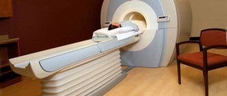
- Closed MRI – used in 90% of clinics. The device is equipped with a retractable platform on which the patient is placed. The platform automatically enters the tomograph. During the examination, the patient remains in a closed space. Closed tomographs have wide functionality and extreme accuracy.
- Open MRI - the patient lies on a platform, magnets are located above and below the area being examined. Such tomographs have a lower magnetic field potential and cannot qualitatively show the structure of some organs. But at the same time they have a number of advantages.
Advantages of open tomographs:
- used for patients suffering from claustrophobia;
- convenient for examining people with increased body weight, more than 130 kg;
- such a device is indispensable if tissue scanning is required during a surgical operation;
- Open-type tomographs are used to conduct examinations for young children who require the presence of their parents.
MRI of the elbow joint should be performed using closed-type machines. Open tomographs cannot qualitatively visualize all structures of the elbow joint.
Treatment of joints with expensive pacifiers: rheumatologists deceived patients across the country for 12 years... Read more>>
How to prepare for diagnosis
Preparing for a tomography does not require much effort. If after the initial study no contraindications to the procedure are found, then the person simply needs to lead a normal lifestyle and eat the same way as before.
Doctors recommend coming to the examination in loose clothing without iron elements. If the clothing does not meet the stated parameters, the person will be given special disposable underwear to wear during the scan.
It is recommended to check with the doctor who prescribed the MRI about the nuances of the procedure. The specialist, who will be present throughout the entire scan, will advise him in detail before placing the subject inside the tomograph.
Timely diagnosis will facilitate treatment of the elbow joint
What a diagnostic MRI examination shows:
- osteoarthritis;
- synovitis, bursitis;
- the nature and severity of the injury;
- presence of neoplasms;
- nerve damage, neuropathy;
- osteochondropathy;
- osteomyelitis;
- the presence of inflammatory diseases in soft tissues, etc.
If you decide to have an MRI done in one of our centers, you will be able to reliably find out which disease is causing you discomfort. Addresses and prices are listed on our website. Our prices will help you undergo diagnostics at optimal material costs.