#!KTna4alo!#
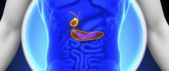
CT scan of the gallbladder
often performed when diagnosing diseases and as an additional examination in preparation for surgery in abdominal surgery. The method allows you to take a series of layer-by-layer images of the area under study, which clearly shows the internal structure of the gallbladder.
Is it possible to do a CT scan of the gallbladder?
The computed tomography method does not always detect gallstone disease. Therefore, this study is prescribed when it is not possible to resort to other diagnostic procedures or in order to clarify the diagnosis. The build and anatomical features of the patient are the determining factor when choosing examination tools.
There is a restriction on performing a CT scan of the gallbladder, which shows that this method is unsafe for health. The recommended frequency of the procedure is no more than once every 3 or 4 months. The primary purposes of computed tomography are:
- Diagnosis of stones and their difference from tumor-like processes of benign and malignant nature.
- Determination of the type of jaundice (hemolytic, mechanical or parenchymal).
If the clinical picture of the disease is atypical, then tomography is recommended. This study allows you to accurately determine whether there is thickening of the walls, changes in shape, and see structural changes in the organ better than with ultrasound. However, identifying purulent processes using CT is difficult, since bile and pus have an equal absorption coefficient.
The use of contrast makes it possible to visualize inflammatory changes. However, this examination is additional for cholecystitis and helps to obtain data for the correct choice of subsequent treatment tactics.
Contraindications
The doctor does not prescribe the procedure for everyone.
The following are not allowed for a CT scan of the gallbladder:
- people who cannot overcome claustrophobia;
- pregnant and lactating women;
- people with an allergy to contrast agent (iodine);
- obese people;
- children under 6 years old;
- patients with certain serious chronic diseases.
If a person has recently undergone fluorography or x-rays, then he should refuse the procedure for several months.
In addition, if the patient is not conscious, then his relatives must sign a document authorizing the CT scan.
If breastfeeding women decide to undergo a CT scan, then the baby should be breastfed no earlier than 2 days after the procedure.
Despite these contraindications, the procedure, carried out no more than once every 4 months, does not have a negative effect on the body of a healthy person.
Indications
Computed tomography of the gallbladder is carried out along with studying the condition of the pancreas and liver. This procedure is prescribed for the following reasons:
- secondary changes in the abdominal organs caused by impaired blood flow;
- detection of neoplasms of various origins and metastases;
- pancreatitis in acute and chronic form;
- identifying the extent of damage after abdominal trauma;
- hepatomegaly (exceeding the normal size of the liver for unknown reasons);
- obstructive jaundice (to identify the causes of the disease);
- pathologies affecting the structure of the liver (hepatitis, fatty liver, cirrhosis, hemochromatosis, cysts);
- cholelithiasis (presence of stones in the cavity of the bladder itself or ducts);
- painful sensations after eating on the right in the iliac region;
- preliminary examinations before surgery and controls after the intervention;
- parasitic diseases;
- monitoring the productivity of treatment of the tumor process.
To identify pathological foci in the liver, including tumor processes, tomography is the best study. This method is complemented by computer cholangiopancreatography, which allows one to obtain images of the bile ducts and bladder. When diagnosing, you can trace the presence of bile and its movement. Thanks to this, it is possible to determine the narrowing of the lumen of the bile ducts, anomalies in their structure, and diagnose the presence of stones and sclerosing cholangitis.
Contraindications
Since the procedure is performed using X-rays, there are contraindications to its use. The study is not carried out if:
- breastfeeding;
- pregnancy;
- multiple myeloma;
- tumors of any nature;
- bronchial asthma;
- claustrophobia;
- allergies to iodine and drugs used as contrast;
- using iodine and barium in the previous 5 days as a contrast agent.
In childhood, tomography is performed only if absolutely necessary; it is recommended to replace it with more gentle diagnostic methods (MRI and ultrasound). Children under 6 years of age are given sedatives or anesthesia to prevent mobility during this procedure. The doctor must tell the patient about all restrictions, contraindications and consequences of radiation in the process of preparing for the session.
Stages of implementation
When a patient is ready for a CT scan, a series of steps are performed:
- First, remove all metal objects, including hearing aids, as they will distort the results.
- The patient is dressed in special clothes.
- He is then placed on his back on a special couch in the tomograph.
- After this, the person finds himself inside a machine that takes pictures of the organ, while moving back and forth.
- Sometimes the patient may be asked to hold their breath.
- The diagnostic results are recorded on disk, and the doctor makes a diagnosis.
During a CT scan, the patient should not move and should always listen to the radiologist's instructions. If this point is not observed, the pictures may turn out unclear.
As noted, CT scans only show the presence of solids in the body. Therefore, if a CT scan does not show stones in the gall bladder, but they are clearly visible on the ultrasound results, this means that you can get rid of them with the help of drugs without resorting to surgery.
To find stones in the organ, it is necessary to perform a CT scan in cholecystography mode.
If both procedures did not show the presence of stones in the gall bladder, but there are changes in the tests, this may indicate the presence of polyps in it. Computed tomography helps determine the cause of new formations and their types. The procedure does not last long: from 15 to 30 minutes.
Preparation
A CT scan of the gallbladder requires certain conditions to be met. Preparation consists of following a diet for 2 days before the procedure. Restrictions are imposed on products that contribute to gas formation:
- vegetables containing coarse fiber;
- carbonated drinks;
- dairy products;
- bakery products.
The evening before the test, you must avoid eating. Otherwise, the information content of the method will be questionable: the images will show darkening and spots from food lumps, which will be impossible to differentiate. Before this event, you should drink a laxative or do an enema. On the eve of the session, medications that block x-rays may be prescribed as a contrast agent. A few hours before the diagnosis, it is recommended to drink still water (2-3 l).
A mandatory condition for admission to such a diagnosis is the absence of metal jewelry and piercings on the body, since these items can distort the results of such an examination.
Contrast method
Sometimes, to obtain more accurate results, a CT scan is performed with the introduction of a substance that stains the bile. It has a natural color that has contrasting properties.
This method is not often used because bile is reflective. However, it can be used in relation to patients with non-standard positions of organs or a special structure of the body.
The contrast agent enters the body in two ways:
- Half an hour before the procedure, it is injected into the blood using a catheter.
- 8–12 hours before the CT scan, take iopanoic acid and eat a high-fat meal 20 minutes before the procedure.
It is possible to detect the presence of stones in the gall bladder without introducing a dye only when their density does not coincide with the density of bile.
A more modern type of CT is spiral computed tomography (SCT). The emitter rotates along a spiral trajectory, while the tomograph itself makes translational movements. The result is tomograms that show the structural features of the organ in various planes. The thickness of the sections is adjusted by the doctor depending on the goals being pursued. In addition, there is multislice CT, which has a number of advantages.
- clearer image;
- the surface under study is larger;
- radiation exposure is less than with the standard procedure;
- the ability to create a 3-D model of an organ.
Thus, there are different subtypes of the same procedure that can be prescribed to a patient.
How is diagnosis done?
A CT scan of the abdominal cavity to determine the density of stones, if any, is performed by trained personnel. The procedure lasts from 20 minutes to half an hour. The results obtained are interpreted by a radiologist, who issues a final description. This medical document is subsequently provided to the attending physician.
Before the session, the patient must remove clothes, jewelry and shoes and remain in underwear. Some centers offer to wear a disposable gown.
The tomograph is a bulky device with a round hole. During diagnostics, the table on which the person lies moves slowly in the annular zone of the device. The equipment makes clicking sounds and takes pictures in different projections.
It is forbidden to move during scanning, otherwise it will not be possible to achieve accurate images. The staff monitors the progress of the study from another room and maintains contact with the patient using a special device. This allows you to control the process and guarantees timely assistance if necessary.
CT with contrast
A CT scan of the gallbladder with contrast is necessary to obtain accurate information. Since bile is reflective, the method is used more often for patients with non-standard placement of internal organs. The introduction of a special substance is carried out in one of the following ways:
- Intravenously. The drug is administered 30 minutes before the test.
- Orally. 12 hours before the session, iopanoic acid is taken. A third of an hour before the procedure, eat fatty foods.
Detection of stones is possible without taking specific medications if the density of the stones does not coincide with the density of the bile. In most cases, the images show stones containing calcifications. It is difficult, but possible, to establish the presence of formations with a different chemical composition. Determining the presence of stones and polyps depends on the professionalism of the doctor.
Computed tomography of the biliary tract with contrast is not performed in the presence of individual intolerance to the drug. If there is a history of diseases such as type 2 diabetes mellitus or thyrotoxicosis, then the administration of iodine-containing drugs should be avoided. Otherwise, metabolic disorders may develop.
Basic methods for diagnosing such pathologies
The difficulty of making an accurate diagnosis of gallbladder diseases is associated not only with their asymptomatic course at an early stage of development. The matter is further complicated by the fact that the clinical picture of such diseases is often very similar (for example, with stones and with gallbladder polyps), and without instrumental differential diagnosis it is not possible to determine an accurate diagnosis.
In addition, modern methods of instrumental research allow doctors to accurately determine not only the nature of the pathology itself, but also the stage of its development, location and condition of the organs surrounding the bladder.
The main symptoms by which one can suspect the development of diseases of the biliary system are:
- pain in the right hypochondrium, which can radiate to the area of the right shoulder blade, right shoulder and even the left side of the abdomen; they can be of varying intensity, occur in paroxysms (the so-called biliary colic) or be permanent;
- bitter taste in the mouth;
- heartburn;
- belching air;
- change in the color of stool (urine darkens and feces lighten);
- dyspeptic disorders (diarrhea and constipation);
- in advanced cases, yellowing of the skin and sclera of the eyes is possible.
Read also: How are the liver and gallbladder connected?
If you notice such negative symptoms, you should seek medical help as soon as possible.
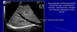
The main method for detecting such diseases is ultrasound - ultrasound examination of the internal organs of the abdominal cavity. This diagnostic technique is widespread and accessible, and its results allow the doctor to fairly fully assess the course of the pathology, the stage of its development and location.
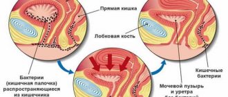
CT and MRI diagnostics of gall bladder stones (arrows)
To clarify the diagnosis, endoscopic examination is also used, in which the equipment installed on the endoscope is delivered to the organ itself and gives a more detailed picture of the course of the disease.
In difficult cases, the doctor may prescribe an MRI (magnetic resonance imaging) procedure, which in the case of the gallbladder is called magnetic resonance cholangiography. This modern technique gives the most detailed picture of the course of the pathological process and allows you to assess the condition of the bile ducts.
It is worth saying that the use of such techniques in addition to ultrasound is not always used. They are needed as additional studies in cases where the doctor has certain doubts about the accuracy of the diagnosis made using ultrasound or when it is necessary to accurately establish the location of the pathology (for example, where are the gallstones or polyps, are they in the biliary tract or no, and also what size they are and how many there are).
Another modern method for diagnosing diseases of this organ is CT scanning of the gallbladder. Let's take a closer look at this study.
Research results
Computed tomography of the gallbladder provides accurate information about the location, structure and presence of stones. The study allows us to establish compaction of the walls of the biliary tract ducts, which indicates an inflammatory process in this area. Using this method, it is possible to detect acquired and congenital anomalies of the abdominal organs.
Tumors that have affected the gallbladder are identified on CT scans as areas with altered density. Accumulations of masses enclosed in a capsule are a sign of a purulent process (abscess). The consequences of traumatic injuries look like this:
- Hematomas in the image are visualized as areas with a homogeneous structure without a capsule, the boundaries of which are the internal tissue of the organ.
- Duct ruptures are characterized by the release of contrast agent beyond their boundaries.
The presence of foreign bodies and their location are determined by tomography with high accuracy. The speed of issuing a medical report depends on the operating regulations of the medical center and can take from several hours to 2 days. The patient is provided with the following kit:
- A CD containing all the pictures taken;
- a doctor’s report, handwritten or printed with a detailed description of the existing deviations;
- a group of photographs on film.
Advantages and disadvantages of the CT method
Diagnostics using a tomograph has a number of advantages:
- possibility of obtaining sections 0.5 cm thick;
- absence of unpleasant sensations for the subject;
- the production of three-dimensional images increases the information content of the method;
- low time spent on examination;
- use of contrast if necessary;
- minimal radiation exposure to the body;
- accurate identification of factors that influenced the blockage of the duct and determination of the location of the pathological process;
- good image quality allows you to detect the slightest changes;
- determining the presence of stones and their density;
- exclusion or confirmation of acute cholecystitis;
- non-invasive;
- Possibility of combination with other examination methods.
The disadvantages of the method include:
- the harm of X-ray radiation limits the possibility of use;
- using tomography it is impossible to judge how an organ functions;
- this method has lower accuracy compared to MRI or ultrasound;
- the procedure can be repeated only after 120-160 days;
- people with a high degree of obesity cannot be tested;
- the high cost makes the method inaccessible to people on a limited budget.
You shouldn’t limit yourself to just CT. It is recommended to undergo a comprehensive examination.
Features of computed tomography
Computed tomography of internal organs, including the gallbladder and biliary tract, is a type of x-ray diagnostics, carried out using special equipment - a tomograph. The method is distinguished by maximum accuracy (up to 98%), information content and reliability.
CT is based on the ability of tissues of different densities to absorb X-rays at different rates. X-ray beams penetrate the organ from different sides with a certain concentration and to the required depth, in order to obtain the most accurate result.
The thickness of the sections reaches 0.5 cm. The pictures, two-dimensional or three-dimensional images, display the entire cavity of the gallbladder in the form of black and white shades, showing all kinds of anatomical, physiological or pathological changes.
The CT technique, depending on the type of tomograph, is divided into types:
- step-by-step (highest radiation dose, accurate result);
- spiral (the radiation dose is much less, even more accuracy and information);
- multispiral (the safest, most accurate, informative).
Some experts equate step-by-step research with information content to ultrasound. The choice in favor of CT is made only by the doctor based on diagnostic feasibility.
What are the analogues of this method?
MRI and ultrasound are alternatives to computed tomography. In terms of content and accessibility, ultrasound diagnostics takes first place. Since ultrasound allows us to identify pathologies of this organ (neoplasms, cholecystitis, stones), it can be called optimal.
Magnetic resonance imaging does not pose a health risk because it uses an electromagnetic field instead of X-rays. MRI makes it possible to diagnose all organ abnormalities, but the high cost of the study limits accessibility for some citizens.
CT scanning is recommended when making a diagnosis to select the correct treatment tactics.