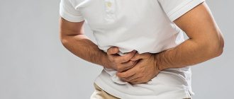
Cholelithiasis is a fairly common pathology of the hepatobiliary system, associated with impaired cholesterol or bilirubin metabolism and the formation of stones in the gall bladder. The disease is widespread in industrialized countries, where people pay little attention to their diet, preferring fried, fatty and spicy foods.
This disease is difficult to treat conservatively, therefore, in the presence of stones, many experts recommend surgical intervention, the “gold” standard of which is laparoscopy of gallstones and cholecystectomy. However, before moving on to therapeutic tactics, it is necessary to study the mechanism of stone formation.
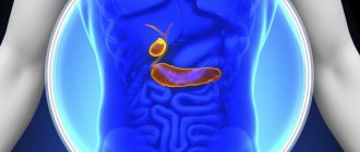
Anatomical features of the gallbladder
The gallbladder is a small organ that is hollow inside and resembles a sac. It is localized under the liver. The bladder has a body, a small narrow end (neck) and its continuation - a duct that connects with the same hepatic one. They merge into one common one - the common bile duct, which flows into the small intestine. At the junction of the ducts there is a valve that regulates the infusion of bile.
The top of the bladder is adjacent to the liver, the bottom is adjacent to the peritoneum and is covered with a connective film. In the middle part of the organ there are muscles that help push out accumulated bile. The inside of the bladder is protected by a mucous membrane. The bottom of the organ is adjacent to the wall of the abdomen. The ducts vary in length and number.
The main function of the bladder is to accumulate bile. Once the bolus of food is in the stomach, the substance is released into the small intestine. The bubble is emptied reflexively. Without this organ, you can exist calmly, but the quality of life is noticeably reduced.
Where do gallstones come from?
Liver bile is a special liquid that resembles plasma in its composition. It has such important components as water, cholesterol, bilirubin and bile acids. While these components are in balance with each other, this liquid promotes the binding of fats with water and their breakdown, the absorption of fatty acids and cholesterol in the intestines, prevents the development of putrefactive processes in the final sections of the digestive tract, stimulates its peristalsis (unidirectional contractions in order to promote the food bolus) .
If the secretion of cholesterol into bile increases or the concentration of bile acids decreases, as well as the contractility of the gallbladder (GB), stagnation and crystallization of its contents occur with the formation of large and small stones.
Predisposing factors to stone formation and the development of cholecystitis are:
- High body mass index.
- Insufficient physical activity.
- Eating foods rich in cholesterol and low in fiber.
- Anomalies in the development of the gallbladder, for example, congenital bending of its neck.
- Elderly age.
- Female.
- Pregnancy.
- Endocrine disorders.
- Chronic infectious diseases of the biliary tract.
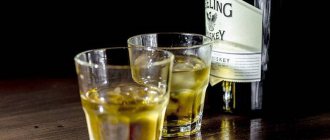
- Alcohol abuse.
- History of surgical interventions on the stomach and intestines.
Proper nutrition and an active lifestyle are excellent prevention of stone formation
Laparoscopic cholecystectomy: general description
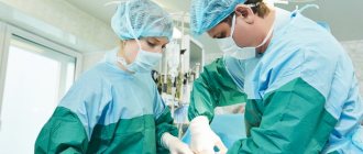
Laparoscopy of the gallbladder is the surgical removal of the organ. Sometimes the same term is used to remove formed stones. The main feature of laparoscopy is that the surgeon performs all manipulations through punctures into which the necessary instruments are placed. Visibility inside the peritoneum is provided by a laparoscope. This is a small mini video camera on a long rod, equipped with a bright flashlight.
The laparoscope is inserted into the punctured hole, and the image is transmitted to an external screen. The surgeon is guided by it during the operation. Various manipulations are carried out by trocars. These are small hollow tubes into which the necessary surgical instruments are placed. Trocars have special devices. With their help, instruments are manipulated - cauterization, pinching, cutting, etc.
How is surgery performed?
Laparoscopic surgery is performed under general anesthesia. First, carbon dioxide is injected into the abdominal cavity through a special needle, which raises the anterior abdominal wall and creates space for surgeons to work. Then, trocars, which are hollow tubes with valves, are inserted through small incisions.
Through them, various surgical instruments can be inserted and removed into the abdomen, an important part of which is the laparoscope (optical system). Next, the gallbladder is directly separated from other anatomical structures and removed through a small incision at the xiphoid process or near the navel.
The laparoscope allows you to display the image on the screen, as well as enlarge and reduce the image for the convenience of surgeons.
After removing the gallbladder, surgeons suture the surgical openings made and remove a special drainage to remove liquid contents from the abdominal cavity that may accumulate there as a result of inevitable injury to soft tissues during the intervention. The length of the operation is, on average, 45 minutes, but its duration can vary within certain limits depending on the extent of the pathological process and the anatomical characteristics of a particular person.
Advantages of laparoscopy compared to laparotomy
During a laparotomy, a cut is made in the abdominal wall so that the surgeon can see the desired organ. This operation is called laparotomy. Laparoscopy has many advantages over it:
- slight postoperative short-term pain;
- instead of incisions, punctures are made, which minimally damages the tissue;
- a hernia occurs extremely rarely;
- scars or seams are barely noticeable, sometimes not visible at all.
Also, laparoscopic surgery to remove the gallbladder has a short recovery period. A person begins to walk within six hours. He stays in a medical facility from 1 to 4 days. Working capacity is restored very quickly. Laparoscopy and laparotomy have the same step-by-step operation scheme. Both follow standard steps.
Postoperative period
After the surgical removal procedures are completed, the postoperative period begins. It involves staying in the intensive care unit for 3-4 hours. Intensive care staff closely monitor the condition of the admitted patient, in particular, how he recovers from anesthesia. After reaching a satisfactory condition, the doctors in the intensive care unit transfer the operated person to the ward. Here he will spend the next few days.
For the first 4-5 hours, the patient should lie in absolute peace, preferably without moving. Drinking, eating and trying to get up is prohibited. After about 6 hours, the patient is allowed to drink plain water without gas or additives. The liquid should enter the body in small portions, that is, you can drink in small sips. The interval between drinks is 7-10 minutes. You are allowed to start eating only one day after surgery.
On the second day after the procedure, the patient is allowed to stand up. This should only be done under the supervision of hospital medical personnel. After 2 days, you are allowed to take liquid food and walk on your own.
Types of laparoscopic operations
There are two types of laparoscopy of the gallbladder - excision of the organ or washing out of stones from it. However, the second option is now almost never used for a number of reasons:
- If there are a lot of stones in the bladder, then the bladder must be removed, since it is so deformed that it cannot perform its functions. In addition, the organ will regularly become inflamed, which entails the appearance of other pathologies.
- If the stones are small or few in number, then other methods of eliminating them are preferable - with the help of drugs or ultrasound.
Stone removal is also called laparoscopy if performed through punctures. However, they are not enucleated; the entire organ is removed.
Indications and prohibitions for bladder laparoscopy
Laparoscopy is done for all types of cholelithiasis or its complications. Indications for surgical intervention are:
- cholecystitis – calculous, not stone-like, asymptomatic (in case of acute cholecystitis, surgery is performed in the first days);
- polypous formations;
- cholesterosis
Laparoscopy of the gallbladder is contraindicated if:
- pancreatitis;
- cicatricial deformities in the neck of the organ;
- cholecystitis: gangrenous, “porcelain”, perforated;
- oncology or suspicion of it;
- intrahepatic localization of the organ;
- fistulas;
- respiratory pathologies;
- installed pacemaker;
- abscess;
- heart pathologies;
- unclear localization (or abnormal location) of organs;
- blood clotting disorders;
- after previous laparotomy operations on the peritoneum.
Laparoscopy of the gallbladder is not performed in the 3rd trimester of pregnancy, with portal hypertension, inflammation of the abdominal wall, or severe obesity. If it is possible to remove the stones in another way or eliminate the pathology with medication, then the operation is temporarily postponed.
Preparation for laparoscopic surgery
Preparation for laparoscopy of the gallbladder begins half a month in advance. First, OAM and OAC, biochemistry are taken, the blood type and its Rh are determined and coagulation is checked. A coagulation and electrocardiogram is performed. The blood is checked for syphilis, all types of hepatitis and HIV infection. A swab is taken from the vagina. If the tests are normal, the person is allowed to undergo surgery. To exclude complications, additional diagnostic methods (for example, ultrasound, CT, etc.) may be performed.
Seven days before the procedure, you need to stop taking medications that affect blood clotting. The day before gallbladder laparoscopy, you must begin to adhere to the diet recommended by your doctor. On the eve of the operation, they have dinner until midnight, then an enema is done (the procedure is repeated in the morning).
Preliminary studies
Before any operation, it is necessary to undergo a comprehensive examination and pass all the necessary tests. Only after this will specialists be able to assess the condition of the gallbladder and its ducts, which will affect the method of surgical intervention. Even if a more gentle laparoscopy is first prescribed, during the examination the specialist may change the recommendation to cholecystectomy.
In most cases, the patient is prescribed the following tests:
- complete urine analysis;
- detailed blood test;
- blood for ESR;
- a full range of biochemical studies;
- tests for HIV, hepatitis, syphilis;
- determination of Rh factor and blood group;
- hemostasiogram.
Additionally, it is necessary to obtain general opinions from the dentist and therapist. The patient will be allowed to undergo surgery if all the obtained indicators are within the physiological norm. In case of deviations, preliminary therapy will be required to normalize the results of the indicators.
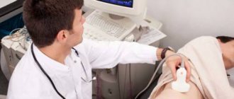
Ultrasonography
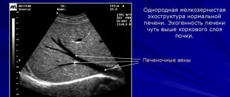
To conduct a complete analysis and assessment of the state of the digestive organs, it is necessary to undergo an ultrasound. With its help, a specialist will identify where and how the stones are located and what size they are. With the help of magnetic resonance imaging, you can obtain not only more accurate information about the stones, but also identify other possible problems with the gallbladder - narrowing of the ducts, the presence of scar formations, inflammatory processes.
In controversial situations, a computed tomography scan is additionally required. Thanks to this study, specialists receive the missing information about the general condition of the organ, the development of adhesions and peri-vesical tissue. Also, before the operation, the patient will have to undergo fluorography and electrocardiography to understand whether the body is ready for serious surgery and general anesthesia.
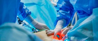
Gallbladder removal technique
After the anesthesia has taken effect, a thin tube is pushed into the stomach. It removes the contents of the organ. The probe remains in it until the operation is completed and prevents gastric contents from entering the respiratory tract.
After inserting the device, the patient's face is covered with a mask leading to the artificial respiration device. This is a necessary condition, since carbon dioxide pumped into the peritoneum compresses the lungs, which disrupts their function.
A small incision is made in the navel. Through it, (usually carbon dioxide) gas is pumped inside so that the peritoneum swells, which ensures maximum access of instruments to the necessary organs, while neighboring ones are not injured. A trocar with a video camera is inserted into the hole near the navel.
Three more punctures are made in the abdomen (right). Trocars are inserted into them, into which the necessary instruments are inserted. The location of the bubble is determined. If there are adhesions nearby, they are removed to free the organ. Then the degree of filling of the organ with bile is determined.
If the bubble is overstressed, then one wall is cut. Some of the liquid is sucked out through the hole. A clamp is then applied to the incision. The common bile duct is located and cut, the artery associated with the bladder is released. It is clamped with two staples, and the vessel is cut between them. The edges are then sewn shut.
The bladder is cut off from the liver. Vessels that have begun to bleed are cauterized with electric current. The bubble is then carefully separated from the rest of the tissue supporting it and pulled out through the hole in the navel. A laparoscope is used to examine the peritoneum from the inside to see if there is any bleeding, bile or altered tissue left in it. If present, they are removed and the vessels are cauterized. Then a liquid antiseptic is injected into the peritoneum to rinse the cavity, then the liquid is sucked out.
All trocars are removed from the punctures, the holes are sutured or sealed. If drainage is required, one hole is left. The tube remains in the body for a couple of days to remove any remaining antiseptic substance. If there is no need, drainage is not installed.
The duration of laparoscopic surgery is 40-90 minutes. In case of severe bleeding, injury to organs adjacent to the bladder, or other difficulties that cannot be corrected through punctures, the peritoneum is cut and a regular abdominal operation is performed.
Removal of stones
Removing stones from the bladder is almost no different from laparoscopy of the organ. The operation is performed under general anesthesia, the person is on artificial respiration until the end. Then all steps are repeated until the trocars are inserted. If adhesions are detected, they are removed.
Then the wall of the organ is incised and a tube is inserted to suction out the contents. When the procedure is completed, the incision is sutured. Then the peritoneum is washed from the inside with an antiseptic solution. The trocars are removed and the punctures are sutured.
Recovery after laparoscopy
After laparoscopy of the gallbladder, the patient gradually recovers from anesthesia. For six hours he is completely at rest. Then you can begin to move, rise and roll over (without sudden movements). For a few more days, the normal diet is gradually restored.
| Day | Medical recommendations |
| First | You are prohibited from eating for 24 hours after surgery. You can drink plain water or unsweetened rosehip infusion. |
| Second | Drink 1.5 liters of fruit compote or low-fat kefir. Drinks should be consumed every one and a half to two hours, half a glass. |
| Third | The diet is supplemented with cottage cheese, weak broth, fermented milk drinks and foods. The menu can include lean ground meat and fruits. |
| Subsequent | Starting from the 4th day, a person returns to a normal diet, but until the tissues are completely healed, he abstains from salty, peppery foods, and black bread. |
Usually the drainage is removed already on the 2nd day after laparoscopy. If the wounds left from the punctures do not heal, they hurt, or bother you, then in order to avoid complications you should consult a doctor. The first part of the rehabilitation period ends after the sutures are removed.
Then for another month you need to avoid lifting heavy objects and physical activity (especially not straining the abdominal muscles). This may cause suture ruptures or hernia. However, the body needs movement and light jogging or fast walking over short distances, and morning exercises are recommended.
In the absence of complications after 2 months. You can start doing physical exercise. Walking helps to cope with bile stagnation. After laparoscopy, you should only wear underwear made from natural fabrics. Full rehabilitation occurs after six months.
However, for the first three months you cannot lift more than three kilograms, for the 6th month - only up to 5 kg. For seven days after the operation, painkillers (for example, Ketonal, Ketanov and others), vitamin complexes (Multi-Tabs, etc.) are taken.
Analgesics are usually required only in the first days, after which the person no longer experiences pain. If it persists, this indicates complications and the need for medical consultation. When pain occurs again after months, it indicates new diseases have appeared.
Life after laparoscopy
After laparoscopy of the gallbladder, diet No. 5 is followed for six months. It supports the normal functioning of the liver, which produces up to 800 milliliters of bile in 24 hours. The fluid is immediately sent to the small intestine. This release of the substance disrupts the functioning of the gastrointestinal tract and provokes the appearance of various pathologies.
To protect yourself from excessive bile production, a person needs to follow the fifth diet for a couple of years (preferably for life). List of prohibited products:
- fresh bread;
- marinades;
- offal;
- smoked meats;
- mushrooms;
- conservation;
- pickles;
- green peas and vegetables (raw);
- spicy dishes;
- chocolate;
- roast;
- baked goods;
- fat;
- pastries, cakes, etc.;
- coffee;
- alcohol.
The menu consists of:
- white (but stale) bread;
- thermally processed vegetables;
- low-fat fermented milk products;
- liquid porridges;
- soups with weak secondary broth;
- lean types of meat and fish;
- fruits and berries (can be eaten fresh);
You can eat honey and jam with tea. You need to eat five to six times daily, in small portions. Dishes are stewed, baked, boiled or steamed. Should only be served warm; cold and hot are excluded. You are allowed to use 60-70 g of vegetable oil or 50 g of butter per day. Bread is allowed no more than 250 g, and granulated sugar - up to 25 g. Before going to bed, it is advisable to drink 200 g of low-fat kefir.
Among the drinks, preference is given to weakly brewed tea, juices, dried fruit compotes, and rosehip infusion. If bile is secreted too often, then you need to drink less fluid. Fish and meat pieces, raw vegetables are added to the menu only after three to four months. This diet is followed for two years, then you are allowed to eat any food.
Certificate of incapacity for work
After discharge from a medical institution, a person is issued a sick leave. It is issued from the first day of hospitalization. After the operation, the certificate of incapacity for work is extended for another 10-12 days. The total sick leave period is 13-19 days (including hospital stay). The period can be extended until the wounds are completely healed. This is determined on an individual basis.
Prevention of complications
To avoid the development of complications, the following preventive methods should be followed. Within 1.5 months it is necessary to remove physical activity. To avoid bacteria and infections, do not shower for 2 weeks. Swimming pool and bath are allowed every other month. Staying in the sun, visiting the bathhouse, sauna is allowed after 3 months.
Sexual intercourse is prohibited for 4 weeks. You should not eat hard-to-digest foods to prevent stretching and tension in the abdominal cavity. In order for laparoscopic sutures to heal without problems, all doctor’s recommendations should be followed. Thus, the healing process proceeds without any complications.
Consequences of laparoscopic surgery
After excision of the bladder, unexpected releases of bile substance may occur directly into the small intestine. This is accompanied by flatulence, heartburn with a bitter taste, and vomiting. Diarrhea and abdominal tenderness may occur. Negative symptoms are treated with Duspatalin and No-Shpy. Before returning to normal, you must follow diet No. 5.
Possible complications
Complications after laparoscopy of the gallbladder are the appearance of small seals, oozing discharge from punctured holes. In this case, treatment continues. The punctures are lubricated with iodine and should not be soaked for five days. Other complications include:
- Temperature increase.
- Leakage of bile into the peritoneum.
- Inflammation and redness of the tissues near the navel or at puncture sites.
- Peritonitis.
- Hernia in the scar area. When pinched, it is painful. Most often it occurs due to non-compliance with the ban on physical activity. The appearance of a hernia is accompanied by vomiting and constipation.
During laparoscopy of the gallbladder, the gastric walls, blood vessels, or organs adjacent to the gallbladder may be accidentally damaged. For example, the integrity of the intestines, liver, etc. is compromised. If such complications arise, the abdominal cavity is dissected and then the operation continues.
With proper preparation for laparoscopy, following all the doctor’s recommendations before and after removal of the bladder, surgical intervention is not accompanied by complications, the wounds heal quickly and the body recovers in the shortest possible time. The diet should be followed for at least 6 months after surgery, ideally up to two years.
- Laparoscopy of ovarian cyst
- Fallopian tube laparoscopy
- Laparoscopy is a new method of diagnosis and surgery
- Pregnancy after laparoscopy: when is conception possible?
- Ultrasound of the abdominal cavity: norms and interpretation of results
When do stitches heal?
Many people are interested in when sutures are removed after laparoscopy. They are usually removed 7 days after surgery. However, there are situations when sutures are removed after 2 weeks. This most often occurs in people who are overweight.
Today, self-absorbable threads are often used. They disappear on their own a week after the abdominal threads dissolve. If everything goes without complications, the scars will become invisible after 2–3 months. During pregnancy, scars after laparoscopic surgery become brighter and form stretch marks.
Materials required for processing