Detectable pathology
Injuries and wounds
- CT allows you to assess the integrity of the bladder wall, its shape, and its relationship to neighboring structures.
- Wall defects (partial and complete) may be detected.
- Rupture and perforation of the wall are accompanied by the release of the contents of the bladder beyond its boundaries.
- To detect defects, the bladder is filled with sterile contrast.
CT scan shows foreign bodies: catheter, wire, knife, bullet, fragment, etc.
Inflammatory diseases
Acute cystitis can manifest itself as intense contrasting of the mucous membrane. Chronic cystitis is manifested by deformation of the bladder and fibrosis of surrounding tissues.
Against this background, stones can be found in the cavity of the bladder, mostly dense, displaceable, 3-15 mm in size.
Anomalies
- Anomalies of position, shape, quantity, and size are rare.
- Sometimes a patent umbilical-vesical duct is found.
- Anomalies of the bladder are combined with disorders of other pelvic organs. Pathological anastomosis between the bladder and neighboring organs may be detected.
Degenerative changes in the wall
A diverticulum is a bulging of the wall of an organ due to its weakening or thinning. Bladder diverticula are a problem primarily in older people.
Sometimes diverticula can reach a huge size. The wall of the diverticulum may rupture with the development of urinary peritonitis.
Foreign bodies
They are divided into those introduced through the urethra, ureter, and those penetrated through the wall. Sometimes objects are found placed in the urethra during perverted sexual practices. Rarely does a ureteral stent enter the bladder cavity.
- Postoperative foreign bodies: needles, bandages, threads, instruments, napkins, etc.
- Traumatic: bone fragments from pelvic fractures, other wounding objects.
Tumors
Bladder neoplasms are divided into primary and secondary. Primary ones arise in the wall of the organ, secondary ones - in the surrounding structures:
- genitourinary system;
- prostate;
- seminal vesicles;
- ducts;
- rectum;
- in women, the uterus and ovaries.
The tumor may manifest itself as diffuse wall thickening in combination with enlarged lymph nodes. It may look like a polypoid formation growing into the lumen of the organ or outward.
Post-operative changes
The bladder may be partially resected, for example after trauma. Its deformation and changes in size are detected.
In patients with urinary problems, an incision is made above the pubis (epicystotomy), into which a catheter is installed to allow the unimpeded flow of urine.
Using Contrast
Contrast is used to visualize the bladder cavity in case of trauma and wounds, and the wall in case of tumors.
- Sterile iodine-containing drugs are used: urografin, non-ionic - omnipaque, ultravist.
- Contrast can be injected through the urethra to assess extravasation.
- Contrast can also be injected into the elbow vein (0.4-0.5 ml per kg of body weight) manually, scanning is performed 10-15 minutes after injection.
To contrast the wall, contrast is used in a dosage of 1-1.5 ml per kg of weight. Contrast is injected into the vein as a bolus using an injector, scanning is performed in three phases: arterial, venous-parenchymal, and delayed.
When should you do a CT scan of the bladder?
special preparation is required for CT examination of the brain, spine, joints, chest, or soft tissues (with the exception of contrast tomography).
CT scan of the abdominal cavity is performed on an empty stomach, i.e. You should stop eating and drinking 5-6 hours before.
A CT scan of the pelvis is performed in a state of average bladder fullness. To do this, you should not urinate 1 hour before the test, and also drink a glass of pure water 30-40 minutes before the test. Some medical centers recommend using the contrast drug “Urografin” according to the following scheme: 1 ampoule is diluted in 1 liter of water. On the day before the test, the patient should drink 1/2 of the solution, and 2 hours before the CT scan - the remaining half. Taking Urografin should be agreed with your doctor to avoid adverse reactions.
Before CT coronary angiography, the patient must refrain from eating for 4 hours. If the patient is taking beta blockers, they will need to be taken with them to the study. If the heart rate is more than 60 beats per minute, the patient will need to take a beta blocker to slow the heart rate, since otherwise the reliability of CT coronary angiography will be low.
CT colonoscopy of the intestine requires complex preparation for 3 days before the study. Since the informative value of virtual colonoscopy depends on good bowel cleansing and intestinal distention, the patient must follow a toxin-free diet for 3 days before the scan, i.e. exclude from your diet legumes, fresh vegetables and fruits, brown bread, baked goods, fermented milk products, carbonated drinks, mushrooms, etc. In addition, the patient must cleanse the intestines with the help of the drug "Fortrans", which is taken according to the following scheme: 1 package of the drug dissolves in 1 liter of water, 1 glass per 15 minutes for 1-2 hours, you need to drink this solution in the afternoon throughout all 3 days of preparation. On the day of the scan, the patient must refrain from eating and drinking.
CT contrast studies
Important: Do not forget to take a referral for a computed tomography scan, which will indicate the need for examination and the area for CT scanning; it will be issued by your attending physician.
Please note that during the examination it is better to wear comfortable, loose clothing that can be easily removed if necessary. Before starting the examination, we recommend removing glasses, watches, jewelry, hearing aids, and other metal objects.
How to prepare properly
Before contrast, a blood test for endogenous creatinine is required. If the indicator is elevated (120 µmol/liter or more), the risk of kidney damage increases (contrast-induced nephropathy).
CT urography requires bladder filling. Before the study, you should not urinate for 1-1.5 hours. You need to drink water to keep your bladder full.
You must take with you the conclusions from previous studies: ultrasound of the pelvic organs, kidneys, CT, MRI.
CT scan
Preparation for the procedure
Before undergoing a CT scan, the patient may have to remove some clothing, accessories, jewelry and metals as they are often opaque and can interfere with the scanning process and the quality of the results. On the day of the study, the patient should not eat anything; the day before the procedure, the patient is advised to eat light food that will not lead to gas formation. CT scan is performed with a full bladder.
CT may be contraindicated in children and pregnant women due to the radiation exposure used during the examination.
There are several more contraindications for performing a CT scan of the bladder with contrast. These include severe renal failure and allergies to contrast agents. In some cases, it may be difficult to examine overweight patients, since some devices are limited in permissible weight.
How the research works
Upon arrival at the clinic, the patient signs an informed consent. You need to remove all metal objects from yourself, remove your trouser belt, suspenders, etc.
Then the patient goes into the office where the tomograph is installed. You need to lie on the table with your back, head to the ring of the device.
- In the case of bolus contrast, a wide bore needle is inserted into the vein. An injector is connected to it.
- During CT urography, contrast is administered with a conventional syringe in a volume of 20-30 ml by hand.
- In retrograde urography, contrast is administered through a catheter in the urethra.
The scan then starts. Tomograms are performed in three stages for tumors, in a delayed phase for CT urography and once for retrograde urography. CT urography also includes examination of the kidneys. The radiologist analyzes the information received and writes a conclusion.
Interpretation of CT results of the urinary system
After the information from the sensors has undergone computer processing and appears on the monitor screen in the form of images, the doctor begins to study it. The radiologist who conducted the examination interprets the results of a CT scan of the urinary system. When describing, he takes into account the complaints with which the patient sought help. Each layer is considered separately. The photographs are printed.
The radiologist will confirm or refute the suspected diagnosis and report the presence or absence of stones or tumors. The doctor will detect the affected areas, describe the nature of the pathology, the degree of damage caused to the tissues. If a tumor is found, it is indicated whether it is malignant or benign. This information will be indicated in the conclusion, which the patient receives in his hands along with photographs, a copy of the contract and a check.
The radiologist does not make a final diagnosis, he only gives a recommendation as to which specialist should be contacted with the results of the examination. The diagnosis is made by a specialist doctor. He also prescribes treatment.
Other diagnostic methods
Computed tomography cannot be performed always, not everywhere, and not for everyone. This method can be replaced and (or) supplemented:
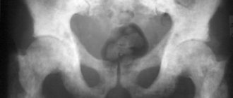
- classical excretory X-ray urography;
- retrograde X-ray urography;
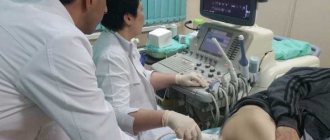
- ultrasound examination;
- MRI with and without contrast;
- nuclear medicine methods.
CT or MRI: what to choose?
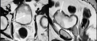
MRI has higher resolution for soft tissue and can distinguish fine structures that are not visible on CT. There is no ionizing radiation.
Computed tomography of the urinary system is preferable for identifying dense stones (calcifications), which are poorly visible on MRI. For bladder trauma, CT is also preferable.
MRI or CT: which diagnostic method is better for examining the bladder?
Magnetic resonance imaging and computed tomography are considered fundamentally similar research methods. One and the other provide the specialist with multilayer images when scanning the bladder. Pictures can be recorded on digital media after computer processing.
An MRI is recommended if there is a suspicion of bladder cancer. This diagnostic method visualizes the prostate, bladder, seminal vesicles, and pelvic lymph nodes. MRI allows you to assess the extent of tumor spread. CT is preferable in diagnosing urolithiasis. But not all stones are visualized by this diagnostic method. Magnetic resonance imaging is prescribed to detect uric acid, urate, and xanthine stones.
CT and MRI can be used simultaneously; they complement each other. Only a specialist decides which one to prescribe in your specific case. If you have been prescribed a CT scan and then an MRI or vice versa, do not refuse the second research method. Perhaps this will allow us to clarify the diagnosis and see the extent of the lesion inside the organ.
Contraindications
There are cases when the benefit of conducting research is less than the risk:
- The study cannot be carried out while pregnant or while breastfeeding.
- If you have problems with the liver, heart or kidneys, a CT scan is not recommended.
- If you are allergic to iodine, then the procedure with contrast cannot be performed.
- Disorders of the thyroid gland, as well as a history of diabetes mellitus, are contraindications to this method.
- You cannot move during the examination, so tomography is not performed on very young children.
When should it be done?
Typically, this diagnostic method is prescribed to the patient by a urologist, nephrologist or therapist. They are prompted to take this step by suspicion of damage to the patient’s bladder, which is manifested by the following symptoms :
- constant aching or sharp cutting pain in the lower abdomen;
- no urination for the last 24 hours;
- changes in the concentration of urine, its color, the appearance of green, brown shades;
- the patient has pain when urinating;
- false urge to urinate;
- the appearance of blood in the urine (even in small quantities);
- urinary incontinence during the day or night;
- problems with the patient’s bladder due to significant weight loss and contact with toxic substances;
- changes in urine odor;
- suspicion of congenital pathology of the urinary tract;
- periodic pain in the lower abdomen against the background of increased temperature and inflammatory changes in the blood.
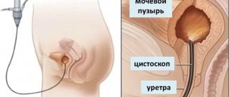
Of particular importance is conducting a CT scan of the bladder in difficult diagnostic cases when it is necessary to confirm the data and conclusions of an ultrasound examination. Tomography also serves as an alternative diagnostic method when the patient or his relatives refuse cystoscopy.
A study is usually prescribed after a detailed history collection from the patient, a general and special physical examination, general blood and urine tests, as well as an ultrasound of the kidneys and excretory system. But modern diagnostic recommendations include performing a computed tomography scan immediately after a patient with a traumatic injury is admitted to the hospital if the doctor suspects a rupture or bleeding in the bladder.
CT technique
Before the examination, the patient must remove his metal jewelry. You will need to lie on your back for about 15 minutes, motionless, so you should take the most comfortable position.
The patient lies down on the table and a special ring begins to rotate around him. It houses beam sensors. The ring will be located above the organ that is being examined. The tomograph hums quietly during operation, which the patient will hear. The procedure will not cause any other inconvenience.
A microphone is built into the device so that the patient can communicate with the diagnostician, who is in the next room and monitors what is happening from there.
Device for computed tomography of the bladder. Source: avaclinic.ru
What does MSCT of the kidneys show?
There are dozens of diseases that can be detected by MSCT diagnostics. The procedure is prescribed for the following purposes:
- To confirm or refute the diagnosis of renal cell carcinoma;
- To determine the stage of tumor development in the kidneys and ureters;
- To identify abscesses and hemorrhages;
- After injuries, the procedure allows you to identify organ bruises;
- To detect occlusive processes in the renal vessels;
- To diagnose polycystic disease, kidney stones, and identify the cause of renal colic.
Progress of the procedure
The procedure may take 20-30 minutes, and if a contrast agent is used, about an hour. CT is no different from other similar examinations, with the exception of noise effects and the round shape of the device itself. It is these features that cause difficulties with the procedure for some people - those with mental disorders or those suffering from claustrophobia. However, if research is necessary in this group of people, in the first case, you can recommend taking a mild sedative, and in the second, find a clinic where there is an open-type device.
After the patient removes all metal objects, he lies down on the couch, and the specialist pushes it inside the device.
Since the tomograph makes a lot of noise during operation, the patient is shown a button so that in case of emergency he can contact the diagnostician.
Next, the specialist leaves the room, but at the same time constantly monitors the patient through a special window. The patient should lie on the couch without moving. After the procedure is completed, the patient takes the results and can go home or work.