Magnetic resonance imaging of the hip joint is a diagnostic technique that uses electromagnetic waves. This is a high-frequency, precise method that produces images of the area of interest.
The interpretation of the result depends on the qualifications of the doctor. With the help of modern equipment and an experienced diagnostician, it is possible to promptly identify severe pathological conditions and direct efforts towards elimination.
Magnetic resonance imaging determines the condition of the hard and soft structures of the hips and iliac bones. The device visualizes:
- injuries;
- tumors;
- developmental pathologies;
- inflammatory, degenerative, autoimmune processes.
There is no radiation exposure, and the contrast agent improves visualization of the pathological focus.
What is the hip joint made of?
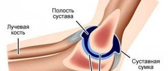
The hip joints are the articulation of the femoral bone tissue, which carries out movements: rotational, flexion, extension, oblique.
This is a support with which people move, perform strength exercises, and move the hip to the side, forward or backward. Joint structure:
- A spherical head that is fixed in the acetabulum.
- Linings of cartilage tissue with gel-like lubricant.
- A periarticular bursa surrounded by synovial fluid with shock-absorbing properties.
The joints are covered with an articular capsule - ligaments with a supporting effect to prevent dislocations.
Ligaments connect the edges of the acetabulum to the hip heads. If the functioning of the hip joint is impaired, pain occurs, motor activity becomes difficult, up to complete immobility.
Diseases of the hip joints: general diagnosis
Hip pathologies are caused by factors:
- overweight;
- menopause;
- lifting weights;
- stress;
- metabolic disorders;
- genetic predisposition;
- hypovitaminosis.
Pain and discomfort occur under the influence of:
- inflammatory processes: arthritis, arthrosis, bursitis;
- degenerative changes;
- congenital pathologies;
- infectious diseases;
- traumatic injuries;
- necrosis;
- tumors;
- diabetes mellitus;
- autoimmune diseases.
If you experience limited mobility or pain, it is recommended to consult a doctor.
Arthritis
They conduct an oral interview, a face-to-face examination, palpation, and a biochemical blood test. Additionally recommended:
- goniometry - determines the range of motion of joints;
- MRI;
- Ultrasound;
- ;
- radiography.
The treatment protocol is selected taking into account the results of complex diagnostics. If conservative measures are ineffective, hip replacement is indicated.
The product is made of titanium or alloys of this metal, the patient can undergo regular scans even after surgery. If ferromagnetic metals were used, the procedure is contraindicated.
Who should use MRI?
Magnetic resonance imaging monitors the dynamics of progressive diseases: tumors, arthrosis, degenerative changes. The study is indicated for patients with:
- injuries, dislocations, fractures, falls;
- arthritis;
- inflammatory processes affecting the synovial bursa;
- dysplasia;
- rheumatoid lesions (Lupus erythematosus, ankylosing spondylitis);
- tumors;
- aseptic necrosis of the femurs;
- osteochondropathy (necrosis of the femoral heads);
- pinching of nerve endings, tendons;
- complaints of chronic pain in the hip joint (at rest, after physical activity).
The examination is prescribed before surgery for patients with contraindications to X-ray or CT.
You need to take with you a referral, a map, the results of the previous scan, certificates and documents related to the disease.
What does it show
If the joint is normal
and suspicions of pathology are not confirmed, the image will show:
- shape and size of the articular head,
- capsule with joint fluid,
- ligamentous apparatus,
- muscle tissue,
- vascular structures.
Otherwise
The MRI image will show:
- consequences of injuries (joint dislocation, sprain, ligament rupture, cracks and fractures),
- the presence of inflammatory processes in the soft tissues and joint structure,
- rheumatic deformities,
- indicating diseases
- development of the tumor process.
If the hip joint has
cyst or malignant tumor, the device will indicate the exact location, size and shape of the tumor.
Contraindications
Hip examination is contraindicated for people with allergic reactions, renal failure, metal structures, pregnant and lactating women.
If the patient cannot remain in a motionless position for a long time, other diagnostic methods are prescribed. Relative contraindications: claustrophobia, psychoemotional illness, mechanical heart valves. In this case, MRI is performed in the presence of objective indications, in life-threatening conditions.
Absolute contraindications include:
- I trimester of pregnancy;
- the presence of an endoprosthesis (material taken into account), an implanted nerve stimulator, an electronic implant, a bullet, a pacemaker, an insulin pump, a vascular stent;
- tattoos made using metal-containing dyes.
If the patient's body does not have devices made of a ferromagnetic alloy, magnetic resonance imaging is allowed. The study is carried out on people with braces, veneers, and modern dentures.
CT or MRI, which is better?
Computed tomography and magnetic resonance scanning of the hip joint are completely different research technologies. It is not entirely correct to compare them with each other.
To objectively evaluate the methods, it is necessary to evaluate the following parameters:
- The principle of influence and capabilities of devices.
CT uses x-rays to visualize the internal structures of the body, so the equipment is most informative in the study of bone tissue: the diameter of the articular head, fracture or dislocation, the presence of bone fragments or foreign objects. MRI relies on the effect of magnetic resonance, which causes vibrations of hydrogen atoms in cells, so the study has the advantage of determining the condition of soft tissues: synovial fluid, ligaments, tendons, feeding blood vessels, muscle fibers. To compile a complete clinical picture, the doctor may prescribe both research methods. - Security and restrictions
. Exposure to a magnetic field during MRI can damage electronic implants and cause discomfort and even pain if there are ferromagnetic implants in the body. In this case, the patient is examined on a computed tomograph. However, the number of procedures is limited to 1-2 studies per year to avoid excessive radiation exposure. CT scanning is also contraindicated for young children and pregnant women. MRI can be performed from the first days of a child’s life. There are also no restrictions on quantity. Magnetic resonance imaging can be repeated as many times as needed. - Duration of the procedure.
MRI of the hip joint requires from 20 to 40 minutes, depending on the technical characteristics of the device and the complexity of the study. A CT scan takes 1-2 minutes.
| CT scan of the hip joint | MRI of the hip joint |
| 3D model |
Preparation and carrying out the procedure
During the preparation process you will need:
- inform the doctor about possible allergic reactions to medications, a history of chronic and recent diseases;
- remove metal jewelry, including piercings;
- if the patient is not sure that he will be able to maintain a motionless position for a long time or there is a fear of closed spaces, sedative medications are used.
When using contrast, the following are excluded: the possibility of allergic reactions, pregnancy, and impaired renal function. It is recommended to refrain from eating and drinking 4-5 hours before the MRI.
Progress of the procedure: the patient puts on comfortable clothes or a disposable cape, which is provided by the clinic, the patient is asked to remove metal jewelry, electronic devices are left outside the machine before the MRI.
If the patient is pregnant or has other contraindications, the doctor must be informed about this. The procedure is carried out in a specially equipped room:
- the patient is positioned on a retractable table that slides into the MRI unit, the limbs are fixed;
- To eliminate the noise that the device makes, use headphones and earplugs;
- It is recommended to relax and remain still;
- the patient can contact medical personnel during the study period;
- pain, fear, inconvenience - indications for using the “panic button” with which the device is equipped.
People with claustrophobia and excess weight are recommended to go to a clinic with a functioning open-type MRI machine. For comparison, you can provide the results of the previous scan.
Open type (C-shaped)
Use of contrasting materials
To take high-quality images, contrast agents are used. They are administered intravenously to better visualize the internal relief of the area being examined.
Depending on the weight, up to 20 ml of a substance is injected, which the body will eliminate in a day. Contrast materials contain gadolinium derivatives, which are better tolerated than iodine. If the drug provokes an allergic reaction, antihistamines are used.
In severe cases, the contrast causes difficulty breathing, abnormal bowel movements, and a feeling of discomfort in the abdominal area. It is recommended to consult your doctor for the selection of symptomatic therapy.
Preparing for an MRI
Tomography of the hip does not require special preparation, with the exception of MRI with contrast. The introduction of a special substance enhances the contrast of tumor formations and helps analyze the blood circulation in them. The main indication of MRI with contrast is the diagnosis of tumors.
When prescribing an examination, the doctor will make sure that there are no contraindications: pregnancy in the first trimester, metal prostheses, pacemaker. Before starting the procedure, the patient must remove jewelry, watches, items of clothing with metal elements, and clothing from the area being examined.
Conditions and diseases that magnetic resonance imaging shows
Tomography is a modern diagnostic method, a clarifying study. What MRI shows: the presence of infectious lesions, pathological changes, pinching.
Visualizes the structure of bones and soft tissues. Scans indicate development:
- enostosis - deformation of bone tissue, which is similar to a tumor;
- trochanteritis - inflammatory processes affecting ligaments and tendons;
- synovitis - inflammatory processes affecting the synovial membrane. Will show the degree of wall thickening, increased fluid synthesis;
- bursitis;
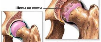
- coxarthrosis - pathological conditions in which the cartilaginous surfaces of the joints are slowly destroyed. At the initial stages there are no symptoms, the disease is detected by chance;
- malignant and benign neoplasms, their metastases.
Bursitis
To confirm the diagnosis, the scan results are sent to the doctor and additional tests are performed.
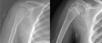
X-ray or MRI, which is better?
X-ray and MRI of the hip joint are different research technologies. Their comparison is not entirely correct.
To give an objective assessment of diagnostic methods, it is necessary to analyze the following indicators:
- Operating principle of the equipment.
An MRI machine operates on the principle of magnetic resonance, which creates vibrations of hydrogen atoms in tissue cells. It does not expose the body to radiation. X-ray equipment uses the ability of x-rays to travel differently through tissue of different densities. - Equipment capabilities.
A magnetic resonance imaging device visualizes bone structures and adjacent soft tissues, which makes it possible to most accurately determine the condition of the hip joint. On an x-ray, bone tissue is visible, while soft tissue is practically not reflected. The method is suitable for diagnosing fractures and dislocations when delay is unacceptable. - Projection quality
. MRI images have high resolution and detailed detail. The reliability of the study is 95%. Possibility of detecting the disease at an early stage of formation. The X-ray image has low resolution. Informative for identifying injuries and formed pathologies. - Availability and cost.
X-rays are included in the list of free services under the compulsory medical insurance policy. This can be done at your local clinic or emergency room. MRI is provided free of charge to patients in regional inpatient departments. In an outpatient setting, MRI examinations are performed for a fee. If you compare prices for procedures, the cost of an X-ray will be 2-3-4 times cheaper than an MRI.
| X-ray | MRI of the hip joint |
How safe is MRI?
MRI is based on the activity of magnetic fields that do not cause pain, discomfort, or damage to tissues and internal organs.
Unlike radio beams and ionizing radiation, magnetic fields up to 4 Tesla are safe.
To obtain accurate results, follow the doctor’s recommendations in the process of preparing and conducting the examination.
After the scan is completed, the patient returns to his normal lifestyle. The results are received in hand after decoding in electronic form. The conclusion is drawn up by a diagnostician within 30-60 minutes.
Ultrasound or MRI, which is better?
Ultrasound examination of the hip joint and MRI of this area are fundamentally different research technologies. There is no clear answer to the question: which method is better.
To give an objective assessment, it is necessary to study the following parameters:
- Technology of influence on the body
. Ultrasound of the hip joint is based on the ability of ultrasound waves to reflect from denser tissues. For imaging, MRI uses the conversion of vibrational pulses of hydrogen atoms in tissue cells. - Device capabilities
. Ultrasound is not 100% informative, since it can only examine superficial abnormalities in the structure of the joint. MRI creates cross-sectional projections, demonstrating the condition of the joint cavity from the inside. This makes it possible to detect pathologies in the early stages of formation. - Security and restriction.
The magnetic field of the tomograph affects electronic and metal implants, can damage a pacemaker, and disrupt the functioning of the cochlear apparatus or insulin pump. For this category of patients, diagnosis is carried out using ultrasound. Otherwise, both technologies are safe for the human body at any age. - Availability and cost of research
. Ultrasound diagnostics can be done under the compulsory medical insurance policy at the clinic at your place of registration. MRI examinations are available free of charge to hospital patients. In outpatient settings, examinations are carried out only on a paid basis. The price list of commercial clinics or the price list of municipal medical institutions that practice seeing patients by self-referral on a paid basis shows a difference that is 2-4 times higher for MRI than for ultrasound. - Duration of the procedure.
An ultrasound is performed in 5 minutes, while you need to stay in the MRI machine for 20 minutes or more.
| Ultrasound | MRI of the hip joint |
Diagnostics of children
MRI is suitable for examining children. If a small child does not control motor activity, sedatives are used. The medicine is administered by an anesthesiologist. Scanning is painless and safe, suitable for babies from the first days of life.
If contrast is necessary, contraindications to the use of the drug are taken into account. Parents report possible allergic reactions and the presence of concomitant diseases.
General anesthesia is indicated for a child under 5 years of age. The doctor and parents talk to older children. Modern machines are equipped with monitors that show cartoons during the scanning period.
Which doctor should I contact?
At the first signs of a disorder, contact a rheumatologist, traumatologist, orthopedist, or oncologist. Consultation with specialists in related fields may be required:
- in case of traumatic lesions, contact traumatologists;
- arthritis, arthrosis - require examination by a rheumatologist;
- in case of complex traumatic lesions, the patient will be taken to a neurosurgeon;
- for malignant tumors - see an oncologist.
The obtained diagnostic results are provided to the attending physician, awaiting further instructions regarding the selection of a treatment strategy.
Price
The cost of the procedure is from 6,000 to 20,000 rubles, scanning and decoding are included. The price depends on:
- need for contrast material;
- type of clinic;
- equipment quality;
- doctor's qualifications.
They take into account the time of day (cheaper at night), the patient’s conditions of stay (hospital, outpatient clinic).
Magnetic resonance imaging - diagnosis of tumors, degenerative-inflammatory processes, congenital malformations.
It is recommended to go to clinics with modern equipment and competent, experienced diagnosticians. When preparing, follow the doctor's instructions to get accurate results.
To kid
MRI of the hip joint has no age restrictions.
It is prescribed to infants with suspected dysplasia, birth trauma, or developmental defects. Children of preschool, primary school age and adolescents are referred for MRI of the hip joint for injuries, developmental defects, and problems with the spine.
To create a high-quality MRI projection, you must remain still. It is difficult for small children to withstand 20 minutes in a tomograph without moving. In this case, the doctor may suggest performing the procedure under superficial anesthesia. Not every commercial clinic has been licensed to conduct research under anesthesia. They set the age limit from 7 years. At this age, children are able to follow the doctor's instructions.
The second factor that complicates MRI scanning for young children is the fear of closed spaces and the noise of the scanning capsule. An alternative solution is to conduct the study in an open tomograph. The parent can be nearby, support the child, and make sure that he does not move his legs.
The list of contraindications for MRI in children is the same as for adult patients.