Radiography, as a method of radiation diagnostics, is widely used in many areas of medicine, including otolaryngology. The examination is carried out without damaging the upper tissues, that is, non-invasive, does not take much time, and is painless.
X-ray of the nose and paranasal space allows you to reliably determine existing changes in bone structures and pathology of the air cavities (sinuses). Clear visualization of organs on an x-ray allows the doctor to objectively assess their condition, make the correct diagnosis, and prescribe appropriate treatment. According to the decision of the ENT doctor, a survey or targeted radiography is performed:
- survey x-ray, displays the bones of the nose and sinuses as a whole, and is often performed during the initial diagnosis;
- targeted radiograph to examine individual anatomical structures in more detail. It is carried out to monitor the treatment of a previously diagnosed disease of a particular nasal area.
How often can this procedure be done? The frequency of examination has not been officially established. The dose of X-rays during a single examination is not dangerous to health, but taking into account the fact that radiation tends to accumulate in the body, X-rays of the nasal sinuses are not recommended more than twice a year.
Features of x-ray of the larynx
Radiology is a science that helps doctors make correct diagnoses. With the advent of X-ray machines, the number of diagnostic errors has decreased by an order of magnitude. And modern devices make it possible to obtain the most accurate images and thereby ensure a speedy recovery for the patient.
An X-ray image helps medical personnel in establishing an accurate diagnosis and, accordingly, prescribing the correct treatment. X-ray of the throat allows you to more accurately and more closely assess the condition of the soft tissues of the cervical spine, as well as bones. The X-ray also shows the entire structure of the cartilage. Reflects bone calcifications and tissue changes that occur with age.
X-ray is a procedure for obtaining a diagnosis, which today has no alternative (it is understood that no other procedure can surpass the quality and accuracy).
In medicine, there are ways to conduct x-rays of the larynx - direct or lateral projection (used to obtain information and detect pathologies on both sides).
When you can't do without radiography
In addition to the fact that the patient will be sent to the X-ray room if a fracture or fissure of the nasal area is suspected, there are other reasons for referral for such an examination. A striking example of this is periostitis, or any other disease in the acute stage of its course or in a chronic form.
Other common reasons for using radiation for diagnostic purposes include:
- foreign bodies;
- sinusitis;
- deviated nasal septum;
- benign and malignant neoplasms;
- osteomyelitis;
- polyps;
- cyst.
People who are scheduled for surgery on the face or sinuses cannot do without a trip to the X-ray room. A control image will allow you to clarify anatomical features that could become an obstacle to surgical intervention according to the standard procedure.
One of the most important advantages of radiography is the lack of any serious preparation. The only requirement for diagnosis is the need to first remove jewelry and other metal objects, the presence of which can negatively affect the clarity of subsequent results of assessing the condition of the nasal area.
Separately, you need to warn the doctor if a person has embedded metal parts in the area being examined. Sometimes, in order to avoid unnecessary stress on problem areas, doctors recommend replacing classic x-rays with a gentle alternative.
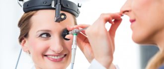
The manipulation itself lasts no more than a couple of minutes. The laboratory technician will sit the victim in front of the protective screen so that he rests his nose and chin directly on the shield of the device.
Some ordinary people believe that such proximity to a source of radiation will negatively affect their well-being in the future. In fact, the development technicians calculated the dosage of incoming rays, which is within acceptable limits for an adult. This means that at one time the patient will receive radiation, which in terms of power will not critically exceed the daily dosage in their natural habitat.
Advantages and disadvantages
X-ray of the throat has a significant advantage - the form of diagnosis is accessible to everyone, is quickly performed and has virtually no contraindications. An important point is also that the patient does not need to prepare for a long time for the study. The doctor processes the results quite simply and quickly. The study takes place in any premises (hospital wards, specialized diagnostic centers, operating rooms).
However, there are some disadvantages:
- Radiation exposure is a primary factor, which makes this procedure inaccessible to pregnant patients and nursing mothers.
- Despite the large amount of information that can actually be obtained from an image of this organ, the picture of the disease is not always fully revealed.
- Lack of information about the condition of soft tissues, which becomes a significant obstacle to obtaining a complete diagnosis.
Even if there are shortcomings, doctors recommend x-rays as the most reliable way to obtain information about the problem and make the correct diagnosis. In some cases, in order to avoid an error, an alternative procedure (for example, MRI) is prescribed in addition. But the data obtained from other sources is used as a supplement to the basic information obtained through x-rays.
X-ray degrees of enlargement of the nasopharynx with adenoids
There are 4 degrees:
- At this degree, enlarged tonsils occupy 1/3 of the nasopharynx. In such a situation, the child begins to snore at night. A prolonged runny nose due to pathology is practically not treated with medications.
- In the second degree, enlarged tonsils occupy 1/2 of the nasopharynx. The child snores while sleeping. The baby's conversation becomes incomprehensible. X-rays at this stage can reveal enlarged tonsils.
- The tonsils occupy the entire lumen of the nasopharynx. The condition makes it difficult to breathe through your nose or speak. Clinically, this condition is accompanied by a decrease in hearing and smell. Doctors recommend surgical treatment for stage 3 of the disease.
- Note that there is no 4th degree. If a radiologist assigns a 4th degree of pathology, he does not have sufficient qualifications.
X-ray of the nasopharynx in children: the location of the adenoids is indicated
What does an x-ray show?
The patient, looking at the x-ray, will see the general picture, but the specialist pays attention to many important points. The main thing that an x-ray will show:
See also
What to do if the lymph nodes on the right or left neck are swollen, how to treat?
Read
- The lumen, which looks like a curved strip of clearing and is a continuation of the pharynx, passes into the trachea. Visualized on lateral projection images.
- The cartilages (epiglottis, cricoid, thyroid), laryngeal ventricles, epiglottis and hyoid bone are visible.
- On direct projection x-rays, the thyroid cartilage and subglottic space are clearly visible. In some cases, it is possible to see the upper and middle parts of the larynx.
- In some cases, when performing a study with functional tests, the doctor is given the opportunity to assess the mobility of the vocal cords and the size of the glottis.
X-ray examination makes it possible to study the morphological state of the organ and identify narrowings and deformations along the path of the air column.
In the photographs it is possible to notice such throat diseases as whooping cough, tracheal stenosis, diphtheria and others.
Contraindications for X-raying the nasopharynx in a child
An X-ray of the nasopharynx is rarely prescribed to a child, only if there is no possibility of a standard medical examination, and the situation requires clarification. Before prescribing a test, the doctor weighs the pros and cons. However, as a rule, the harm from an advanced stage of illness or blindly prescribed treatment is much greater than from a small dose of radiation. It is advisable not to do x-rays of the nasopharynx in children under one year of age, who are especially susceptible to radiation. Due to their small size, their organs are located very close to each other. In this regard, the effect of radiation on neighboring organs during the study increases by 2-3 times. It is worth noting that the lowest dose of radiation is provided by the most modern equipment, which, in addition, is also more informative.
Is there an alternative to examination?
An alternative to an X-ray of the nasopharynx for a child can be echosinusoscopy - a method of ultrasound diagnostics of the nasal sinuses, which opens up a view of the nasopharynx area. However, this study, which combines ultrasound and endoscopic methods, is not suitable for a child who cannot remain motionless for a long time and tolerate discomfort. MRI of the nasopharynx
also requires remaining motionless for a long time. Therefore, despite being highly informative, these examinations cannot replace an x-ray of the nasopharynx for your child.
#!RentgenVRA4!#
Indications for use
The patient can have an x-ray of the larynx without a doctor’s prescription, at his own request. If the doctor has prescribed a procedure, then there is a suspicion of the following pathological conditions:
- Traumatic injury to the neck organs.
- The presence of a foreign body in the lumen of the larynx or trachea.
- Chemical or thermal burns of the larynx, pharynx, upper esophagus and trachea.
- Chronic laryngitis.
- Paresis and paralysis of the larynx.
An X-ray diagnosis of the larynx is prescribed by a doctor if the patient suspects various injuries - external or internal. For burns, x-rays are sometimes prescribed. The most valuable method for diagnosing paresis and paralysis.
As an auxiliary study, x-rays of the larynx are used to diagnose chronic pathologies of the larynx with inflammatory processes, as well as tracheal stenosis, malignant tumors, whooping cough, and diphtheria.
X-rays also play a key role in diagnosing laryngeal cancer. The study is used in tandem with other types of diagnostics to obtain an accurate result.
Maxillaryography
X-rays of the maxillary sinuses in cases of complicated sinusitis are performed using a contrast agent. Maxillography is performed as prescribed by a doctor, and only for adult patients. The methodology includes:
- use of local anesthesia;
- puncture of the sinus wall;
- cavity rinsing;
- injection of contrast into the studied sinuses;
- taking sequential photographs in several projections.
A photograph from an x-ray of the paranasal sinuses with a diagnosis of sinusitis clarifies the degree of filling of the sinus with contents
Removal of the contrast agent occurs spontaneously within 24 hours after the examination. Particular attention is paid to this disease and its diagnosis due to the danger of severe complications. Untimely detected sinusitis goes into a chronic stage, during which inflammatory processes may develop in the bronchopulmonary system and the brain.
Preparation for the procedure
No preparatory steps are required from the patient for radiography. The main condition is to remove your jewelry before the examination. The conditions for the procedure are described below.
In some cases, to improve image clarity, the organ being x-rayed is “shaded” or “illuminated” with medications. This option requires more thorough preparation (for example, refusing to eat a few hours before the procedure).
How to conduct an examination
To take a correct picture of the larynx, the patient must take a position lying on his stomach. When placed sideways, the image shows the lumen of the larynx. In some difficult cases, radiopaque agents are used, which are sprayed on.
The procedure proceeds as follows:
- The patient is placed on his side.
- A specialized film is applied to the back of the neck opposite the recording device.
- A focal distance of about 60 centimeters is maintained.
- The patient holds his breath while the image is taken. This is necessary to obtain the clearest image.
See also
Treatment of inflammation of the lymph nodes with chickenpox in children and adults
Read
In rare cases, a second x-ray is taken. To do this, the patient is numbed with drugs to the area being filmed, and the film is placed in the area of the larynx. The X-ray beam is directed clearly at the area of the disease focus, which makes it possible to accurately reflect the nature of the disease.
Decoding the results
The clarity of the image in the image, as well as the correct decoding of the available data, largely depends on the type of equipment on which the study is carried out. Modern digital devices transmit information more accurately than their analog predecessors.
All interpretation of the results should be carried out by the attending physician. This is necessary so that the patient does not prematurely and independently make an erroneous diagnosis, which will lead to negative consequences.
An erroneous diagnosis of a patient leads to typical self-medication, which ends in unforeseen complications. It is critically important at any stage of the disease or diagnosis to focus on the recommendations of a specialist and act in accordance with the instructions.
Survey area
The picture is taken in black and white, which clearly shows the bone skeleton of the nose, the size of the nasopharynx, the structure and contours of the air cavities (aka sinuses). Using the image, the doctor analyzes the integrity of the bones and cartilage tissue, the condition of the hollow formations adjacent to the nose:
- frontal (frontal) sinuses, located in the frontal bone of the skull;
- lattice labyrinth;
- the main sphenoid sinus, divided by a septum;
- paired maxillary (maxillary) cavities;
- the lower part of the temporal bones with a cellular structure (mastoid process).
According to the norm, in the absence of pathologies, the outline of the bones should be smooth, the pneumatization (airiness) of the sinuses should not be impaired, that is, there should be no neoplasms or fluid or foreign objects
Contraindications
Before going to a radiologist for a picture of the larynx, you need to consult an otolaryngologist. Only the attending physician prescribes the procedure based on the indications and can take into account possible negative consequences. The only contraindications are pregnancy and breastfeeding, as mentioned above.
When conducting contrast diagnostics, there are a number of additional contraindications:
- patient intolerance to drugs containing iodine;
- problems with the thyroid gland;
- patients suffering from active tuberculosis;
- liver and urinary tract problems;
- decompressed diabetes mellitus.
X-ray is a procedure that involves exposure to radiation. Therefore, people who do not suffer from acute diseases of internal organs should resort to it. If the patient has, for example, a cold or open bleeding at the time of the procedure, it is better to reschedule the x-ray.
Features of examination of the nasopharynx and sinuses in children
We already wrote above that any radiography (including the sinuses) is possible for children over 7 years old. It is not recommended to conduct research before this age, since soft tissues and bones are in the process of active growth. There is no clear answer to the question of how often you can do an X-ray of a child’s nose. This should be determined by the attending physician, taking into account the patient’s condition and the possibility of alternative diagnostics.
In emergency situations, such as a fall from a height, a car accident, serious domestic injuries, when the question of saving the baby’s life is raised, x-rays are used as an urgent diagnostic method without taking into account the child’s age.
X-ray of the nasopharynx is a simple procedure that does not require special preparation of the child for the examination. Just like adults, you just need to remove metal jewelry, as it can affect the quality of the photo.
Examination of the sinuses and nasopharynx in children can be prescribed in various projections. It is also permissible to conduct research using a contrast agent. An X-ray of a child’s nose is usually prescribed by an ENT specialist.
Similar diagnostic methods
Since x-rays of the larynx classify the functionality of the organ, there are a number of methods aimed at the same:
- Valsalva maneuver. The procedure is performed when the patient exhales with the glottis closed and the muscles of the anterior abdominal wall tense after a deep inhalation.
- Carrying out the study while taking a long breath or pronouncing the vowel sounds “i”, “o”, “u”, “e”.
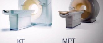
- CT scan.
- MRI.
Despite the number of alternative diagnostics, x-rays of the larynx remain an effective and affordable diagnostic method. Even when several diagnostic methods are used, X-ray readings remain decisive in making a diagnosis.
Possible harm
Such a study will not cause any significant harm to the body, unless this person undergoes x-rays several times in a short period of time. Moreover, this applies to the study of various parts of the body, as well as the use of computed tomography. Also, the reason for refusing to frequently use this diagnosis and changing its date to a more distant period may be the weakened immunity of the patient due to a recent serious illness or psycho-emotional stress. But this contraindication is relative.
This is what doctors call restrictions, which can sometimes be ignored if the benefit from the result turns out to be an order of magnitude greater than the possible harm caused.
But pregnancy is an absolute contraindication. Moreover, the ban applies to all periods of an interesting situation, as it poses a threat to significantly harm the normal course of fetal development. In the future, this threatens to lead to mental or physical pathologies in the child due to exposure to radiation during fetal development.
If alternative methods such as ultrasound are not possible, then pregnant women still have to take risks in situations where their lives depend on the results of the examination.
To be on the safe side, they are given special lead aprons to cover their stomachs.
Any other visitor to the diagnostic room receives the same protection to cover particularly vulnerable areas of the genitals and thyroid gland.
Due to the fact that the devices are usually configured to deliver the maximum permissible dosage designed for an adult, children under 15 years of age are not recommended to be exposed to such a load. The only exception is a suspicion of pneumonia, or a foreign body caught in the area of the nasal passage.
It is believed that equipment that is more than twenty to twenty-five years old requires “write-off” and cancellation of use for diagnostic purposes. The reason for this is the increased radiation effect on the body. Digital similar machines do not have such a strong effect on the body, using identical techniques and even providing images of improved quality.
The average permissible radiation dose is 5 mSv per year. Despite the fact that the standard set aside for examining the bow has low radiation levels, the annual standard includes taking into account:
- fluorography;
- X-rays of other organs;
- computed tomography;
- natural radiation.
Because of this, people who, for some reason, often have to undergo X-ray monitoring for the condition of the nasal septum or sinuses, it is better not to exceed two acceptable times a year. All other times, it is better to use the services of magnetic resonance imaging, which works on a different principle and does not use x-rays.
In general, in private clinics, doctors record each x-ray case on a special card called the “Dose Load Sheet”. But most clinics do not do this, leaving everything to the control of the patients themselves.