In what cases are leg x-rays taken? This procedure is prescribed to the patient when there is a suspicion of any pathology in the human musculoskeletal system. As a rule, pathological processes of this nature are accompanied by pain in the foot area. In addition, externally you can notice its deformation.
It is worth saying that x-rays of the legs are considered an effective diagnostic tool and are available to many categories of citizens. This is due to the fact that this method of examination does not require special preparation from the patient and is done for little money. Also, using x-rays, the doctor can make an accurate diagnosis and outline a treatment plan.
X-ray: purpose
In what cases is a patient prescribed a leg x-ray? The foot examination is carried out in a special room, which is equipped with the necessary instruments. X-rays of the legs are prescribed in cases where the patient reports the following concerns:
- The patient experiences pain in the foot. An x-ray of the leg will have to be taken in cases where the person has not had any physical activity, for example, walking for a long time, or wearing shoes that are not very comfortable, etc.
- There are cases when the patient experiences visual changes in the shape of the foot.
- If a person has injuries, such as sprained feet, sprains, or fractures.
- A person who is at risk for musculoskeletal disorders is also prescribed an x-ray. This category includes athletes, overweight people and those with poor heredity.
Preparation and technique
No special preparation is required for the examination unless a contrast agent is used. Then it is determined in advance whether the patient has an allergy. X-rays can be performed in different ways, depending on what part of the limb is being examined. For the ankle and foot, diagnostics are carried out under load.
The leg is freed from clothing and placed on a small stand. The other limb is bent at the knee so that the weight is transferred to the one being examined. In this case, the cassette is positioned along the leg and is fixed at the bottom with a weight. The photographs are taken in a number of projections - from behind, from the front, from the back and from the side.
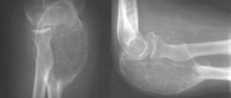
The knee joints are removed from the front and sides. For accurate results, the leg is straightened. If possible fractures, ruptures or deformation of the ligaments are suspected, the procedure is performed under load; in other cases, conventional films are performed. X-rays are taken in a standing position.
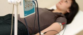
Before diagnosing blood vessels, the patient is given anesthetics and sedatives (using a dropper). Then local anesthesia is given and a catheter is inserted into the vein through a small incision. A contrast agent is poured through it. X-rays are then taken. If blood clots are detected, the issue of their removal is decided. The catheter is then removed and a bandage is applied to the incision site.
In what cases are foot x-rays taken?
There are a number of problems in which a person is given a referral for this examination.
- If the patient is visualized with a disturbed position of the bone tissue, then he is sent for an x-ray. This type of examination is also prescribed for rupture of soft tissue.
- To obtain data about the condition of the limb, if there are any deformities.
- X-rays are also prescribed to examine the organs of the hip joint in cases where a person complains of pain.
- The patient must be examined by x-ray in order to obtain data on the condition of the knee joint, if there is an indication for this.
- If a person’s leg is swollen, then he is prescribed an x-ray of the limb. If any deformities are present, it is also necessary to undergo this examination.
- Injuries such as a bruise or sprain are also examined using x-rays. This is necessary so that the doctor can see a complete picture of the condition of the damaged area.
- An X-ray of the leg fracture is also taken (a photo of its results is presented above for clarity). This study allows you to accurately determine the diagnosis. For arthritis and arthrosis, you also need to undergo this examination. An X-ray of the feet allows you to see the condition of the fingers.
- An examination is usually prescribed to see how the joint reacts to stress. X-rays of the legs are taken in several projections.
- It is worth knowing that in case of damage to the legs such as frostbite, this examination is also prescribed. This is necessary so that the doctor can find out to what extent the limbs are damaged. If leg dysfunction is metastatic, then x-ray is the main way to study this disease.
X-ray has great potential for studying certain injuries. But this method of studying the body has contraindications. Namely, it is contraindicated for those women who are pregnant and people in serious condition.
A person does not require a preparatory stage before undergoing an x-ray. The exception is pictures of the spine and tailbone. In this case, the person must come to the study with an empty intestine.
Indications for diagnosis
Indications for the study:
- injuries of the lower limb (open and closed fractures, cracks in the leg bones, dislocations, sprains);
- reactive arthritis;
- autoimmune inflammation of the joints (to identify the X-ray stage of rheumatoid arthritis);
- swelling of the lower limb in a separate area;
- severe bruise with severe pain;
- deformation of leg tissues (pathological curvature of the joint, displacement of bone fragments, and so on);
- frostbite or burn;
- pain in the leg when moving or at rest;
- flat feet;
- valgus or varus deformity of the legs;
- gouty manifestations (an X-ray of the big toe is often performed);
- cancer of bone structures;
- osteomyelitis.
X-ray for heel spurs
Surely many people have heard about this type of defect called a heel spur. There is a modern way to treat this disease. It's called X-ray therapy. Modern technologies have reached such a level that radiation does not have a negative impact on human health.
The modern method of treatment using x-rays is that the device is automatically adjusted. Through it, he distributes the radiation influence in such a way that it reaches a person in the dosage required for his condition. With automatic distribution, the possibility of patient exposure is eliminated.
X-ray treatment brings good results. This is due to the fact that malignant formations are destroyed through rays. Due to the high degree of effectiveness of this method of treatment, it has become widespread. X-rays can also be used to stop formations such as papillomas and warts.
Another advantage of this method is that the rays are directed only to the damaged area. Healthy cells of the body do not experience any pressure. There are a number of indicators due to which an x-ray of a person’s leg is considered a highly effective method of examination. Namely:
- a small list of contraindications;
- absolutely painless method of exposure;
- radiation is produced only at the site of tissue damage.
In order to get an x-ray of your foot, you do not need to go to the hospital. This procedure is performed on an outpatient basis.
What can be detected with radiation therapy?
Diagnostics allows you to accurately identify foot pathologies that require immediate treatment. She shows:
- osteomyelitis;
- deforming osteoarthritis;
- dislocations;
- fractures;
- Paget's disease;
- tendinitis;
- endarteritis (a very dangerous vascular disease);
- sarcoma;
- metastases;
- microcracks;
- peripheral edema.
The list continues: improper fusion of bones, flat feet, abnormal bone structure, bursitis of the big toe, arthritis, heel spur, sprain, avascular necrosis. This also includes osteoporosis, gout of the foot, hallux valgus, false joints, Morton's neuroma, ligament rupture, and clubfoot.
Diagnosis of a broken heel bone fracture using x-ray
To diagnose a calcaneal fracture, an x-ray is prescribed. You should know that this examination has its own characteristics.
The peculiarity is that a picture is taken of two legs at once, namely the heel bones. This is necessary to accurately understand what deformations occurred in the injured leg. Thanks to the image, the doctor will prescribe the treatment method that will be most effective in a particular case.
Can X-rays be used during pregnancy?
As a rule, pregnancy excludes this method. But there are cases when an x-ray needs to be taken. For example, a fracture of a finger or some other organ.
You should be aware that the fetus is sensitive to x-rays. The fact is that ionizing rays penetrate to it through cells that are destroyed inside. In this regard, nucleic acids are destroyed, and the likelihood of a malfunction in the deoxyribonucleic acid chain increases.
Because of this, mutations and pathologies may occur in the unborn child. But if it is necessary to take an x-ray of the toe, then it will not have a harmful effect on the fetus. But an X-ray of the pelvis, abdominal cavity or back is considered dangerous for the unborn child. Therefore, if it is possible to avoid this method of examination, then it is not prescribed.
Also, pregnant women should not have X-rays of their lungs. This is very dangerous, both for the health of the woman herself and for the unborn child. Therefore, the doctor needs to be one hundred percent sure of the diagnosis, since an X-ray of the lungs may be unjustified. If the doctor nevertheless prescribed such a procedure to a pregnant woman, then there is no need to worry, since modern technology minimizes the harmful effects of x-rays on the body.
X-ray benefits, reliability and alternative methods
Radiography is one of the available tests. Almost all clinics are equipped with an X-ray machine. The advantages of this type of diagnosis include:
- high detail of images;
- low-cost procedure (even in multidisciplinary centers);
- minimal contraindications.
Alternative diagnostic methods may include ultrasound, MRI, and CT.
Ultrasound will help assess the condition of ligaments, menisci, and soft tissues. MRI will give a more detailed assessment of soft tissue, and CT will give a more detailed assessment of bone tissue.
The reliability of x-rays depends on how the x-ray of the leg is taken. If the position is correctly chosen and the diagnostician’s instructions are followed, the reliability of the study reaches almost one hundred percent accuracy.
The patient must show the result of the x-ray to the doctor, who will decide on the need for treatment.
Where to get tested?
Where can I get an x-ray of my leg? It is worth knowing that it can be done in different medical institutions. It is necessary to pay attention to the devices that are located in these places. As a rule, clinics and emergency rooms have old equipment for carrying out such a procedure. But in paid medical institutions there are more modern x-ray machines.
If a person goes to a paid clinic, then he should find out what type of X-ray machine is available in this clinic. Next, you can familiarize yourself with the price list and find out its technical characteristics. It will be better to take an x-ray in an institution where modern medical equipment is available. The quality of research and treatment will be much better with modern equipment. What indicators of the device should you pay attention to:
- on the radiation dose that affects the human body;
- while the images are being taken;
- Is it possible to select a better quality image?
- is it possible to make the study area larger;
- on how much weight the device can support.
Objects under study
X-rays of bones and joints are taken:
- femur;
- knee joint;
- shin bones;
- ankle joint;
- feet.
If necessary, X-rays of the fingers (big toe, little toe) and heel bone are taken separately on the foot. This targeted examination helps reduce the radiation dose and increase the contrast of the examination in the desired area.
Despite the fact that soft tissues are poorly visualized on x-rays, acute inflammation, joint sprains can be detected, and transudate effusion can be judged by the expansion of the joint space. Indirect signs allow us to judge the nature of inflammation.
Leg fracture: x-ray
Fracture of the foot is less common than other parts of the body. But under no circumstances should you take it lightly. The fact is that a fracture in the foot can lead to a person being unable to walk. First of all, it is worth saying that with such an injury you should not self-medicate. It is necessary to contact a medical institution for professional help.
What does a leg x-ray show? Different types of fractures, namely:
- displaced fracture;
- damage to the metatarsal bone;
- fracture of the scaphoid bone.
The test can also show whether there is a fracture of the cuboid bone. In all of the above cases, an x-ray of the foot is taken to make a diagnosis.
It is also worth knowing that the rehabilitation period is important. You should not neglect it, since rest is an important component of treatment.
There are statistics that a foot fracture occurs in 3 or 10 percent of cases of the total number of fractures. The peculiarity is that when one element is damaged, dysfunction of the entire foot is created. This happens because all elements are interconnected. It often happens that damage to the foot leads to complications such as arthrosis and flat feet. All bones are connected to each other through ligaments and joints. When different bones of the foot are damaged, a person develops certain symptoms.
X-ray diagnostics of leg bones: why it is performed, advantages of the procedure
X-ray of the leg is a radiation diagnostic method that is used for the primary diagnosis of bone pathologies. The advantages of diagnosis are painlessness, simplicity and low cost. X-ray equipment is widespread and generally available, so it is still very popular today, despite the advent of CT and MRI.
Carrying out a diagnostic technique
Diagnostics are carried out in a specially equipped room in which an X-ray machine is located, and the walls are insulated and protect the outside world from excessive radiation. The patient is brought in or carried on a stretcher; the examination can be carried out even on a seriously ill patient who is unconscious.
The laboratory assistant or doctor places the limb on a special stand, inside of which there is a sensing device.
The area required for diagnosis is cleared of clothing and objects that could distort the x-ray image of the leg.
Specialists correct the position of the limb in relation to the stand and position the tube so as to obtain the required angle. Healthy areas of the patient's body are covered with a lead collar to protect them from unnecessary radiation.
Depending on the angle of passage of the rays, diagnostics can be carried out in several projections. This is required for better visualization of the pathological focus and determination of the extent of damage.
Objects under study
X-rays of bones and joints are taken:
- femur;
- knee joint;
- shin bones;
- ankle joint;
- feet.
Find out the dose received during an X-ray of the leg using a modern digital device, and compare the indicators with the natural background. Go to “Dosimeter”.
If necessary, X-rays of the fingers (big toe, little toe) and heel bone are taken separately on the foot. This targeted examination helps reduce the radiation dose and increase the contrast of the examination in the desired area.
Despite the fact that soft tissues are poorly visualized on x-rays, acute inflammation, joint sprains can be detected, and transudate effusion can be judged by the expansion of the joint space. Indirect signs allow us to judge the nature of inflammation.
Indications for diagnosis
Indications for the study:
- injuries of the lower limb (open and closed fractures, cracks in the leg bones, dislocations, sprains);
- reactive arthritis;
- autoimmune inflammation of the joints (to identify the X-ray stage of rheumatoid arthritis);
- swelling of the lower limb in a separate area;
- severe bruise with severe pain;
- deformation of leg tissues (pathological curvature of the joint, displacement of bone fragments, and so on);
- frostbite or burn;
- pain in the leg when moving or at rest;
- flat feet;
- valgus or varus deformity of the legs;
- gouty manifestations (an X-ray of the big toe is often performed);
- cancer of bone structures;
- osteomyelitis.
What is discovered using radiation research techniques?
As a result of the diagnosis, the radiologist gives a conclusion in which he describes in detail the pathological changes, their location and the nature of the damage. This helps the attending physician gain a complete understanding of the patient's condition and create a treatment plan.
X-ray images can reveal:
- fractures (describes the exact location, extent, levels of displacement, the presence of fragments and splinters, the degree of damage to soft tissues);
- cracks (look like areas of clearing);
- “green stick” type fractures (detected in childhood, when the periosteum is preserved);
- osteoarthritis of the joints (detect narrowing of the joint space, degeneration of articular surfaces, inflammatory changes in soft tissues in the form of thickening and compaction);
- ankylosis (fusion of the joint space with the formation of a fixed joint);
- replacement of the center of tubular bones with an inflammatory infiltrate during osteomyelitis, the appearance of osteolysis (melting of the bone structure in the form of blurred contours, clearing of the bone);
- identification of a heel spur (pathological bone spike on the talus bone);
- oncological disease of bone tissue (Ewing's sarcoma, osteosarcoma, chondrosarcoma and others);
- metastases to bone structures;
- bone deformation;
- formation of pathological joints (formation of articular surfaces in atypical places, for example, with improperly healed fractures);
- degree of flat feet.
X-ray diagnostics of the leg helps not only to confirm and establish the diagnosis, but also to outline treatment tactics, including surgical and orthopedic, and also to predict the outcome of the disease.
Thanks to x-rays, the severity of the process is determined: chronic or acute (with a chronic course, the changes are more pronounced, not only the soft tissue component is affected, bone degeneration occurs in the form of osteochondrosis changes - the bone density in the image decreases).
Ilizarov apparatus
Advantages of diagnostics
The advantages of the study are:
- diagnostic accuracy: with digital processing, it is possible to enhance and reduce the contrast of the image, due to which the pathology is well visualized;
- speed of execution: the procedure takes no more than 5 minutes, during which it is possible to carry out diagnostics in several projections;
- the general availability of the technique: X-ray machines of different generations are present even in the most remote corners of the state.
Is it possible to diagnose during pregnancy?
The X-ray technique is contraindicated for pregnant women, since ionizing radiation from the device can harm the baby. X-rays affect rapidly dividing fetal tissue and can cause mutations and developmental abnormalities.
If absolutely necessary, the distal parts of the leg (lower leg, foot) are examined, provided that the woman’s abdomen is protected with a lead apron and the rays are strictly dosed. Since the area of study and irradiation is far away, the risk of deformities in the baby is minimal.
Cost of leg x-ray
The cost of diagnostics is low, depending on the size of the film and its quantity (the need for several projections) that will be required to visualize the damage area. The cheapest cost is an x-ray of the toes (from 300 rubles), and the most expensive is an x-ray of the femur (up to 900 rubles).
This price is significantly lower than the cost of modern tomographic techniques. Therefore, radiography is carried out both for diagnostic purposes and as a dynamic observation during treatment.
Due to the availability, speed and low cost of the technique, radiography will for a long time be the main diagnostic method of research for diseases of the bones and joints of the legs. Modern equipment improves the quality of images and minimizes the radiation dose, which makes the procedure even easier.
Source: https://osnimke.ru/kosti-i-sustavy/rentgen-nogi.html
How is a bone fracture treated?
If a person has a damaged talus bone, the patient undergoes repositioning of the remains. It is worth knowing that if time has passed, then the only way to collect the remains is to open the bones. Skeletal traction is also done. If the posterior process is fractured, a cast is applied for 3 weeks.
The other parts of the talus are immobilized for four or five weeks.
After 3 weeks, the patient needs to remove the splint from his leg. You also need to do special exercises. This is necessary in order to stretch the ankle joint. In addition to the above treatment, the patient with a fracture is prescribed physiotherapy, massage sessions, and special gymnastic exercises. The human body fully recovers after this type of fracture after 3 months. Then you need to take care of the damaged leg and use an arch support for another year. These measures are necessary to ensure that a person’s leg is not re-injured. You should give the body time for the recovery process.