Description of the procedure
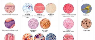
Soft tissues make up a significant part of the body. These include:
- skin;
- muscles;
- subcutaneous tissue.
All of them are available for ultrasound examination. Ultrasound also looks at structures located within soft tissues - vessels, lymph nodes, cartilage.
Ultrasound is reflected to varying degrees from objects with different densities. The lower this indicator, the worse the ultrasound is reflected. The ultrasound image is obtained due to the difference in the speed of ultrasound frequencies.
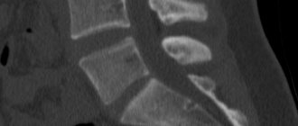
From dense tissues - bones, cartilage - sound is reflected quickly; in the picture they are represented in white. Soft tissues absorb some of the sound and appear dark gray in the photograph, or less often black.
Ultrasound of the hand in Moscow
The specialized medical clinic “Stoparthrosis” will quickly and efficiently conduct an ultrasound examination of soft tissues and joints of the upper extremities at an affordable price. Ultrasound of the hand ligaments, joints and muscles is performed by real professionals using advanced equipment using highly sensitive sensors. Diagnostics can be performed without a queue at any time convenient for the patient in comfortable conditions. The results of the study are issued on the same day. If necessary, in the clinic you can consult with an experienced doctor who will answer your questions, correctly diagnose and prescribe effective treatment.
Indications for use
Based on the definition of soft tissue ultrasound and understanding of what it is, one can judge the indications for the procedure. Ultrasound is prescribed for suspicion of various soft tissue diseases, when external examination is not enough to make a diagnosis.
- Muscle pathologies. These include various injuries - bruises, ruptures, wounds. Parasitic or fungal cysts sometimes form in the muscles.
- Lipoma. This is a wen - a tumor formed by adipose tissue. It appears on the head, back, chest.
- Atheroma. This is the name given to a cyst formed as a result of blockage of the sebaceous gland. Atheromas appear where there is a lot of hair - on the head, chest, and pubic area.
- Malignant tumors. Ultrasound is used to diagnose cancer of the skin, lymph nodes, and muscles. Ultrasound diagnosis of malignant tumors cannot be the only method. To confirm the oncological nature, an MRI is prescribed.
- Hematomas. This is an accumulation of blood between soft tissues, most often muscle. Occurs as a result of injury or in people with impaired clotting.
- Diseases of the lymph nodes. An ultrasound is performed if inflammation of the lymph nodes of viral or bacterial origin is suspected. Also, nodes change as a result of blood diseases or malignant tumors.
- Postoperative examination. Ultrasound is performed to assess the quality of the surgical intervention and the condition of the sutures.
- Pathology of joints. Ultrasound is used to detect sprains and ligament tears. Tendon calcifications that appear due to infections or metabolic disorders are also detected.
- Hernias. This is a protrusion of internal organs through separated muscles. An ultrasound can diagnose inguinal, umbilical or hernias of the white line of the abdomen.
Ultrasound diagnostics is highly accurate. Additional examination methods are rarely required. If there is doubt about the diagnosis, computed tomography or magnetic resonance imaging is prescribed.
Watch a video about ultrasound examination of body tissues:
Preparing for the examination
A feature of soft tissue ultrasound is often called the absence of the need for any preparation for the procedure. In most cases, the study does not really require maintaining a diet, and there is no need to drink a lot of liquid immediately before the test.
An exception is ultrasound of the soft tissues of the abdomen. When preparing for it, it is necessary to limit the consumption of foods that increase gas formation. Patients suffering from constipation are advised to cleanse the intestines: do an enema. More than 6 hours should pass between the study and the last meal.
It is not recommended to conduct an examination if the patient had a gastroscopy or colonoscopy the day before.
To perform the procedure, the patient is asked to remove clothing from the area that needs diagnosis. A gel is applied to the skin to increase wave conductivity by preventing air from entering between the device’s sensor and the skin.
Organ examination technique
The examination of each part of the body has its own characteristics. Since the skin on almost the entire body is covered with hairs, an air gap forms between it and the sensor. It makes diagnosis difficult. To eliminate it, use a special sound-conducting gel.
- Face. Here, with the help of ultrasound, the cheeks, lip area, and parotid lymph nodes are examined. The skin on the face is thin and devoid of coarse hair. Therefore, it is not necessary to use gel here. The doctor moves the sensor over the desired areas and may ask the patient to turn or tilt his head.
- Neck. Here the condition of the lymph nodes is assessed. The skin is treated with gel. The sensor is passed along the lateral, front and back surfaces of the neck.
- Hands. The lymph nodes in the shoulder and armpit area are examined. Sprains and suppurative processes can be found on the forearm. Hands are most susceptible to diseases and injuries. The limb is treated with gel, the sensor is passed along the back and palmar side of the hand.
- Chest area. The lymph nodes are examined and the condition of the subcutaneous tissue is assessed. The gel is applied and the sensor is passed along the front and back surfaces of the chest.
- Stomach. The soft tissues of the abdomen are muscles, peritoneum, and subcutaneous tissue. The skin is treated with gel, and the entire surface of the abdomen is examined with a sensor.
- Small of the back. On the back and lower back, the subcutaneous tissue is most susceptible to diseases. After applying the gel, all parts of the back are examined with a sensor.
- Taz. Here the perineal organs and lymph nodes of the groin area are examined. If you plan to have your pubic area examined, the hair will need to be shaved off. The examination also includes examination of the gluteal muscles. Requires application of gel.
- Legs. The condition of the lymph nodes, muscles, and ligaments is assessed. Parasitic cysts can form in the soft tissues of the thighs. The upper part of the shin is the site of sprains.
During the examination, the doctor asks the patient to turn around, change body position, and bend a limb. This is necessary for a comprehensive examination of pathological formations.
In the following video, a specialist will tell you how ultrasound helps identify pathology:
Anatomical features of the abdominal wall
The abdominal wall is a complex anatomical structure in the human body. It consists of front, side and back parts. The first two of them are considered the most important when performing ultrasound diagnostics. The anterior abdominal wall consists of the rectus abdominis muscles, which are connected in the middle by the linea alba. They are divided into separate sectors using tendon jumpers.
Throughout its entire thickness, the anterior abdominal wall consists of the following layers:
- skin;
- fatty tissue (thickness from 3 cm depending on the person’s physique);
- fascia;
- muscles (this layer is absent in the area of the white line);
- preperitoneal tissue.
Under the muscle layer is the peritoneum. It has an extremely small thickness, so when performing ultrasound diagnostics it is practically invisible. The only exception is that when performing respiratory movements, it is noticeable that the internal organs are slightly mixed relative to the abdominal wall.
Obtained results
Ultrasound is aimed at identifying various diseases. To determine them, you need to know the differences between healthy soft tissues and pathologically altered ones.
Soft tissue health criteria:
- muscle tissue has a homogeneous structure, whole fibers can be traced, attached to the tendons on both sides;
- fatty tissue with a homogeneous structure, without compactions;
- lymph nodes have a round shape, size 1-1.5 cm;
- the tendons are straight, without breaks.
Diseases of soft tissues are diverse. They arise as a result of injuries, infections, metabolic disorders. What the ultrasound shows depends on the type of disease.
- Cysts. These are cavities filled with air or liquid. They are formed against the background of parasitic infections, as a result of metabolic disorders. Cysts are found in muscles, near joints. In the picture, the cyst appears as a dark spot with a white rim around it.
- Hematomas. Hemorrhage in the tissue in the picture is represented by a dark, almost black spot. It has a density less than the surrounding tissue.
- Tumors. Found in muscle, adipose tissue, lymph nodes, and ligaments. Benign tumors have clear contours, oval or round shape. Sarcoma, carcinoma and other malignant tumors are characterized by irregular shape and blurred borders.
- Inflammatory processes. Infiltrates often form in muscles after bruises and infections. In the photo they appear as areas of darker color. An abscess is a purulent inflammation, represented by a cavity with liquid inside. Such ulcers appear in muscles and adipose tissue due to a bacterial infection.
- Sprains. They occur due to strong extension of the limb in the foot, knee or hip joints. The image shows swelling of the tendon and its elongation.
- Bruises and muscle tears. In case of bruises, the image shows a darkening of the muscle area. The rupture is characterized by a violation of the integrity of the muscle fiber.
- Inflammation of the lymph nodes. They are enlarged and have unclear boundaries. Various inclusions are identified inside.
- Hernias. Some internal organs are identified outside the body cavity. If the hernia is not strangulated, movement of the organs is noticeable.
The soft tissue ultrasound protocol includes a description of the size of the pathological formation, its contours, structure, and the presence of additional inclusions. The conclusion template is filled out by a specialist. The description is given to the patient. It is deciphered by the attending physician, taking into account other examination methods.
Additionally, watch the video to see what the formations look like:
Decoding the results: what is visible on an ultrasound
When deciphering the image, the diagnostician evaluates the structure, blood supply of tissues, the presence or absence of neoplasms and cavities. Normally, there should be no inclusions or “bags” of liquid. The standard size of lymph nodes is from 8 to 10 mm, they are almost indistinguishable from healthy tissue. Size and density change with the appearance of pathologies. Most often, ultrasound of soft tissues reveals diseases:
- enlarged lymph nodes due to inflammation, infection or metastasis;
- lipoma (benign neoplasm);
- myositis (inflammation in the muscles);
- pathologies of connective structures;
- abscesses;
- cancerous tumors;
- phlegmon (purulent inflammation of connective tissue);
- atheroma (an elastic formation like a tumor that appears due to blockage of the sebaceous glands, with clear contours);
- hygroma (a dense formation in the tendons, resembling a cyst, having a cavity with liquid contents);
- postoperative complications;
- hematomas (formations consisting of blood clots located in muscle tissue);
- hemangioma (benign tumor with blurred contours and heterogeneous structure);
- lymphostasis (swelling due to fluid stagnation, as a result of which the nodes rupture);
- tendon rupture;
- chondroma (benign tumor in cartilage tissue).
Ultrasound of soft tissues is the simplest and most accessible procedure. Due to the absence of harmful effects, it can be carried out regardless of age, from the first day of birth of the child. As a result, many soft tissue diseases can be prevented at the very beginning and the development of the pathological process can be prevented.
- Mammography or breast ultrasound: which is better?
- Ultrasound of neck vessels
- What will a heart ultrasound show? What diseases will it detect?
- Ultrasound of the intestines
- Ultrasound of the abdominal cavity - which organs are checked
Contraindications
Ultrasound is the safest diagnostic procedure. It can be performed without fear on newborn children, pregnant women, and the elderly. The frequency of the procedure also does not matter. Ultrasound is performed as many times as required to make a diagnosis.
Relative contraindications include:
- inadequate condition of the patient;
- the presence of wounds, burns, inflammations at the site of the proposed examination.
For emergency diagnosis, contraindications may not be taken into account.
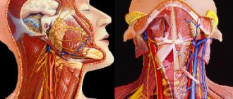
Ultrasound examination of the maxillofacial area
Ultrasound scanner HS50
Affordable efficiency.
A versatile ultrasound scanner with compact design and innovative capabilities.
The ultrasound research method has become firmly established in general diagnostic practice; its role can hardly be overestimated. A modern approach to diagnosing diseases in the clinic of internal medicine is unthinkable without ultrasound examination of the abdominal organs (including the retroperitoneum and pelvis), thyroid gland, mammary glands, heart and blood vessels.
Compared to the above-listed areas of application of echography, ultrasound examination of the maxillofacial area is performed much less frequently. This is due, on the one hand, to the clinical isolation of dentistry and maxillofacial surgery, which does not allow general practitioners of ultrasound diagnostics to gain sufficient experience in research in this area, and on the other hand, to some conservatism of dentists and maxillofacial surgeons, who consider the main diagnostic tool for them to be X-ray examination method. Their skepticism regarding ultrasound examination is based on the fact that almost all soft tissue structures of the maxillofacial region are accessible to palpation, and the skin and mucous membranes are accessible to inspection.
However, paying tribute to the history of the development of ultrasound diagnostics, it is necessary to mention that the object of the very first (then still one-dimensional - in A-mode) echographic studies performed by a group of researchers led by D. Howry in 1955 were the parotid glands.
Ultrasound of soft tissues of the face and neck in its modern version does not require the use of any special ultrasound scanners or sensors and can be performed on equipment designed for studying peripheral structures: linear sensors with an oscillation frequency of 5.0-7.5-9 are quite sufficient .0 MHz. Transcutaneous echography is quite informative and generally satisfies the needs of clinicians: almost all parts of the face and neck (including the body and root of the tongue) are accessible to echographic examination using external sensors. Only the upper parts of the peripharyngeal space and the pterygomaxillary space, shielded by the branch of the lower jaw, are inaccessible.
There are no age restrictions or special preparation of the patient for an ultrasound examination.
For an ultrasound diagnostic physician, the maxillofacial area can be of great professional interest, since diseases of all nosological groups are found here (from inflammatory, autoimmune and degenerative-dystrophic to tumor), as well as various malformations (angiodysplasia, lymphangiomas, congenital cysts). Differential diagnostic difficulties increase due to the fact that the maxillofacial area is a zone of massive infection and the existence of primary non-inflammatory diseases is often masked by the addition of an inflammatory process with the entire spectrum (from erased to clinically pronounced) of its signs.
The complexity of the anatomical structure of the maxillofacial region creates additional difficulties for interpreting the results of ultrasound examination. At the same time, anatomical detail is of great importance, since determining the organ affiliation of the pathological process and clarifying the topographic and anatomical features of its distribution are one of the most important diagnostic tasks, along with identifying the nosological form of the disease. This point becomes especially relevant if we take into account that during operations specifically on the maxillofacial area, surgeons are especially faced with the task of finding a compromise between choosing the optimal access to carry out the maximum possible radical intervention and causing the least possible aesthetic damage to the patient’s face.
Particular diagnostic issues
Currently, thanks to the introduction of ultrasound diagnostic technologies into obstetric practice, the maxillofacial area becomes an object of medical interest even before the birth of the child. This makes available the intrauterine detection of clefts and other malformations of the fetal face and neck, a number of syndromes with facial signs (Down syndrome, Turner syndrome, Goldenhar syndrome, etc.), as well as recognition of teratomas, hemangiomas and lymphangiomas of the fetus.
Timely detection of these changes forces in some cases to reconsider the approach to pregnancy management tactics or to provide for the need to carry out certain organizational, tactical and therapeutic measures in the perinatal and neonatal periods. This concerns, in particular, the expansion of the obstetric team with the involvement of maxillofacial surgeons to provide specialized care as early as possible.
Ultrasound examination makes a significant contribution to the diagnosis of diseases of the major salivary glands.
In inflammatory diseases of the parotid glands, echography allows for differential diagnosis of various forms of mumps, identifying sialodochitis - inflammation in the ducts of the salivary glands, recognizing inflammation of the intraglandular lymph nodes (lymphadenitis) and clarifying its stage. All this is essentially a distinction between surgical and non-surgical pathology of the parotid glands (Fig. 1-7).
Rice. 1.
Right-sided acute parotitis.
Rice. 2.
Left-sided chronic parenchymal parotitis.
Rice. 3.
Sialodochitis of the left submandibular gland.
Rice. 4.
Acute serous lymphadenitis in the left parotid gland (in two scanning planes).
Rice. 5.
Acute serous lymphadenitis in the left parotid gland with limited periadenitis.
Rice. 6.
Acute serous lymphadenitis with widespread periadenitis (lymphogenous mumps, Herzenberg's mumps) in the left parotid gland.
Rice. 7.
Purulent lymphadenitis in the parotid gland (parotid abscess).
In salivary stone disease, which is most often found in the submandibular glands, ultrasound allows one to identify stones regardless of their location (in the parenchyma of the gland, intraglandular ducts, excretory duct) and the degree of their mineralization, to clarify the presence of sialadenitis - inflammation of the parenchyma of the gland, which can be independent or accompanying salivary stone disease (Fig. 8-11).
Rice. 8.
Salivary stone disease. Calcified calculus in the area of inflection of the excretory duct (typical location) of the left submandibular gland. Associated sialadenitis.
Rice. 9.
Salivary stone disease. Uncalcified calculus in the ampullary part of the excretory duct of the submandibular gland.
Rice. 10.
Salivary stone disease. Calcified stones in the parenchyma of the left submandibular gland. Perifocal edema of the gland parenchyma.
Rice. eleven.
Sialadenitis of the right submandibular gland.
The use of echography in extraorgan inflammatory processes on the face and neck makes it possible to distinguish purulent (abscess, phlegmon) and non-purulent (infiltrate) lesions of soft tissues, and when a purulent lesion is detected, accurately localize accumulations of pus (Fig. 12-14).
Rice. 12.
Infiltration of the left buccal area.
Rice. 13.
Phlegmon of the submandibular region after opening and drainage (drainage is visible).
Rice. 14.
Abscess of the submandibular region.
Visualization of the outer (vestibular) surface of the jaw bones makes it possible to establish periostitis, which is one of the most common causes of inflammatory changes in the soft tissues of the face (Fig. 15).
Rice. 15.
Serous periostitis of the lower jaw on the right. Infiltration in the soft tissues of the right cheek.
The exceptionally developed lymphatic system of the face and neck brings significant specificity to the spectrum of diseases in this area. This applies to both inflammatory and non-inflammatory lesions of the lymph nodes. An echo-graphic study allows one to visualize altered lymph nodes and, based on a number of reference signs (the number of altered nodes, their size, shape and proportions, the nature of the contours, the presence or absence of inclusions and their distribution in the node, the degree of decrease in echogenicity) to reconstruct with a high degree of reliability what is happening in the lymphatic node processes.
In the structure of inflammatory diseases of the soft tissues of the maxillofacial region, inflammatory lesions of the lymph nodes occupy one of the first places. An echographic examination allows one to reliably distinguish reactive (inflammatory) hyperplasia of the lymph nodes from their true inflammation - lymphadenitis (Fig. 16-18).
Rice. 16.
Acute reactive (inflammatory) hyperplasia of the lymph node of the neck.
Rice. 17.
Chronic reactive (inflammatory) hyperplasia of the lymph node of the neck.
Rice. 18.
Acute serous lymphadenitis of the neck.
The key point in choosing a treatment method for lymphadenitis is to determine the nature (stage of development) of the inflammatory process: serous or purulent lesion of the lymph node. Ultrasound examination, even in B-mode, makes it possible to resolve this issue with a high degree of reliability, and the use of blood flow visualization techniques (color Doppler mapping, power Doppler mapping) makes it possible to identify the process of purulent melting at its earliest stage (Fig. 18-21).
Rice. 19.
Purulent lymphadenitis of the neck with periadenitis.
Rice. 20.
Purulent lymphadenitis of the submandibular region.
Rice. 21.
Dopplerographic dynamics of the process of purulent melting of the lymph node (over 3 days).
All this makes it possible to rationally approach the combination of methods of conservative therapy and surgical treatment, to choose adequate treatment tactics, avoiding the use of unjustified treatment methods. In everyday practice, metastatic lesions of the lymph nodes of the neck are often encountered, as well as their involvement in the pathological process in lymphoproliferative diseases. An echographic examination makes it possible to distinguish these groups of diseases with a high degree of reliability and differentiate these types of lesions from inflammatory changes in the lymph nodes.
In case of injury from glass, wooden and plastic objects, as well as after gunshot wounds and road traffic accidents, foreign bodies often remain in the soft tissues of the face and neck, the timely detection of which allows for the most effective primary surgical treatment of the wound and reduces the risk of inflammation.
After gunshot wounds, multiple and, as a rule, metallic foreign bodies usually remain in the soft tissues, the high radiopacity of which allows traditional X-ray examination to see even the smallest of them and determine their skeletotopy. In these cases, echography, of course, is inferior to x-ray examination in determining the number and size of foreign bodies, but ultrasound examination is certainly necessary to clarify their organotopy in relation to soft tissue structures - vascular bundle, muscles and fascia.
In cases where there are glass, plastic or wooden foreign bodies in the soft tissues, echography is essentially the only method that allows you to study the infiltration zone, obtain an image of foreign bodies, determine their number, size, location and organotopy (Fig. 22- 26).
Rice. 22.
Foreign body in the soft tissues of the left parotid-masticatory region (metal ball - air gun bullet).
Rice. 23.
A foreign body in the subcutaneous tissue of the left cheek is a glass fragment.
Rice. 24.
A foreign body in the left cheek area is a splinter.
Rice. 25.
Foreign bodies in the keloid scar of the lower lip are small stones and asphalt crumbs (after a traffic accident).
Rice. 26.
A foreign body in the submandibular region is a fragment of a plastic catheter.
A significant proportion of diseases of the maxillofacial area are congenital cysts of the neck and oral cavity. An echographic examination makes it possible to visualize them, clarify their structure and organotopic characteristics, on the basis of which differential diagnosis is based.
Among congenital cysts of the neck and oral cavity, there are: thyroglossal (middle) and branchial (lateral) cysts and fistulas, retention cysts of the sublingual gland, as well as dermoid cysts.
The urgent need for distinctive recognition of the group affiliation of congenital cysts and fistulas is dictated by the necessity of their radical excision in order to avoid relapse. And if dermoid cysts and retention cysts of the sublingual gland are characterized by a clear demarcation and absence of fistulous tracts, then thyroglossal cysts usually have a connection with the hyoid bone and the root of the tongue, and branchial cysts are often connected by a fistulous tract with the lateral wall of the pharynx (Fig. 27-30).
Rice. 27.
Thyroglossal (median) cyst of the neck. There is a connection with the hyoid bone and the root of the tongue.
Rice. 28.
Branchial (lateral) cyst of the neck. Relationship with the vascular bundle of the neck (compression of the internal jugular vein and displacement of the common carotid artery).
Rice. 29.
Dermoid cyst of the floor of the mouth.
Rice. thirty.
Retention cyst of the right sublingual gland.
Clinicians are well aware of the difficulties in distinguishing between festering cysts and abscessing lymphadenitis. The specificity of the echographic picture makes it possible not only to solve emerging differential diagnostic difficulties, but also to determine the severity of scar changes around the cyst.
Congenital cysts of the neck and oral cavity often have to be differentiated from the cystic form of lymphangioma, which is essentially a malformation of the lymphatic vessels. Cystic lymphangioma is more characterized by multiple chambered cavities or multiple cyst-like formations, as well as significant prevalence. All these signs are clearly identified echographically. In cases of single-chamber and limited prevalence of lymphangioma, differential diagnosis is significantly difficult (Fig. 31).
Rice. 31.
Lymphangioma of the left parotid gland.
In the face and neck area, pathological formations from blood vessels are often found - so-called “hemangiomas”, which in most cases (up to 95-97%) are vascular hyperplasias and vascular malformations (angiodysplasias). True vascular tumors occur only in 3-5% of cases.
In this case, either only the vascular periphery - capillaries, or only larger vessels with the formation of arteriovenous communications (stomachs, fistulas) can be involved in the pathological process; a combination of these options is possible. Dysplastic changes can be localized only in the venous part of the vascular bed; this type of lesion is referred to as “venous dysplasia” (Fig. 32).
Rice. 32.
Venous dysplasia of the right parotid gland.
Vascular formations can have a diffuse distribution in soft tissues or be clearly demarcated, and also have their own hemodynamic characteristics (Fig. 33).
Rice. 33.
Vascular hyperplasia of the right buccal region.
All these factors entail the need for an individual approach to the choice of treatment method and tactics. Ultrasound examination, allowing to determine the main morphological and hemodynamic parameters of vascular formations of the face and neck, is an effective method for their diagnosis.
A very significant role belongs to echography in recognizing tumors of the salivary glands. The possibility of a detailed assessment of the contours of the neoplasm and its internal structure (echostructure) makes it possible to distinguish benign tumors of the salivary glands from malignant tumors with high reliability (Fig. 34-36).
Rice. 34.
Polymorphic adenoma of the left parotid gland.
Rice. 35.
Lipoma of the left parotid gland.
Rice. 36.
Malignant tumor of the right parotid gland (histologically: undifferentiated carcinoma).
Of great importance in clarifying the nature and extent of the tumor process is also the assessment of the condition of the regional lymph nodes, in which echographic examination is a recognized leader (Fig. 37).
Rice. 37.
Malignant tumor of the left parotid gland (histologically: adenoid cystic carcinoma). Metastatic lesions of regional lymph nodes.
The abundance of lymphoid tissue in the parotid glands determines the high frequency of their damage by benign and malignant lymphoproliferative diseases, which also have their own characteristic echographic signs (Fig. 38-39).
Rice. 38.
Damage to the left parotid gland due to lymphoproliferative disease (histologically: lymphogranulomatosis).
Rice. 39.
Damage to the lymph nodes of the neck due to lymphoproliferative disease (histologically: lymphosarcoma).
Without in any way contrasting ultrasound of the maxillofacial area with the traditional clinical examination of the patient, there is every reason to assert that the use of echography is certainly indicated not only in diagnostically unclear cases, but even with an already established diagnosis, when the method allows us to identify individual characteristics of the course of the disease , which can be significant when planning a patient’s treatment.
The non-invasiveness and harmlessness of ultrasound examination makes it possible to carry it out many times to monitor the dynamics of the pathological process and evaluate the effectiveness of treatment measures.
The high information content of an echographic examination (reaching 95-98% in general) makes it possible to limit the use of radiological techniques (traditional radiography, sialography, angiography), significantly less often use invasive diagnostic interventions (soft tissue biopsy) and more rationally approach the use of such expensive studies as computer and magnetic resonance imaging.
Ultrasound scanner HS50
Affordable efficiency.
A versatile ultrasound scanner with compact design and innovative capabilities.
Examination of leg tissues
During an ultrasound, the doctor assesses the condition of soft tissues, muscles, tendons, and also looks at the condition of the nerve processes. Indications for
management:
- presence of ankle injuries;
- tendon ruptures;
- Crick;
- for tumor diagnosis;
- the presence of a Baker's cyst, which is filled with fluid;
- it is possible to view growths on the heels;
- After fractures, you can monitor how well the bones heal.
Ultrasound of neck muscles
Sometimes patients complain of discomfort in the neck area. In such cases, specialists prescribe an ultrasound scan of the neck, since such an examination can give a reliable picture of the disease, most often expressed in changes occurring in the soft tissues, impaired blood flow or dysfunction of the neck organs.
Ultrasound of the neck is a common test that can be performed at an outpatient clinic or hospital, in paid clinics upon a doctor’s referral or at your own request.
Examination of limb tissues
An examination of the soft tissues of the extremities is carried out with the following indicators:
- if the patient has a hernia of the hip joint;
- the presence of neoplasms;
- unclearly visible fracture of fragile bones;
- unexplained pain;
- after bruises, injuries and fractures;
- for complications of joint function. The complex also performs ultrasound of the joints.