If a woman who dreams of becoming a mother closely monitors her health and controls her menstrual cycle, then from the first days of the delay she can hope that pregnancy has occurred. As a rule, they rush to do the test, which can be purchased at any pharmacy.
But remember that hCG levels rise differently for everyone, so the test strip may give a false negative or false positive result. When a rapid test gives a positive result, some pregnant women, of their own free will or on the recommendation of a doctor, in some controversial cases, are sent for ultrasound diagnostics.
At what stage does an ultrasound show pregnancy is a fairly popular question among expectant mothers. In addition, those who are especially suspicious may be interested in when will an ultrasound show pregnancy outside the uterus? Can an abdominal ultrasound determine pregnancy at the earliest stage, or in this case is only a vaginal ultrasound relevant?
First scheduled ultrasound
During a normal pregnancy, a woman undergoes only three scheduled examinations during the entire period. The first of them is prescribed at 11-12 weeks. By this moment, the fetus already has the rudiments of all organs and systems. The embryo grows so much that it can be clearly examined with the help of a device and possible developmental abnormalities can be identified.
At this stage, a diagnosis is carried out to determine whether the child has Down syndrome or some other genetic pathologies. This ultrasound examination is called screening. Its results are supplemented by the results of blood tests for chromosomal dysfunctions.
Women should have their first ultrasound examination approximately 7-8 weeks after the delay, when the implantation of the embryo is already confirmed and beyond doubt. To make a diagnosis, it is enough to do a blood test for hCG and undergo an examination by a gynecologist. However, there are cases when an ultrasound is necessary to determine pregnancy in the early stages.
Transvaginal ultrasound
Ultrasound examination using a transvaginal sensor allows you to assess the development of pregnancy at very early stages. In the process of performing it, you can identify all sorts of deviations and problems, consider the fertilized egg, its location in the uterus and other nuances. As a result of the examination, the doctor determines the presence of physiological pregnancy and possible problems.
The transvaginal technique involves inserting a special sensor into the pregnant woman’s vagina, onto which a condom is placed. By the 5th obstetric (3rd gestational) week of gestation, the yolk sac has already been formed, which provides the hematopoietic function and nutrition of the embryo and the amnion (amniotic sac with an embryo inside). On ultrasound they can be clearly seen at this stage.
Thus, in the third gestational week of pregnancy, a fetal egg is visible on ultrasound, although its size at this time is very small. It turns out that if a woman has already been pregnant for 6 days, and she does not know at what stage pregnancy can be determined by ultrasound, then you can safely contact your gynecologist, who will refer her for such a diagnosis.
As a rule, not everyone is prescribed ultrasound for such a short period of time. This requires serious reasons:
- a woman has undergone an IVF procedure - it is determined how well the fertilized egg has implanted in the uterus;
- there is a serious suspicion of ectopic implantation of a fertilized egg;
- Pregnant women have complaints of nagging pain in the lower abdomen, as well as various vaginal discharge;
- unsatisfactory test results for hCG or other hormones;
- the woman has been diagnosed with miscarriages, miscarriages, or ectopic pregnancies in the past.
Transvaginal ultrasound can cause spontaneous miscarriage
As a rule, ultrasound itself does not provoke bleeding or vaginal discharge. But if a pregnant woman experiences stress, this can lead to uterine tone and there is a serious threat of miscarriage. If, after an ultrasound, a woman notices various types of vaginal discharge, including blood, then one cannot remain silent about it - a consultation with an obstetrician-gynecologist is necessary.
Examination of a pregnant woman using a vaginal sensor is effective until the uterus extends beyond the pelvis (up to 12 obstetric weeks). At later stages of intrauterine development of the fetus, the use of standard ultrasound (through the anterior abdominal wall) is practiced.
Reasons and goals of early ultrasound diagnosis during pregnancy
Before the first scheduled examination, a woman is recommended to visit an ultrasound specialist in several situations:
- previously there were problems with pregnancy (miscarriages occurred);
- with positive tests and elevated hCG levels, the doctor does not see pregnancy during the examination;
- According to calculations, the period has already been several weeks, and the second strip of dough is very pale;
- after a long delay, brownish discharge appeared, not similar to menstruation;
- there is nagging pain in the lower abdomen;
- When pregnancy is confirmed by a doctor, bloody discharge suddenly appears.
Quite rare, but there are cases when the fertilized egg does not reach its final destination, but is fixed in the tube. This pathology is called an ectopic pregnancy. It can pose a danger to a woman's life. In the absence of timely measures taken, the risk of infertility is high. Ultrasound examination can identify a dangerous situation and help avoid serious consequences.
A hardware examination will allow you to find out whether there is a threat of miscarriage due to pain in the lower abdomen and bloody discharge. An ultrasound can be a lifesaver for the fetus. An ultrasound diagnostic method can detect the cause of a long delay when pregnancy is not confirmed. This could be a cyst, fibroma, or a very dangerous disease called “hydatidiform mole”, in which all the signs coincide, but conception has not occurred.
If there is an ectopic pregnancy, it can be seen on ultrasound
When the zygote is fixed outside the uterus, it is quite difficult to determine this by ultrasound.
Even the most sensitive apparatus is not able to identify the zygote in the fallopian tubes or other organs of the reproductive system. Suspicion of an ectopic pregnancy arises when the following conditions are simultaneously met:
- positive test, rising hCG level;
- absence of embryo in the uterus;
- abdominal discomfort.
4-5 weeks after conception, the doctor may detect an enlargement of the organ in which the embryo is attached. In the first 2-3 weeks after fertilization of the egg, it is impossible to see a frozen pregnancy on an ultrasound. A fertilized egg can implant anywhere: the fallopian tube, ovary, or directly on the peritoneal tissue. This is a serious pathology that requires surgical intervention and can threaten a woman’s life.
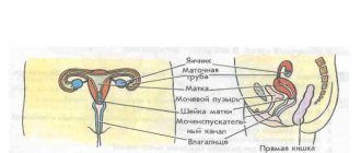
Types of ultrasound examinations
There are four types of pelvic ultrasound that are practiced in gynecology. Brief description of each:
- The transrectal view involves inserting a sensor through the rectum. It is carried out for girls who are not yet sexually active. Not used to determine pregnancy.
- The transvaginal view involves inserting a sensor through the vaginal tract to the uterus. It is most accurate in detecting pregnancy in the early stages.
- The transabdominal view involves examination using a special probe without penetration. The device is inserted into the body through the anterior wall of the abdominal cavity. When determining pregnancy in the early stages, the method does not guarantee an accurate result.
- A combined examination involves transabdomial and vaginal ultrasound.
The optimal way to detect pregnancy in the early stages is the transvaginal method. However, it cannot always be used. There are contraindications and risks of complications.
Inaccuracies and complications
And yet, concerned women are interested in whether an ultrasound will show an ectopic pregnancy in the early stages in all cases? Or are there errors? No one can give a 100% guarantee due to the following reasons:
- Too short a pregnancy does not physically allow one to determine whether there is an embryo in the uterine cavity or not.
- Limited range of ultrasound machine capabilities.
- Imperfection of the equipment used and its possible malfunction.
- The specialist performing the diagnostic procedure did not have sufficient qualifications and experience.
Sometimes an accumulation of fluid or a blood clot in the uterus looks like a fertilized egg and the specialist mistakenly determines intrauterine pregnancy. The woman calms down accordingly, and if the internal bleeding is insignificant, then a pathological pregnancy can proceed hidden for a long time and at the same time not have pronounced symptoms.
A frequent option for further developments is rupture of the fallopian tube. This happens suddenly and is accompanied by the following symptoms:
- A sharp pain is felt in the lower abdomen, mainly on the side where the fertilized egg was attached to the tube. The pain radiates to the rectum area, as well as the right collarbone.
- Often there is a false urge to defecate or loose stools appear. The abdomen is swollen and painful on palpation.
- Decreased blood pressure, severe weakness, even fainting. With severe bleeding, hemorrhagic shock develops.
- Pale skin and mucous membranes, shortness of breath, cold sweat.
- Apathy, lethargy, rapid, weak pulse.
Treatment in this case involves immediate surgery. The damaged fallopian tube is removed laparoscopically, and if hemorrhagic shock occurs, then by performing a laparotomy (open access to the abdominal cavity).
Ultrasound with the introduction of a vaginal sensor is considered the most accurate diagnostic method in determining ectopic pregnancy. But you shouldn’t expect a 100% guarantee from him either. Not in all cases this pathology will be visible and the correct diagnosis will be made. It is advisable to combine ultrasound diagnostics with other methods for greater accuracy.
At what stage can ultrasound detect pregnancy?
Conception occurs on the day of ovulation (the release of an egg from the ovary). The egg is then sent through the tubes to the uterus. The egg implants in it after a few days. However, it is not yet possible to examine the embryo due to its insignificant size. At what stage does an ultrasound show pregnancy? Approximately on the 20th day from the moment of conception. If you count from the last day of menstrual flow - in the 5th week.
At this stage, the embryo begins to develop a pulse. This can already be recorded by equipment. You can see the fertilized egg a little earlier, but there are cases when it turns out to be empty and the pregnancy is false. It is initially possible to recognize in the early stages whether there is an embryo and whether it is alive only by the beating of the heart.
In the third week from conception (approximately the fifth or sixth day of a missed period), only a vaginal sensor can detect the appearance of a fetus. The abdominal one will cope with this task only 14-21 days later, that is, at the 7th, 8th obstetric week.
The reason for this is the abdominal wall, which prevents the device from identifying the tiny embryo and detecting its heartbeat. Sometimes it is possible to determine a multiple pregnancy at the 6th obstetric week. However, more often it is possible to do this closer to the end of the first trimester - after 2.5 months.
At what week after conception can pregnancy be seen in the uterine cavity?
Conception occurs after ovulation, when the egg is released into the uterine cavity. This usually happens 14 days from the beginning of the cycle; it is during this period that fertilization is possible. On ultrasound, the birth of a new life can be seen only when the zygote attaches to the wall of the uterus, which occurs only 7-10 days after conception. Often a doctor sees a fertilized egg in the uterus in the short term, but does not observe an embryo in it. This may indicate a frozen pregnancy or too early contact with a specialist.
The absence of an embryo in the fertilized egg requires a mandatory repeat of the study after 7-10 days to see whether the pregnancy develops. It is not recommended to make premature conclusions before 6-7 weeks.
To accurately determine the presence of an embryo, it is recommended to apply for an ultrasound no earlier than 4-5 weeks into the obstetric period, which corresponds to the period from the first day of the last menstruation. It was during this period that attachment has definitely occurred, and the doctor will see changes in the uterine cavity.
How to prepare for research
If an ultrasound examination is not performed on an emergency basis, then it is necessary to prepare for the procedure in advance. A couple of days before the examination, it is advisable to avoid foods that can cause increased gas formation:
- cabbage;
- legumes;
- baked goods;
- carbonated drinks;
- grapes;
- pears
An increased amount of gases in the intestines will interfere with the examination, which is already a rather complicated procedure in the early stages. Before a transvaginal ultrasound, the drinking regime can remain normal. However, your bladder must be empty during the procedure.
Transabdominal examination, on the contrary, is carried out when the stomach is full. To do this, three hours before the procedure you need to drink at least a liter of liquid and you cannot go to the toilet. If this condition is not feasible for a pregnant woman, then it is allowed to reduce the time period for filling the bladder to an hour.
The day before a vaginal ultrasound examination, it is recommended to refrain from having sex. Any type of ultrasound diagnostics does not require any further preparation.
What period does ultrasound show: obstetric or from the moment of conception?
Obstetricians-gynecologists use terms such as obstetric and gestational (embryonic) terms of pregnancy in their work. There is a slight difference between these concepts. By obstetric period we mean the number of weeks that have passed since the beginning of the last menstruation. The gestational (embryonic) period is the period that begins from the moment of fertilization of the egg.
The period determined by ultrasound is considered embryonic. In obstetric practice, the first concept is widely used. That is why, in order to avoid confusion, experts convert the gestational period to the obstetric period, adding 2 weeks to it.
Progress of the procedure
Transvaginal ultrasound is somewhat different from transabdominal ultrasound in terms of the procedure. In the first case, the woman takes off everything below the waist, lies face up on the couch, and spreads her legs. An oblong-shaped sensor is inserted into her vagina, onto which a condom is placed before the examination begins.
During a transabdomial ultrasound examination, you should only expose your stomach and lower your panties a little. The procedure is also performed in the supine position. First, the stomach is lubricated with a special gel. Then the doctor begins to move the sensor along it. After the procedure is completed, the gel applied to block air access is wiped off with a disposable napkin.
Ultrasound examination in both cases lasts only a few minutes. During this time, the specialist receives comprehensive information about the location of the embryo, its size, heart rate, etc. During the procedure and after its completion, the woman does not experience any discomfort. However, when the bladder is full, the pressure from the sensor may increase the urge to go to the toilet.
How and why are the weeks of the cherished nine months counted?
And yet, why does the study suddenly set the period to be 2 weeks less or more than the monthly period, and how is it correct? There are several methods for this in medical practice.
The simplest of them is based on the fact that on average pregnancy lasts no more than 40 weeks or 280 days. This is the so-called “obstetric period”. This is how long it usually takes from the start of your last period to the birth of your baby. The first question that a future mother is asked at the antenatal clinic is: “When was the last time your period started?”
To find out the expected date of birth of the heir, you need to count three months ago from this day, and then add 7 days.
This “obstetric” formula was developed by the French gynecologist F.K. Negele. However, it is only suitable for women with a regular 28-day menstrual cycle. It is imperative to keep in mind that it is impossible to predict the specific date of birth. It is only assumed. This is a period of ± 10-12 days. After all, for each woman everything is strictly individual.
Determination of the embryonic period
Another method is used to calculate “embryonic life.” It is not counted by menstruation, but from the day of conception, which, as a rule, coincides with ovulation. A woman's egg matures by the beginning of the third week of the menstrual cycle. Modern doctors know that fertilization can occur after ovulation for another two days. The activity of male sperm lasts longer - four days. Thus, conception can occur within about six days. The “embryonic period” thus differs from the “obstetric period”, which is approximately fourteen days longer.
As a rule, during ultrasound diagnostics, more calculations are used based on the “obstetric period”, because it is easier to find out from patients when they had their periods than to find out the exact date of conception. Almost everyone finds it difficult to name it.
Other methods of determining the period are also used. For example, by the size of the uterus or by the movement of the fetus. However, these criteria are purely individual in nature for each woman in labor and, because of this, are less accurate. Indeed, with the same time intervals of gestation in different women, uterine parameters vary over a very wide range, which makes it impossible to estimate the period with weekly accuracy in each specific case.
Intrauterine movements of the fetus are also felt very subjectively, this is influenced by the sensitivity threshold, which is different for all women. So, for example, one expectant mother begins to feel the baby kicking from the inside from the eighteenth week, and another - only from the twenty-second. And this despite the fact that in reality the activity of the fetus manifests itself already from the second month, unnoticed by the mother.
Possible errors in ultrasound results
Despite the fact that ultrasound is considered one of the most accurate ways to determine pregnancy for a period of five obstetric weeks, there are cases when the device does not see the embryo or diagnoses a conception that does not exist. The reasons for the errors in the research results may be:
- Excess weight of a woman during transabdomial ultrasound. A large fat layer prevents the sensor from gaining full access to the object being examined.
- Broken or outdated equipment.
- Insufficiently qualified specialist conducting diagnostics.
- The presence of neoplasms (polyps, cysts) in the uterus, which the doctor may mistake for a fertilized egg.
A common situation is when the date of the last menstruation indicates five obstetric weeks, but the embryo is not visible on ultrasound. This happens in women with long menstrual cycles. The actual period at the time of examination turns out to be too short, and the sensor is not able to see the embryo.
It is believed that errors in determining pregnancy at its very beginning using ultrasound occur in one out of ten cases. Therefore, you cannot trust the results of the equipment 100 percent. You need to continue to monitor your condition and try other methods. If there are doubts about the quality of the equipment that was used or the competence of the specialist, you can undergo a re-examination.
Table of pregnancy dates according to ultrasound
| weeks | KTR | Average Ø ovum | BPR | Average Ø yolk sac |
| 5 | 2 | 18 | ||
| 6 | 5 | 22 | 3 | |
| 7 | 9 | 24 | 4 | |
| 8 | 16 | 30 | 6 | 4,4 |
| 9 | 23 | 33 | 8,5 | 4,6 |
| 10 | 31 | 39 | 11 | 5 |
Here you can see when an ultrasound can detect pregnancy - from the 5th week.
Evaluation of the fetus according to such parameters as indicated in the table is called fetometry. From the 3rd month, “age” is calculated using fetometry:
| A week | Biparietal size | Fronto-occipital size | Head circumference | Abdominal girth | Hip | Shin | Shoulder | Forearm |
| 11 | 17-21 | 63-73 | 51-62 | 5,6-7,8 | ||||
| 12 | 21-24 | 71-84 | 61-72 | 7,3-10,6 | ||||
| 13 | 24-28 | 84-96 | 69-80 | 9,4-11,8 | ||||
| 14 | 27-31 | 97-110 | 78-90 | 12,4-15,8 | ||||
| 15 | 31 | 110 | 90 | 16,2 | ||||
| 16 | 34-37 | 45-49 | 124-136 | 102-116 | 20-23 | 18-21,0 | 18-21 | 15-18 |
| 17 | 38-42 | 50-54 | 135-149 | 112-131 | 24-28 | 21-25 | 21-25 | 18-21 |
| 18 | 42-47 | 54-59 | 146-161 | 124-144 | 27-31 | 24-28 | 24-28 | 20-23 |
| 19 | 45-49 | 58-63 | 158-174 | 134-154 | 30-34 | 27-31 | 27-31 | 23-26 |
| 20 | 48-53 | 62-68 | 170-186 | 144-164 | 33-37 | 30-34 | 30-34 | 26-29 |
| 21 | 51-56 | 66-72 | 183-200 | 157-177 | 36-40 | 33-37 | 33-37 | 28,0-32 |
| 22 | 54-60 | 70-76 | 195-212 | 169-190 | 39-43 | 35-39 | 35-39 | 30-34 |
| 23 | 58-64 | 74-81 | 207-224 | 181-202 | 41-45 | 38-42 | 38-42 | 33-37 |
| 24 | 61-67 | 78-85 | 219-237 | 193-224 | 44-48 | 40-44 | 40-44 | 35-39 |
The most common questions about fetal ultrasound measurement
What stage of pregnancy does an ultrasound show?
There are two main ages of the fetus:
- obstetric – determined from the first day of the last monthly bleeding (the date of birth for this period is calculated as follows: from this date minus 3 months and plus 7 days)
- embryonic – from the moment of conception (this date is taken as the day of ovulation). It turns out shorter than the first by 2 weeks.
Ultrasound does not directly calculate gestational age. With the help of this study, an analysis is made of how many weeks (obstetrics are taken as a basis) the fetal parameters correspond to.
Is it worth doing an ultrasound to determine pregnancy?
It is not for nothing that the first planned ultrasound examination is prescribed at 11-12 weeks. During this period, the embryo has already developed so much that enough information can be obtained about it. However, it is not too late to terminate the pregnancy if serious abnormalities in the development of the fetus are detected.
Women who get ultrasounds earlier still return to the specialist's office a few weeks later. The negative impact of ultrasonic waves on an unborn child has not been proven by science, but it has not been refuted either. You should not risk the health of your unborn child and resort to the procedure in the absence of indications more often than necessary.
It is also important to know that the vaginal method, which is considered the most accurate in the early stages, can cause a miscarriage. This method is not recommended for couples who have been planning a pregnancy for many years, as well as women at risk. It is better to wait a couple of weeks when you can resort to the traditional method - without penetration.
To eliminate risks, it is better to undergo the procedure when an ultrasound shows 100 percent pregnancy. Before an ultrasound examination, you should consult a gynecologist. He will help you choose the optimal time for the examination and the type of procedure.
- The test is positive, but the ultrasound does not show pregnancy
- Ultrasound during pregnancy
- Gynecological ultrasound of the pelvic organs
- Standards for screening in the second trimester of pregnancy.
- First ultrasound during pregnancy
Why do ultrasound examinations and what is its importance?
At the first signs of fertilization, the girl should contact the antenatal clinic to confirm her status and register. After all, medical supervision is very important during this period. For various reasons, pregnancy may not be confirmed: the test may show false data, or the cycle may simply go wrong for any other reason. The doctor, after examination, will confirm or deny this fact, but the presence of a fetus will definitely be shown by the first ultrasound procedure.
Why is it important to carry out this procedure after a delay? Let's look at the reasons in more detail:
- In case of prolonged absence of menstruation, it is possible to find out the true cause of the pathology if fertilization is not confirmed. After all, such a development of events can signal the development of a gynecological disease.
- In the early stages, pregnancy can be determined using ultrasound.
- Using an ultrasound, you can check whether the fetus is well attached, then assess its condition and rule out the development of any kind of anomalies.
- The procedure allows you to detect an ectopic pregnancy and protect the woman from the risk of harm to her health and life.
- Ultrasound examination is the only reliable source with which you can see fetal fading or a failed miscarriage.
- An ultrasound can save the child by showing in advance the threat of miscarriage due to increased tone or another reason. The doctor will prescribe the necessary therapy in a timely manner.
Ultrasound examination is an absolutely safe procedure, both for the unborn baby and for the young mother. All prejudices about its harmfulness are a complete myth.
Carrying out the procedure
Pelvic ultrasound during pregnancy does not have any peculiarities for the woman being examined. The patient undresses, exposing the groin and pubic areas. A special gel is applied to the skin over the area under study to ensure the necessary glide and contact of the ultrasound sensor with the skin. Next, the specialist passes the sensor to different departments, and all the information is sent to the monitor in real time. Thanks to this, it becomes possible to determine the configuration, structure, echogenicity, and size of the anatomical structures under study. After the diagnosis is completed, the woman is given a disposable dry wipe to wipe off excess gel.
After that, all that remains is to wait for the results of the examination.
Ovarian examination
Ultrasound diagnostics of the ovaries makes it possible to determine their size, anatomical position, confirm or refute the development of neoplasms, ovarian atrophy, inflammation, etc.
Bladder
During an examination of the bladder, it is possible to identify neoplasms, diverticula (pathological protrusions) and pseudodiverticula, and polyps.