Ultrasound examination is today one of the most popular types of diagnostics of any part of the body. Doctors love this method for its information content, patients love it for its accessibility and painlessness.
Therapists and neurologists recommend that certain types of ultrasound be performed routinely, and one of these examinations is the diagnosis of the cervical vertebrae . This is where the thinnest and most fragile vertebrae are located, so you need to treat them with special care.
In what cases is ultrasound examination of the neck recommended?
A doctor may prescribe an ultrasound examination of the cervical vertebrae if the following symptoms occur:
- dizziness and frequent headaches that are difficult to respond to drug therapy;
- limited neck mobility;
- numbness of the face or part thereof, as well as hands or fingers;
- pressure changes;
- decreased vision or hearing;
- a sharp deterioration in mental abilities;
- neuroses and insomnia.
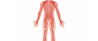
The neck, as a connecting link between the head and body, is the center of vital arteries.
Anatomically, the neck is divided into 4 areas:
- Front.
- Rear.
- Two sterno-mastoid-clavicular.
- Two side parts.
Vessels supplying the brain pass through the neck - which is why pathologies of the cervical vertebrae are often accompanied by pain in the head, deterioration of mental and cognitive functions, as well as limited head mobility.
Interpretation of the Results of Ultrasound of the Vessels of the Child's Neck
Of course, having received the results, parents want to know if everything is fine with their child. But what awaits them is only a set of numbers that are difficult to understand. There are the diameter of the vessels, the speed of blood flow in them, the degree of stenosis of the vessels, and other pathologies.
In order not to get confused in all the terms and draw the right conclusions for yourself, you need to consult professionals. This is either a vascular surgeon, or a neurologist and pediatrician. His consultations will practically not complicate the patient’s situation in any way, but they can also improve it in any way they can.
Features of the study of the cervical spine
The structural features of the spine are that each vertebra is associated with a specific organ:
- The first vertebra from above is responsible for pressure.
- The second is associated with the visual and auditory nerves, as well as the eyes.
- The third is responsible for the functioning of the facial nerve, ears and dental health.
- Disturbances in the functioning of the fourth vertebra will cause hearing problems, hypertrophy of the adenoids and malfunction of the thyroid gland.
- The fifth vertebra is responsible for the functioning of the ligaments; if there are disturbances in its functioning, tonsillitis, tracheitis and laryngitis will become “regular guests”.
- The sixth is associated with the muscles of the neck and forearm.
- Seventh – regulates the functioning of the thyroid gland and affects the mobility of the upper limbs.
Few people know about these nuances, and therefore it happens that the symptoms do not go away even after treatment. And based on the results of a competent diagnosis of the spine, it turns out that it was necessary to treat, for example, not the thyroid gland, but the neck!
The main purpose of ultrasound examination of the spine is to formulate an idea of the condition of the discs and intervertebral joints , spinal processes and the yellow ligament of each vertebra.
Indications and contraindications
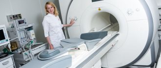
The effectiveness of this procedure is not much different from magnetic resonance imaging - a very informative, but extremely financially expensive method. An ultrasound of the neck provides complete information about the condition of each intervertebral joint, disc and its processes.
Ultrasound examination of the cervical vertebrae can determine the presence of diseases, the symptoms of which threaten the life and health of a person. It must be carried out in the following cases:
- if you have breathing problems;
- if you are worried about memory loss;
- with curvature of the spinal column;
- with an increase/decrease in blood pressure;
- if you often experience dizziness and migraines;
- if there is a feeling of discomfort and pain in the joints and spine;
- when the functioning of the organs of vision and hearing is impaired;
- with a decrease or loss of sensitivity of the skin of the face and limbs.
There are no contraindications to the study of the structures of the cervical spine. It can be done both for newborns and for women preparing to become mothers. You can refuse to examine the cervical spine if the skin in the neck area is damaged. After the x-ray, you need to wait a day, or preferably more, before performing an ultrasound examination.
An examination of the cervical spine reveals any neck injuries, bone tissue, plexuses of blood vessels, nerves, cartilage, and interarticular fluid. Using ultrasound, you can diagnose oncological formations, hernias and much more.
In addition, read the article: Ultrasound examination of the lymph nodes of the neck.
Examination of the cervical spine in children
For children, especially newborns, ultrasound methods have important diagnostic value.
Important! Pronounced symptoms of certain diseases may be absent at an early age, so it is necessary to undergo preventive examinations on time.
And, although pediatricians and neonatologists prefer Dopplerography and neurosonography, ultrasound of the cervical spine in infants remains a valuable technique for diagnosing certain pathologies, especially in cases of birth trauma.
It allows you to find out:
- are there any pathologies or developmental features in the spinal column;
- whether there are damaged arteries or spinal membranes.
Video about why it is necessary to carry out diagnostics in children
Instability of the cervical spine in children:
Causes of birth injuries
During the birth process, the baby has to withstand serious mechanical and physical stress. Nature provides for the baby’s ideal adaptation to stress during passage through the birth canal and until birth. But there is always the possibility of birth injuries in babies.
Risk factors include:
- difficult delivery, with fetal weight above 4.5 kg;
- assisted delivery for children weighing less than 3 kg;
- stimulation of labor in a woman in labor;
- the use of obstetric forceps and other auxiliary methods of delivery;
- unplanned caesarean section.
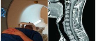
It is worth noting that the birth of a baby with a large weight is almost always accompanied by injury to the spinal column and muscle tissue of the neck. For injuries of this type, radiography as a diagnostic method is poorly effective, since X-ray radiation does not detect damage to soft tissues. In this regard, an infant with suspected spinal cord pathology is prescribed an ultrasound scan of the neck.
What does an ultrasound show?
During the ultrasound procedure, the doctor can diagnose a number of features characteristic of certain diseases:
- With osteochondrosis, a disruption of cellular nutrition processes and an increase in degenerative disorders in the vertebrae will be noted. The degree of damage to cartilage, bone and connective tissue and the level of compression of the coronary vessels supplying blood to the brain are also revealed.
- With protrusion, there is a slight protrusion of the discs (up to 9 mm). If the protrusion exceeds the accepted norm, then we are talking about a hernia .
- If the spinal ligaments are weak, the relative position of the vertebrae is compared without load and during tests with load.
- If the patient has suffered neck injuries, soft tissue bruises, dislocations, ligament ruptures and vertebral fractures may be detected.
- With torticollis, diagnosing the causes of the disease is of particular importance in choosing the right treatment. Hypertonicity of the sternocleidomastoid muscles can cause damage to muscle fibers, calcification or hemorrhages - it is important that the diagnostician correctly determines the cause of the disease.
- If there are anomalies in the development of the arteries, the doctor will note narrowings, turns or tortuosities of the vascular trunks, which can cause unpleasant symptoms.
- In case of occlusion of the vertebral artery, vascular ultrasound will show the absence of blood flow through this artery, and an increase in blood flow occurs through the contralateral vessel.
- With vertebral artery syndrome, a narrowing of the vessel occurs, which not only can lead to fainting or dizziness, but is also fraught with cerebral ischemia.
- With spondylolisthesis, the vertebrae are displaced relative to each other, often damaging the nerve roots. Ultrasound shows a defect in the arch area located between the joints.
Ultrasound is an important diagnostic method
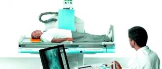
The main methods for diagnosing spinal injuries are x-ray studies. Radiology allows you to determine disorders of bone structures, i.e. promptly determine the presence of injuries such as dislocation or fracture of the vertebrae. However, this method does not make it possible to study the condition of the vertebral arteries, the spinal cord and its membranes. Data about these structures makes it possible to determine a clear clinical picture and predict the development of pathology, while x-rays can only reveal gross orthopedic disorders.
Comparison of ultrasound and MRI
Compared to magnetic resonance imaging, ultrasound has a number of advantages and disadvantages.
Disadvantages of ultrasound:
- Lower rendering quality
- Relative difficulty in interpreting results
- The need to follow a diet before the examination
Advantages of ultrasound:
- The study is carried out quite quickly (about 15 minutes), and the patient does not have to sit in the MRI chamber of the machine, which also eliminates such contraindications for MRI as claustrophobia and weight more than 120 kg; there is no need for sedation for children or people with unstable mental health.
- Ultrasound is not performed automatically, so the patient can move and change his position if necessary (which is also important for identifying functional pathologies). In addition, when placed in an MRI machine, the patient most often lies down (some modern MRI machines are designed to change the body position from horizontal to vertical), which can show herniations and protrusions of intervertebral discs in the early stages, as well as possible transient compression of the spinal nerve roots.
- There are no contraindications in the presence of metal prostheses and implants in the body, such as pacemakers, stents in blood vessels, or osteosynthesis devices.
- There is an opportunity to communicate with a doctor and ask questions.
- There are no other subjective unpleasant sensations during the procedure, such as noise. There is no need to remove clothing with metal, jewelry, or leave personal items.
- For some spinal injuries that are difficult to diagnose using a plain X-ray, it is not possible to put the person down, much less transfer him to a tomography machine.
When is a spinal ultrasound recommended?
The following complaints and symptoms may be indications for an ultrasound of the spine:
- Any unpleasant sensations and back pain, including those that refer to other places - in the chest, lower back.
- Regular numbness in any part of the body.
- Constant, high-intensity headaches, as well as regular dizziness and fainting of unknown origin. The cervical spine is checked first.
- Pain in the neck, pain when trying to tilt/turn your head.
- Visual/hearing/memory impairments of unknown origin. The cervical spine is also checked; in this and the previous case, an ultrasound of the vessels of the brain and neck should first be done.
- Poor posture, in which it is painful/impossible to take the correct position.
- Difficulty breathing and frequent changes in its type (thoracic, abdominal), frequent shortness of breath, blood pressure disorders. Attention! First you need to check the respiratory system and do an ultrasound of the heart.
- Pain in the limbs, pain in the bones, in the sacral region, lumbago, difficulty moving. First, you need to examine the place where the pain is localized.
Ultrasound examination is today one of the most popular types of diagnostics of any part of the body. Doctors love this method for its information content, patients love it for its accessibility and painlessness.
Therapists and neurologists recommend that certain types of ultrasound be performed routinely, and one of these examinations is the diagnosis of the cervical vertebrae. This is where the thinnest and most fragile vertebrae are located, so you need to treat them with special care.
Advantages of ultrasound diagnostics
The main prerogative aspects of spinal ultrasonography (another name for spinal ultrasound) include:
- Availability. Firstly, the procedure is not expensive, and secondly, most medical institutions at all levels are equipped with the necessary equipment.
- Absolute safety. Ultrasonic waves cannot cause harm because they are natural to humans. Passing into the body, they are reflected by an echo signal, on the basis of which a computer program visualizes the organs.
- No need for long-term preliminary preparation. The procedure does not provide any restrictions before it is carried out.
- No contraindications. Women during the perinatal and lactation periods, newborn children, and elderly people are allowed to be diagnosed.
- Comfortable conditions for the patient. Ultrasound does not cause pain or other unpleasant sensations.
- Unlimited frequency of examination. X-rays, due to their negative radiation effects on the body, are allowed to be performed twice a year. Ultrasound can be done as needed.
In addition, unlike MRI, sonography of the back has a shorter time interval (from 15 to 20 minutes).
Cost of the study
Each clinic sets its own price for cervical spine examination. It depends on its location, position in the ranking, how high-quality and new its equipment is, etc. Private medical centers charge a price from 1 to 3 thousand rubles. In municipal institutions, the examination is carried out free of charge, with the exception of payment for the materials used.
In Moscow clinics the price ranges from 1000 to 1500 rubles. In St. Petersburg - from 1200 to 1600 rubles. In other regions from 1200 to 1800 rubles.
Indications for ultrasound
There are different types of ultrasound. To objectively determine the affected area, a two-dimensional study is used, which allows timely identification of the disorder and diagnosis. To clarify data or details needed for an operation, a three-dimensional or even four-dimensional technique is used. It all depends on the symptoms, preliminary diagnosis and localization of pain in the spine. An ultrasound of the lumbar spine is prescribed by a doctor only if there are indications for it. The main ones are:
- The appearance of coordination disorders.
- Pathological changes in posture (scoliosis, lordosis, kyphosis).
- Impaired sensitivity of the limbs.
- Frequent dizziness, nausea, headaches.
- Blocks or stiffness in the back.
- Pain in the joints or in the spine itself.
- Decreased level of performance, rapid fatigue.
A study is also prescribed in a number of other cases, if the doctor considers it appropriate to use the data of such a technique. An ultrasound scan of the vertebrae allows us to identify any deviations from the norm.
How to prepare for the procedure
The examination can be completed in both public and private institutions. The study is carried out free of charge in clinics and hospitals. To do this, you need to get a referral from your attending physician and get on the waiting list.
In private clinics, the cost of the study is 1500–2500 rubles. The price of the service depends on the qualification category of the doctor and the department that is subject to examination.
Prices for ultrasound of the spine in children in the regions:
| Department to be researched | Moscow | Saint Petersburg | Kaliningrad |
| cervical | 1800–2200 | 1800–2500 | 1500–2000 |
| chest | 2000–2300 | 1900–2100 | 1500–2100 |
| lumbar | 1800–2000 | 1800–2000 | 1500–1950 |
| sacral | 1500–2000 | 1500–2000 | 1500–1900 |
Ultrasound of the spine does not require any special preparation. For the procedure itself, you need to take a sheet or towel with you. Before the test, you should not apply creams or lotions to your skin. This may affect the image quality.