Diagnosis of Helicobacter pylori infection is a complex process, since none of the available tests alone can serve as the basis for making a final diagnosis. A person can be a carrier of Helicobacter pylori throughout his life, and the manifestation of clinical symptoms is not necessary.
There is experimental data on the possibility of spontaneous elimination of infection, however, in most cases, the selection of adequate treatment methods under the supervision of a physician is required.
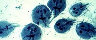
Helicobacter pylori: general information about the microorganism
Helicobacter pylori (Helicobacter pylori) is an opportunistic bacterium of a spiral shape, Gram-stained red (gram-negative). The predominant habitat in the human body is the stomach and duodenum.
The role of Helicobacter pylori in the development of diseases of the gastrointestinal tract (GIT) has been denied for a long time. Only in 2005, Australian pathologist R. Warren and physician B. Marshall managed to prove the medical significance of bacteria, for which they were awarded the Nobel Prize.
Feature: in 90% of carriers, Helicobacter pylori is part of the normal microflora and does not cause the development of an infectious disease. However, there is an opinion that this particular species is the cause of many gastrointestinal pathologies (ulcers, gastritis, cancer, lymphoma).
Relation to opportunistic bacteria means their ability to provoke an infectious process in the presence of certain conditions (factors). For example, long-term use of antibiotics with subsequent dysbacteriosis, decreased immunity and the presence of concomitant pathologies. However, when infected with strains with pronounced pathogenic properties, the presence of the above factors is not necessary.
When is analysis necessary?
The introduction of bacteria into the walls of the gastric region is manifested by certain symptoms. The following indicates an elevated Helicobacter in the blood:
- Pain in the epistragal region. They can vary in intensity and can be felt before, during, and after meals, on an empty stomach.
- Heartburn. This is a consequence of the reflux of acidic gastric juice into the esophagus, which leads to irritation of the latter. Its frequent relapses will indicate a violation of acidity and damage to regulatory processes.
- Feeling of heaviness in the epigastric area. Appears even after a small snack.
- Causeless nausea.
- Sometimes, when the level of Helicobacter pylori in the blood increases, signs of food poisoning are observed - vomiting, sharp, cutting stomach pains.
- Mucus and bloody spots are found in the stool.
- The patient suffers from periodic belching.
- Flatulence, bloating.
- Unreasonable weight loss.
- Various disorders of intestinal emptying - from diarrhea to constipation.
Where does Helicobacter pylori come from and how is it transmitted?
The infection is not transmitted by airborne droplets, since it is a strict anaerobe (dies on contact with oxygen). You can become infected by neglecting the rules of personal hygiene (cutlery and dishes, personal cosmetics and personal hygiene products), as well as by kissing.
Primary infection can occur in childhood (from mother to child). Another route of infection is water and meat that has undergone insufficient heat treatment. Infection through an endoscope, which is used for gastroendoscopy, is possible.
How does infection occur?
Rapid colonization of the mucous membrane of the gastrointestinal tract is ensured due to the high degree of mobility of Helicobacter pylori (using flagella). Specific proteins and lipopolysaccharides on the surface of the membrane help bacteria attach to the surface of cells. The presence of foreign antigens provokes the development of an immune response (the release of specific antibodies to Helicobacter pylori) and initiates inflammation of the mucous membrane.
Bacteria release enzymes into the external environment that dissolve the protective mucus of the stomach. Survival in the acidic environment of the stomach is ensured by the enzyme urease, which decomposes urea with the release of ammonia (neutralizes hydrochloric acid). A side effect of ammonia is chemical irritation of cells followed by their death. Along with this, bacteria release toxins that enhance the process of cell degradation and death.
Symptoms of Helicobacter pylori in adults
In most cases (up to 70%), carriage does not manifest itself in the form of clinical symptoms and is discovered accidentally during a comprehensive examination of the patient. However, pathologies of the stomach and intestinal tract, accompanied by Helicobacter pylori infection, have certain signs:
- feeling of pain in the abdominal area (stomach);
- frequent heartburn and belching;
- unexplained loss of appetite and weight;
- nausea or vomiting;
- heavy coating on the tongue;
- inflammation of the gums;
- putrid odor from the mouth (with the exception of dental diseases);
- feeling of heaviness after eating food;
- increased gas formation.
It was noted that in children the severity of clinical signs is higher than in adults. This situation is especially often observed in the presence of physical or emotional stress, as well as when the diet changes for the worse (replacing soups with sandwiches or eating irregularly).
Patients ask the question: when should you get tested for Helicobacter pylori? A referral for laboratory diagnostics can be issued by a general practitioner, pediatrician, gastroenterologist or infectious disease specialist. Indications for testing for Helicobacter pylori: suspicion or presence of gastrointestinal disease, as well as the manifestation of the above symptoms.
Symptoms of helicobacteriosis
After entering the stomach, the bacterium begins to release waste products that damage the walls of the stomach, causing discomfort.
The presence of infection may be indicated by repeated severe pain in the stomach. They usually occur on an empty stomach and disappear after eating. This means that ulcers and erosions have formed on the walls of the stomach.
Many patients also complain of heartburn and heaviness in the stomach. Symptoms are especially pronounced when eating fatty meat foods. If the disease is left untreated for a long time, nausea and even vomiting may occur.
Helicobacteriosis can manifest itself in different forms:
- The latent form practically does not make itself felt if a person has a sufficiently strong immune system, but in the presence of favorable conditions it makes itself felt. However, even if there are no symptoms, the microorganism gradually disrupts the functioning of not only the stomach, but also the pancreas.
- Acute gastritis can be recognized by pain in the epigastric region and vomiting. As is correct, over time it becomes chronic.
- Chronic gastritis occurs in most of the population. And it manifests itself as periodic pain and a feeling of fullness in the stomach. Heartburn, an unpleasant taste in the mouth, belching, nausea and increased bleeding of the gums are also observed.
- Chronic gastroduentitis is characterized by the penetration of bacteria into the duodenum. In addition to the symptoms of gastritis, constipation, loss and decreased appetite may be observed.
- Ulcers and erosions of the stomach walls appear when the deep layers of the gastric walls are affected. When eating, the disease manifests itself as pain in the upper part of the stomach after eating. In this case, the patient experiences heaviness in the pancreas, as well as symptoms observed with gastritis.
Also, patients with helicobacteriosis often experience acne on the face, as well as bad breath.
The disease gradually progresses, occupying new territories in the stomach until it occupies the entire area. As a result, almost no gastric juice is produced, and the protein is not over-poisoned.
In 80% of cases, infection does not manifest itself in any way.
How to get tested for Helicobacter pylori?
Methods for identifying Helicobacter pylori are different:
- breath (urease) test;
- real-time PCR to detect pathogen DNA;
- enzyme-linked immunosorbent assay (ELISA) to determine the level of antibodies produced in response to infection;
- one-step immunochromatographic method for detecting pathogen antigens in the test material;
- biopsy during esophagogastroduodenoscopy.
Depending on the diagnostic method, the biomaterial studied, the cost and timing of the study differ. It is important that the patient follows the rules for preparing for the analysis; the accuracy and reliability of the results obtained depends on this. Let's take a closer look at each method.
ELISA for diagnostics
To confirm this pathology, several methods are used, including invasive biopsy. The latter method, although accurate, is quite unpleasant for patients, and they try in every possible way to avoid it. An excellent alternative to a biopsy is ELISA (enzyme-linked immunosorbent assay), which allows you to assess the condition of the blood components being studied.
This study is a non-invasive direct method for diagnosing the presence of bacteria in the body. The essence of ELISA is not to search for the bacteria themselves, but to detect in the plasma specific protein compounds of antibodies (AT), which are produced by the body in response to the entry of a pathogenic antigen (AG). AT or as they are also called immunoglobulins (Ig) belong to glycoproteins and are produced by plasma cells formed from B lymphocytes.
What is a gastric biopsy using endoscopy?
AT are located on the surface of B lymphocytes and in the blood serum, acting as membrane-binding receptors. Each of the immunoglobulins has a certain specificity, that is, different antibodies are produced for different antigens. In mammals and humans, there are 5 Ig classes, of which the most significant are A, M, G.
Immunoglobulins A and M indicate an early stage of the development of the disease, when no more than 1–2 weeks have passed from the moment of infection. These are the so-called acute phase proteins. IgG is usually elevated from 3-4 weeks, and can remain at this level for one and a half years after therapy in 50% of patients.
The technique provides the opportunity to conduct two analyzes - qualitative (detection of the presence of antibodies) and quantitative (determination of concentration (titer)). The first confirms or denies the presence of AT, that is, the disease, and the second allows for monitoring during therapy and assessing its effectiveness.
Antibody activity in the immune system response
What is a urease test for Helicobacter pylori?
Detection of Helicobacter pylori using a breath test. The Helicobacter pylori test is increasingly used in routine diagnostic practice. Advantages of the method:
- short time to obtain results (up to several hours);
- low cost;
- painlessness;
- no contraindications;
- no need for expensive equipment.
Disadvantages include the possibility of obtaining a false negative or false positive result. Reduced reliability of the study due to internal bleeding.
In what cases can a urease breath test for Helicobacter show a false negative result? In addition to improper preparation of the patient for the test and errors at the stage of collecting biomaterial, a false negative result can be obtained when infected with strains that do not produce urease. In other words, even if bacteria colonize the patient’s gastrointestinal tract but do not release urease, the test result will be negative.
Preparing for the ureaplase test
For 3 days, alcohol and medications in which alcohol is a solvent are completely eliminated. For 6 hours, food intake is limited, clean unsweetened water is allowed to drink. The minimum interval between the last dose of antibiotics and bismuth-containing drugs is 6 weeks. It is advisable to stop taking any medications 2 weeks in advance, in consultation with your doctor.
Collection of biomaterial (exhaled air) is allowed 24 hours after FGDS (gastroscopy).
10 minutes before collecting air, you should drink juice (grapefruit or orange) in order to slow down the evacuation from the stomach. Then the patient exhales as much air as possible into a special bag.
After which you need to drink a solution of urea labeled with a carbon isotope (50 ml for adults, 25 ml for children under 12 years of age). The solution has no specific taste or smell; it must be prepared immediately before use. After 30 minutes, a control collection of exhaled air is carried out.
Both samples are analyzed on a special device and the carbon dioxide ratio is determined.
Preparing for the test
It is very important that the patient properly prepares for the diagnostic examination, since preliminary preparation can significantly reduce the risk of diagnostically incorrect data, which can complicate the further treatment process. Before taking the test, you will need to exclude all spicy, fatty and overly spicy foods from your diet. You should also stop drinking alcoholic beverages and smoking tobacco products. The consumption of the above foods and alcohol should be excluded from the diet 2-3 days before the test, and smoking should not be done until just an hour before the procedure. All this will increase the accuracy of the results of a serological blood test.
Antibodies to Helicobacter pylori
Infection with Helicobacter pylori infection triggers protective immune responses. Immunoglobulin M (IgM) is produced first, followed by large quantities of IgG and IgA. Blood testing for antibodies to Helicobacter pylori allows one to establish the fact of infection, since IgG is detected in 90–100%, and IgA in 80% of cases.
It should be noted that a blood test for Helicobacter pylori may be an alternative to invasive diagnostic methods (if endoscopy is not possible). This rule does not apply to elderly patients. The strength of their immune response is insufficient, so it is possible to obtain false negative results.
A high titer of IgG indicates recent infection and an active process of infection, provided that the patient has not taken antibiotics. The IgG concentration remains moderately elevated for a long time (up to 1.5 years), so this test is not used to assess the effectiveness of the chosen treatment.
The IgA value allows you to determine the severity of the infectious disease. A low IgA content persists for up to several years; however, the absence of positive dynamics in reducing its value indicates the ineffectiveness of treatment.
How is blood donated for Helicobacter pylori (how is the test taken)? The biomaterial is venous blood from a peripheral vein on the elbow. No special preparation is required for the analysis. It is advisable to donate blood for Helicobacter pylori after 2-3 hours without food; smoking is prohibited for half an hour.
What does it mean if Helicobacter pylori IgG is positive?
If antibodies to Helicobacter pylori IgG are detected in the biomaterial, then a conclusion is drawn about:
- active infection - in the presence of a pronounced clinical picture;
- bacterial carriage.
A decrease in the IgG titer in a blood test for Helicobacter by 25% within six months after completion of treatment indicates the death of bacteria.
Stool analysis for Helicobacter pylori
Feces are examined using 2 methods: immunochromatography (detection of antigens) and PCR (presence of pathogen DNA). Both methods are characterized by high sensitivity and act as complementary methods.
Determination of antigens
Testing stool for Helicobacter pylori antigen is a qualitative method, the accuracy of which reaches 95%. Obtaining positive results 7 days after taking antibiotics indicates the ineffectiveness of treatment. A repeat test is carried out after 1.5 months of therapy, and the absence of antigens in the patient’s stool indicates complete destruction of the bacterium.
The method does not allow one to determine the type of bacteria: H. suis, H. Baculiformis or H. Pylori, since all their biomaterial is foreign (antigen) to humans.
Real-time PCR
The sensitivity of the stool PCR method for Helicobacter pylori infection reaches 95%. The analysis makes it possible to determine infection by unculturable forms of bacteria. Disadvantages include the possibility of obtaining false-positive results after a successful course of treatment, since destroyed bacterial cells (and their DNA) remain in the human body for a long time.
The possibility of obtaining false positive results is excluded, since the specificity of the method reaches 100%. The method is an alternative to the breath test or FGDS for young children.
No special preparation is required for collecting biomaterial for both studies. Feces are collected naturally without the use of laxatives, preferably before starting antibiotics.
Decoding the analysis results
Decoding Helicobacter in the blood is the prerogative of specialists. It should be noted that the norm of bacteria content and deviations from it are secondary information for the attending physician. For the doctor, a positive (the bacteria is in the body) or negative test result is sufficient.
What does a deviation from the norm of Helicobacter pylori indicate? Only about the proliferation activity of the microorganism in the gastrointestinal tract of the subject.
It often happens that the laboratory cannot give an accurate negative or positive result. Then a conclusion is issued about a questionable analysis - you need to donate blood again in a week.
Norm Helicobacter pylori in the blood in numbers
Decoding the blood test for Helicobacter pylori, as well as other data obtained, is the work of the doctor and does not allow the patient to independently interpret the results. The table shows normal values for each diagnostic technique.
| Method name | Norm | Approximate cost (for a private laboratory), rub. | Deadline for receiving results (excluding the day the biomaterial was taken) |
| Breath test | Less than 4 ‰ | 850 | Up to 6 days |
| PCR | Not detected | 500 | Up to 2 days |
| Antigens | 750 | 1 day | |
| Biopsy | 600 | ||
| Antibodies | IgG 0-0.9 IU/ml | 550 | |
| IgA 0 – 13.5 IU/ml | 650 | Up to 8 |
Patients are concerned about the question - what does Helicobacter negative mean? Obtaining such a result indicates the absence of Helicobacter pylori infection or successful therapy with complete destruction of bacteria.