Among the existing methods for identifying bacteria in the gastrointestinal tract, a biopsy for Helicobacter provides high accuracy of the results obtained. A study using a fragment of mucosal tissue makes it possible to establish the stage of the disease in the shortest possible time, which speeds up the process of making a diagnosis and prescribing treatment.
How to determine the presence of Helicobacter in the stomach
It is not possible to see such a tiny organism with a length of only 2-3 microns with the eye, nor is it possible to carry out diagnostics at home.
The patient can only assume the presence of gastritis based on the corresponding symptoms: epigastric pain after eating, heaviness and discomfort in the stomach, heartburn, belching of air or sour, metallic taste in the mouth. These signs of increased acidity very often accompany gastritis associated with a pathogenic microbe.
But it is possible to reliably determine whether the Helicobacter pylori bacterium has settled in the body or not only in the diagnostic department of an outpatient clinic, hospital or laboratory.
There are special methods that make it possible to detect with high reliability both the microbe itself and its metabolic products, as well as the antibodies produced by the body in response to the introduction of the microbe:
- Bacteriological
Detection of a pathogen in smears from a section of the inner wall of the stomach or cultivation of the microorganism on nutrient media.
- Serological
Detection of antibodies in the blood, microbial antigens in the stool.
- Morphological
Detection of N. rulori under a microscope by coating a research sample with special dyes.
- Molecular genetic
Polymerase chain reaction methods.
- Biochemical
Urease test, breath test.
All of the above methods can be classified into two large groups:
- Invasive. Diagnostic methods based on endoscopic examination - FGDS, with taking a biopsy sample. A section of the inner wall of the stomach can then be subjected to cytological, cultural examination, and a urease test.
- Non-invasive. Other methods of detecting infection in which FGDS is not performed.
What is a HELIC test?
The HELIK test is a non-invasive respiratory diagnosis of Helicobacter pylori infection. Suitable for use by gastroenterologists, internists, pediatricians and family doctors. The HELIK breath test is intended for the primary diagnosis of Helicobacter pylori, as well as for monitoring the progress of anti-Helicobacter pylori therapy and checking the effectiveness of therapy already carried out.
Diagnosis with the HELIK test is completely painless, because during the examination only the air exhaled by a person is checked.
In what cases should I be examined?
You should be examined if you are bothered by abdominal pain, heartburn, nausea, early satiety after eating, or belching. The main cause of these complaints may be Helicobacter pylori. The HELIK test is carried out for the primary diagnosis of Helicobacter pylori, as well as to check the effectiveness of already carried out therapy.
Who should take the HELIC test?
Helicobacteriosis is a family disease, so diagnosis of infection should be carried out not only in a patient with gastrointestinal pathology, but also among all members of his family.
Test for Helicobacter pylori - what is it?
Before conducting research and diagnosis, the doctor needs to take biological material from the patient. Such material could be:
- A small area of the gastric mucosa.
A piece of mucous membrane is split off during fibrogastroscopy - a biopsy is performed with a special device directly during FGDS.
Next, the biopsy is subjected to various studies: microscopy, culture on nutrient media, or express diagnostics. The methods have one goal: identifying Helicobacter rulori or its toxins.
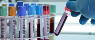
- Blood
A blood test detects not the bacterium itself, but the immunoglobulins that are formed in the body in response to infection: IgA, IgG, IgM.
Prevention
Compliance with hygiene standards helps prevent the spread of bacteria in the body:
- It is not recommended to use shared utensils during meals;
- Before eating and after visiting the toilet, you must wash your hands with soap;
- you need to use only personal hygiene items - a towel, toothbrush, washcloth;
- It is necessary to stop abusing alcoholic beverages and smoking, including passive smoking.
It is almost impossible for only one person in a family to be affected by the microorganism Helicobacter pylori. Therefore, each member must undergo diagnostics and appropriate treatment.
What tests need to be taken for Helicobacter pylori
Currently, there are a great variety of methods for detecting bacteria, its metabolic products and antibodies to it. Each method has certain advantages and disadvantages.
Therefore, the patient is recommended to take several tests to confirm the presence of the microbe in the body; they should be recommended by a doctor, taking into account the specific clinical case and the technical equipment of the institution in which the diagnosis will be carried out.
Each patient must undergo an FGDS with a biopsy. Further study of the biopsied mucosa is possible under a microscope, rapid tests, or by cultivating bacteria on nutrient media.
It would be a good idea to donate blood and feces for serological diagnosis. After all, confirmation of infection can be a high titer of antibodies of the bacterium or its DNE fragments in the feces.
A breath test is an excellent non-invasive way to reliably confirm the presence of bacteria in the body and its activity. And if there is an opportunity to undergo it in a medical institution, you should definitely take advantage of it.
Tests for Helicobacter pylori infection are performed not only to identify the microorganism, but also to monitor cure rates. What kind of research is needed is always determined by the doctor.
Which test for Helicobacter pylori is the most informative?
The exact analyzes are:
- cytological method, when the doctor observes the presence of bacteria under a microscope
- cultural method - growing bacteria on nutrient media
- PCR diagnostic method or molecular genetic method - detection of genes or DNA fragments of bacteria
All these methods are based on the initial taking of a biopsy specimen - a piece of gastric mucosa during FGDS. They are invasive. These methods cannot be performed without “swallowing the tube.”
Unlike the methods of serological blood testing, which detect antibodies that indirectly indicate the presence of a microbe in the body or enzymatic methods that can diagnose the products of its vital activity, cytology reveals the pathogen as a whole, in person.
For this study, biopsy smears of the gastric mucosa are used. It is important to take a biopsy from the most informative areas and suspected habitats of the bacterium - most often the antrum.
The smears are dried, stained with special dyes and examined under a microscope. The bacteria are located in the mucus, they are S-shaped or spiral-shaped, and have flagella at the end of the body. The experienced eye of a diagnostician will not confuse these microorganisms with any others.
The cultural method allows you to grow an entire colony of bacteria on special nutrient media. Pathogens love an environment with low oxygen content (no more than 5%), blood nutrient media are used to grow them.
Under favorable conditions, compliance with the temperature regime during cultivation and anaerobic conditions, after 3-5 days round, transparent colonies of bacteria grow on the medium, which are then identified.
PCR methods for detecting genes and fragments of Helicobacter DNA are informative, but require special equipment and reagents. Not every clinic today has it.
Doctors recommend using three, or preferably four, diagnostic methods to identify H. pylori in order to be as informative as possible: cytological, cultural, PCR, and breath test.
What is the best test for Helicobacter?
The list of tests should be determined by the doctor, taking into account the specific case and purpose of the study.
For preventive purposes, without complaints from the gastrointestinal tract, you can use non-invasive diagnostic methods (without FGDS):
- blood test for serological detection of antibodies to the microbe
- stool analysis for PCR diagnostics of DNA fragments
- breath test
Non-invasive methods are also recommended after treatment in order to clarify its effectiveness, for weakened, seriously ill patients who cannot be given a probe and invasive diagnostic methods.
If there are complaints from the gastrointestinal tract or suspicion of infection, it is necessary to conduct an FGDS followed by taking a section of the gastric mucosa. In this case, cytological, cultural, urease rapid tests or PCR diagnostics of the biopsy are recommended.
There is no “gold standard” for diagnosing a microbe. All methods complement each other, so you need to use several of them. The choice and tactics of diagnosis are the prerogative of the attending physician.
What diseases can a microorganism cause?
Interesting! The scientific community doubted that the bacterium was able to live and develop in the sharply acidic contents of the stomach, but Barry Marshall drank a culture of Helicobacter pylori and developed gastritis, which was confirmed by endoscopic examination. At the next stage of the experiment, the researcher cured gastritis by taking metronidazole and bismuth compounds, which are harmful to bacteria. In 2005, R. Warren and B. Marshall were awarded the Nobel Prize for their discovery.
There are a number of pathologies of the digestive system, if suspected, the gastroenterologist is obliged to prescribe tests for Helicobacter pylori:
- chronic and acute gastritis;
- stomach ulcer;
- malignant neoplasms in the stomach;
- chronic and acute duodenitis;
- combined ulcerative lesions of the gastrointestinal tract.
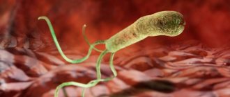
Important: bacteria belonging to the genus Helicobacter are the only microorganisms discovered to date that not only survive for a long time in a sharply acidic gastric environment, but are also able to multiply there, colonizing the mucous membranes of the gastrointestinal tract.
How to test for the bacterium Helicobacter pylori
If they want to diagnose the bacterium itself or its fragments, they take a section of the mucous membrane from the stomach with a special device during FGDS. The doctor determines the puncture site - these are the most hyperemic and swollen areas of the internal gastric wall. You cannot take a piece from the bottom of erosions or ulcers for examination.
If the purpose of diagnosis is a preventive examination or assessment of the effectiveness of treatment, non-invasive methods can be used: take a blood test, stool test, or conduct a breath test.
Before carrying out invasive tests, the patient is required to prepare only for an endoscopic examination - fibrogastroscopy.
Blood for testing is taken from a vein; the analysis does not require any special preparation from the patient. It is advisable to donate blood on an empty stomach; in the evening, a light dinner is acceptable; it is not advisable to overeat or eat fatty foods.
Before stool analysis, you should eat right for 3 days: do not eat foods containing large amounts of dyes and preservatives, coarse fiber foods, do not take medications, or alcohol.
Preparation before taking a breath test is also important. You cannot eat after 22.00 the evening before the test and in the morning. Two days before the test, exclude all gas-inducing foods and liquids that can increase the CO2 concentration in exhaled air: cabbage, legumes, apples, baked goods, soda. Do not drink alcohol, smoke, or use chewing gum.
The quality and results of the tests depend on how consciously the patient prepares for the tests. This means subsequent treatment and overall well-being.
Norm
If there are no Helicobacter pylori strains in the subject’s body, all tests, both invasive and non-invasive, will give a negative result.
Important: individual strains of the Helicobacter genus have been identified in the liver of some mammals. They can cause liver disease in humans.
Plisov Vladimir, medical observer
10, total, today
( 40 votes, average: 4.15 out of 5)
Angiodysplasia of the large intestine: causes, symptoms, treatment
Ultrasound of the hip joints in a child
Related Posts
Helicobacter test for FGDS and gastroscopy
Rapid tests for diagnosing bacteria are becoming widely popular. They are quite informative and allow one to quickly establish infection with H. pylori within a few minutes after FGDS. These are rapid urease tests.
They are based on the ability of the microbial enzyme urease to break down urea, releasing ammonium. Ammonium ions create an alkaline environment and contribute to the color change of the express system indicator.
The one-time express kit includes:
- urea
- pH indicator (initially its color is yellow)
- bacteriostatic agent
When performing FGDS, a section of the mucous membrane is taken. This section is placed on the express dial panel. If this mucous membrane contains a microbe, its urease enzyme begins to actively break down the urea contained in the dough.
Ammonia is released, alkalizes the medium, the indicator reacts to its release and changes its color from yellow to crimson. The test takes from a few minutes to a day. Raspberry coloration will indicate the presence of infection and a positive test.
If the indicator color does not change, or it appears after a day, the result is considered negative. There is no pathogen in the biopsy specimen.
FGDS with biopsy for Helicobacter
It is necessary for persons who:
- Have symptoms of gastrointestinal pathology: heartburn, nausea, discomfort or pain in the epigastrium, metallic taste in the mouth
- Have close contact with persons who have already been diagnosed with this infection, or among family members this diagnosis has been established
- Already have a history of gastritis, esophagitis, ulcer without an established etiology
- Completed a course of eradication therapy for this infection to assess the quality of treatment
- Have skin problems of unknown etiology, immune disorders
- Successfully completed treatment for H. pylori with reliably confirmed laboratory data, to prevent reinfection once a year.
Diagnostic methods
Blood test for antibodies to Helicobacter pylori
Advantages
- available to all patients;
- is not accompanied by severe discomfort and stress during the procedure (injection method of blood sampling);
- taking medications used to treat diseases of the stomach and duodenum does not affect the results of the study.
Preparation
The blood sampling procedure is carried out in the morning on an empty stomach.
- The day before the test, fatty foods are excluded from the diet.
- Stop smoking 8 hours before the test.
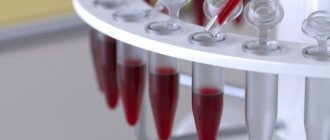
Carrying out the procedure
Blood is collected by venipuncture. The material is collected in a sterile vacuum tube with a clotting activator to quickly separate the serum. The blood is delivered to the laboratory within 2 hours. Microtubes with serum can be stored in the refrigerator at a temperature of 2-8 ° C or in the freezer at -20 ° C for up to 10 days.
Features of the method
To identify bacteria, an enzyme-linked immunosorbent assay (ELISA) is used. This laboratory method involves testing blood serum. Through chemical reactions, doctors determine the presence of antibodies in it - specific proteins of an immunological nature that are released when the human body is exposed to various antigens that are unfamiliar to it (foreign bacteria, viruses, fungi, toxins, allergens, etc.). Helicobacter pylori is an infectious agent of gastropathology. When it enters the gastrointestinal tract, it activates the immune system and causes the production of antibodies - immunoglobulins ( Ig ). ELISA helps diagnose the presence or absence of this infection.
There are situations when detected antibodies in the blood serum are a false positive result. This may be due to prolonged circulation of immunoglobulins in the plasma after the destruction of bacteria due to eradication therapy. Antibodies are present in the blood for a month after treatment, and in some people they remain for life. In another case, it's the other way around. There is an infection, but antibodies in the blood serum are not detected (false negative result). This may be due to the early period after infection. Antibodies do not have time to develop due to a weak immune response. To avoid inaccuracies in the results, it is necessary to further examine the fractions of immunoglobulins of 3 types: M, G, A.
Characteristics of specific blood proteins:
- IgM - determined immediately after infection, before IgG is detected.
- IgG – is produced 3-4 weeks after an infectious “agent” enters the body. The concentration of this class of immunoglobulins directly depends on the start of eradication therapy. A false positive result for antibodies to Helicobacter pylori persists for a month after the pathogen is completely destroyed.
- IgA is a secretory immunoglobulin. Can be detected in blood, saliva and gastric juice. Indicates the severity of the inflammatory process. Appears in the blood 2-3 weeks after infection and can circulate for several years.
Be sure to read: Erosive gastroduodenitis: symptoms and methods of treating pathology
Analysis transcript
There is a qualitative and quantitative determination of antibodies to Helicobacter pylori. In the first case, the presence of specific immunoglobulins is diagnosed, and in the second, their concentration. In a qualitative analysis, total antibodies (IgM, IgG, IgA) are determined. In quantitative research, the norms for the content of specific proteins in the blood are used.
Each laboratory has its own reference values used for a certain type of reagent.
| Type of study | Norm | Pathology |
| Qualitative test for antibodies to Helicobacter pylori | Negative (no antibodies) | Positive (presence of antibodies) |
| Quantitative analysis for antibodies to Helicobacter pylori: | Normal reference values | Exceeding indicators |
| Negative (IgM values are within normal limits) With normal IgG and IgA values there is no infection | Positive (reference values exceed the norm) Determined immediately after infection with helicobacteriosis (detected within 3-4 weeks from contact with the pathogen) |
| A negative IgG may be false negative for 3-4 weeks after infection (IgM positive) | Positive Helicobacter pylori infection May exceed the norm for a month after eradication therapy |
| Negative May be false negative in the early period after contact with the pathogen (within 2-3 weeks from the moment of infection) | Positive The higher the concentration of IgA in the blood serum, the more active the inflammatory process. |
The results of the study may be questionable. In such cases, the test for antibodies to Helicobacter pylori is repeated after 2-3 weeks.
Breath urease test for Helicobacter pylori
Advantages
- helps to identify even low concentrations of the pathogen;
- a safe method, not accompanied by a radioactive load on the patient (when using test solutions of urea labeled with non-radioactive isotopes);
- the percentage of bacteria on the entire surface of the gastric mucosa is assessed;
- convenient for patients (non-invasive method);
- urease breath test with urea solution is allowed for pregnant women and infants;
- is an accurate diagnostic test.
Preparation
- The urease breath test is performed on an empty stomach. Before the examination, you can brush your teeth in the morning without using mouth rinses or mouth fresheners. Chewing gum is not recommended.
- A light dinner is allowed on the eve of sampling. You can’t eat from 10 pm until the start of the procedure; stop drinking an hour before the test.
- 2-3 days before the aerotest, drinking alcohol is prohibited (alcohol vapor distorts test results). Also during this period, foods that cause flatulence (cabbage, legumes, garlic, apples, mushrooms, black bread and others) are excluded.
- Stop smoking 8 hours before the start of the procedure.
- 2 weeks before the study, bismuth drugs (De-nol, Vikalin, Vikair), proton pump inhibitors (Omez, Nolpaza, Nexium and others) are discontinued. These drugs inhibit urease activity.
- A week before the test, stop taking non-steroidal anti-inflammatory and antibacterial drugs.
- 2 days before the procedure, medications from the group of H2 blockers - histamine receptors (Histodil, Primamet, Ranitidine, Famotidine and others) are excluded.
- Drugs that reduce excess production of hydrochloric acid by the stomach are allowed (antacids - Almagel, Phosphalugel, Maalox, Gaviscon, etc.).
Specificity of the method
The urease breath test for Helicobacter pylori is associated with the life activity of the bacterium. During its existence, the pathogen secretes a protective enzyme - urease. This biologically active compound helps the microorganism cope with the aggressive effects of hydrochloric acid released in the gastric juice. During digestion, urease breaks down the product of protein metabolism - urea. As a result, ammonia and carbon dioxide are formed. This chemical reaction became the basis for the aerotest. As the patient breathes, the concentration of carbon dioxide in the exhaled air can be measured.
Types of urease breath test for Helicobacter pylori:
- Aerotest using a test solution or pellet of urea labeled with radioactive carbon (14C). This method is rarely used due to radiation exposure to the human body.
Carrying out the procedure:
The patient breathes through a special tube located deep in the mouth. Breathing should be smooth and calm. The tube is connected to a special gas spectrometer, which measures the percentage of labeled carbon dioxide in the exhaled air. At the beginning of the procedure, 2 background aerotest samples are taken. The patient is then given a test solution to drink or a special pill containing urea labeled with a radioactive isotope. Every 15 minutes (for an hour), an additional 4 samples of exhaled air are taken. It is not permissible for saliva to enter the tube; the test result may become unreliable. In this case, the test will have to be repeated 40-60 minutes after the previous one. To avoid an unpleasant situation, the tube must be periodically removed during the procedure and the saliva swallowed.
- Breath urease test using a stable solution of urea labeled with non-radioactive carbon (13C). This method is the “gold standard” in diagnosing helicobacteriosis (for more information about the method, see here).
Carrying out the procedure:
During the study, 2 samples of exhaled air are taken. The procedure is performed on an empty stomach, in a sitting position. The patient takes a deep breath and holds his breath for a few seconds. Then exhales into a sealed disposable bag, which is immediately tightly closed. The patient is given a test solution of urea labeled with a non-radioactive isotope to drink. After half an hour, exhaled air is taken again. Using a mass spectrometer, the concentration of carbon dioxide in exhaled air is measured. The results obtained are compared and conclusions are drawn about the presence of infection.
- Aerotest using a solution of urea (carbamide). This technique is not as accurate as possible. Recommended for diagnosing helicobacteriosis in pregnant women and young children.
Carrying out the procedure:
The technique is similar to the previous test. 2 samples of exhaled air are taken from the patient. Measure the carbon dioxide concentration before and after applying the urea test solution.
Interpretation of aerotest results for Helicobacter pylori
| Type of study | Norm | Pathology |
| Qualitative determination of urease activity of Helicobacter pylori | Negative aerotest No infection or complete cure after eradication therapy | Positive aerotest Helicobacter pylori infection |
| Quantitative determination of urease activity of Helicobacter pylori (based on the percentage of isotope in exhaled air) | Less than 1% no Helicobacteriosis |
|
Urease rapid test for Helicobacter pylori
During gastroscopy (FGDS), the endoscopist takes a piece of mucous membrane from any part of the stomach. The biopsy is placed on a reagent test strip treated with a urea solution. Under the action of the enzyme urease secreted by the bacteria Helicobacter pylori, the environment becomes alkalized. Urea breaks down. As a result, the indicator strip changes its color - from yellow to crimson. The test is considered positive. The severity of the inflammatory process and the concentration of the pathogen in the biopsy specimen depend on the rate of change in the color of the reagent.
Evaluation of the result obtained:
- Helicobacter pylori (+) – mild infection (the reagent test strip changed color within 24 hours).
- Helicobacter pylori (++) – moderate infectious process (reagent test strip changed color within 2-3 hours).
- Helicobacter pylori (+++) – severe infection (reagent test strip changed color within one hour).
Biopsy with material examination
Advantages
- the most accurate method for diagnosing Helicobacter pylori (specificity up to 100%);
- eliminates the possibility of false positive results and confirms the presence of infection.
Preparation
- The FGDS procedure involves a 3-day diet with the exclusion of foods that irritate the digestive tract (fried, smoked, salty, fatty and spicy foods are not allowed).
- Carbonated drinks, coffee, and alcohol are prohibited.
- A slag-free diet based on an easily digestible menu (porridge with water, boiled meat, yogurt, vegetable salads, etc.). Last meal 12 hours before FGDS.
- 3 hours before the start of the study you must give up cigarettes.
- You should not take medications before an endoscopic procedure.
- Liquid intake is stopped 1-2 hours before the start of the study.
- Before the procedure, you are not allowed to brush your teeth, use mouth rinses or mouth fresheners, or chew gum.
- Gastroscopy is performed on an empty stomach.
Carrying out the procedure
FGDS is accompanied by the introduction of a special probe into the stomach. The procedure is unpleasant due to dyspeptic symptoms. When the probe is swallowed, the receptors in the oropharynx are irritated, and the patient experiences a feeling of nausea and vomiting. The duration of the procedure usually does not exceed 10-15 minutes. During this time, the specialist examines the mucous membrane of the stomach and duodenum, identifies foci of inflammation (erosions, ulcers, defects, etc.) and collects material for subsequent morphological examination.
For bacteriological culture of a biopsy specimen on a nutrient medium, at least 5 tissue samples will be required. They are placed in sterile tubes with transport solution. Then they are delivered to the bacteriological laboratory. Helicobacter pylori is grown on nutrient media for 3-6 days, sometimes longer (up to 12 days).
Histological examination of fingerprint smears obtained from biopsies of the gastric mucosa is the “gold standard” among invasive methods for diagnosing helicobacteriosis. During FGDS, tissue sections are taken from intact epithelium (erosive and ulcerative defects are excluded). Helicobacter pylori lives on the healthy lining of the stomach and duodenum. Then the resulting fingerprint smears are stained (Romanovsky-Giemsa, Gram or other dyes). Perform microscopy. Bacteria are found in gastric mucus and are located freely. Helicobacter pylori has characteristic curved shapes: spiral-shaped, in the form of the letter “S” or the open wings of a bird.
Evaluation of biopsy results
| Type of study | Norm | Pathology |
| Bacteriological culture of biopsy specimen for Helicobacter pylori | Negative (no bacterial growth detected) | Positive (Helicobacter pylori cultures grew on the nutrient medium) |
| Histological examination of biopsy specimen for Helicobacter pylori (qualitative analysis) | Negative (no bacteria in the smear) | Positive (one or more bacteria detected by microscopy) |
| Histological examination of biopsy specimen for Helicobacter pylori (quantitative analysis) | Negative (0 bacteria in the smear, no infection) | 3 degrees of pathogen spread (microscopy at 360x magnification):
|