Probably every visitor to an ultrasound diagnostic room has seen on the monitor a chaotic image of gray shadows changing their position in accordance with the movement of the ultrasound sensor. In fact, the internal organs located in the projection of the ultrasound beam generated by the scanning device appear as “gray shadows”.
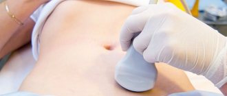
Despite the fact that ultrasound is used to diagnose pathologies of various organs and systems, the pelvic and abdominal organs remain a priority, which does not require the patient and doctor to spend a lot of time preparing, conducting and analyzing the results. Ultrasound of the pelvic organs (PUS) in women is especially relevant, since often the speed of the examination and the high information content of the results play a decisive role in the future fate of the woman.
Deciphering pelvic ultrasound in women is a rather complex process, during which the doctor must not only identify abnormalities and exclude all possible variants of the norm, but also differentiate the detected pathologies by type. Since OMT diseases in women are extremely diverse, the correct interpretation of the results obtained is decisive in the further choice of treatment tactics and imposes great responsibility on the doctor.
Causes
Experts call the main reason for the appearance of uterine fibroids an imbalance of sex hormones in a woman’s body - excess estrogen and lack of progesterone, as well as the development of hypergonadotropism. In addition, the following factors are noted that provoke the development of this disease:
- hereditary predisposition - every fifth woman was diagnosed with uterine fibroids in her family tree along the female line;
- late onset of first menstruation;
- absence of childbirth and breastfeeding in women over 30 years of age;
- previous gynecological manipulations and procedures (abortions, curettage);
- inflammatory and infectious processes in the genital organs;
- lack of meaningful sexual relationships;
- physical overload;
- psycho-emotional discomfort, stress;
- sedentary lifestyle.
Most often, such small myomatous nodes are discovered during gynecological examinations. Only in some cases, as the tumors grow, symptoms of uterine fibroids begin to appear. First of all, a woman experiences uterine bleeding, menstruation becomes heavy, often with blood clots. Many sick women have intermenstrual bleeding. In addition, the woman constantly feels discomfort, nagging pain in the lower abdomen. During menstruation, the intensity of pain increases significantly.
A sign of small uterine fibroids can be a woman’s infertility and frequent miscarriages. This is due to the fact that the myomatous node often interferes with the attachment of the fertilized egg to the wall of the uterus, acting as a kind of “contraceptive coil”.
Deviations from the norm: what the sonologist’s notes say
To correctly interpret pelvic ultrasound in women, you need to know what the specialist’s notes mean.
Deviation of the uterus from normal positions may indirectly indicate the presence of adhesions or inflammation. To the latter is usually added fluid, which is found in the retrouterine space.
If neoplasms are found in the cavity of the reproductive organ, then we can talk about a cyst, fibroid or polyp. The reason for their appearance is hormonal imbalance, frequent surgery, infectious processes, and so on. Often, with neoplasms, the size of the uterus exceeds its norm.
Endometrial thickness exceeding its standard values may indicate hyperplasia. An insufficiently voluminous mucous surface often becomes an indicator of hormonal imbalance.
If during an ultrasound the specialist detects visualized fallopian tubes, this is a sign of inflammation. They can also be detected during an ectopic pregnancy developing in this area.
Blurred areas of the contours of the reproductive organ indicate an inflammatory process, and uneven areas indicate fibroids, cancer or another type of tumor.
If a heterogeneous structure of the cervical canal or its causeless expansion is detected, inflammation or infection that was acquired during sexual intercourse can be suspected. Inflammation of the ovaries is indicated by their distorted contour.
If the size of the organ is too large, then polycystic disease or a single neoplasm can be suspected. All ovarian tumors are functional and non-functional. An ultrasound can with a high probability suggest that the cyst belongs to one type or another.
Diagnostics
The diagnosis is made when the size of the myomatous node is no more than 15 mm, which corresponds to 12 weeks of pregnancy.
Diagnosing uterine fibroids, even small ones, is not particularly difficult. Usually the tumor is detected during a gynecological examination. To clarify the diagnosis, the doctor prescribes an ultrasound examination, during which the localization of myomatous nodes, their size and density are determined.
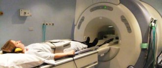
In some cases, a woman is prescribed hysteroscopy, which is used to assess the condition of the endometrium of the uterus. For atypical tumor localization, computed tomography (CT) and magnetic resonance imaging (MRI) are performed. To determine the development prospects and growth rate of myomatous nodes, Doppler examination of blood circulation in the neoplasm is sometimes used.
What is an exocervix smear? Analysis transcript
This mucous layer is located on the vaginal area of the cervix, that is, it is in contact with the microflora of the vagina. Normally, the upper layer of the exocervix is squamous stratified epithelium.
To detect pathologies, the doctor prescribes an exocervix smear to the patient. An inflammatory cytogram may show the presence of columnar epithelial cells, which is an abnormal situation.
There should be a clear boundary between the cylindrical and flat epithelium, located at the site of the external pharynx. If columnar epithelium covers the exocervix, then this will not be the norm. This condition is called cervical ectopia. To avoid complications from this pathology, you need to be treated.
Sometimes this phenomenon is considered an exceptional norm, in particular this applies to the age of girls from birth to puberty. That is, at the time of puberty, clear boundaries between the cylindrical and squamous epithelium should form.
Treatment
Although most cases of uterine fibroids involve surgical removal of the tumor, conservative therapy is often used to treat small fibroids.
Indications for conservative treatment are the size of the myomatous node up to 12 weeks, the asymptomatic course of the disease, and the woman’s desire to preserve reproductive function.
Each myomatous node consists of a center (determining the smallest possible tumor size) and a main growing volume. Conservative therapy correctly prescribed by a doctor can minimize the main growing volume. But at the same time, the center of the tumor can be preserved. Small uterine fibroids can remain in minimal size for a long time without affecting the woman’s well-being. Therefore, the doctor often chooses a wait-and-see approach, in which the woman is under constant medical supervision.
Drug treatment
Drug therapy includes the use of a complex of medications, among which the following drugs can be distinguished:
- antigonadotropins (Gestrinone) - drugs that prevent the tumor from increasing in size, but are not able to reduce its size;
- gonadotropic releasing hormone agonists (Triptorelin, Buserelin, Zoladex, Goserelin) – drugs that help reduce fibroids, prevent the development of uterine bleeding, and eliminate pain; taking these medications leads to a decrease in the amount of sex hormones in the blood, creating an artificial menopause, which is eliminated when you stop using the drugs;
- vitamin complexes – used to stabilize or correct hormonal levels and increase a woman’s immunity;
- sedative (calming) drugs.
Endocervical cysts
Quite often, an ultrasound scan in women can diagnose an endocervical cyst or a Nabothian cyst of the cervix. This is a round-shaped formation that forms when the glands of the cervix are blocked (cervical retention cyst). Endocervical cysts can be located not only on the outer vaginal surface of the cervix, but throughout the entire cervical canal.
Endocervix cysts - causes
Most often, the cause of the appearance of endocervical cysts is the ectopia of the columnar epithelium from the cervical canal onto the outer surface of the cervix or, conversely, the squamous epithelium inside the cervical canal during inflammatory processes, trauma to the cervix during childbirth, cauterization, and surgical interventions. Endocervix cysts are located on the outer surface of the cervix, facing the vagina, when glands from the columnar epithelium are ectopic there from the exocervix. Small endocervical cysts (up to 5 mm) are often found in women who have given birth and can be considered a normal variant.
Endocervical cysts - symptoms
Signs of endocervical cysts can be detected on ultrasound or colposcopy, but the woman does not make any complaints. Sometimes women may complain of bloody or brown spotting before menstruation and these symptoms are mistaken for signs of a cyst, but these may more likely be signs of cervical endometriosis.
Diagnosis of endocervical cysts
Ultrasound remains one of the most informative methods for diagnosing endocervical cysts. Echosigns of endocervical cysts on ultrasound are anechoic (black) round-shaped formations with clear, smooth edges ranging in size from a few millimeters to 1-2 cm. Most often, single endocervical cysts of small sizes are found. But, over time, the cysts can increase in size, deforming the cervix, or multiple endocervical cysts of different sizes appear.
In addition to ultrasound, endocervical cysts can be diagnosed using a routine speculum examination by a gynecologist. Upon examination, round-shaped formations of a whitish color with liquid contents are revealed. But colposcopy under a microscope will be more informative. For differential diagnosis, a cytological examination of a smear and a PAP smear are additionally used, which help to identify precancerous and cancerous changes in the cervix in a timely manner. Additionally, a smear is examined for genital infections so as not to miss inflammatory diseases of the cervix.
Endocervix cysts of the cervix - treatment
After an endocervical cyst is diagnosed, the doctor chooses a treatment method. Before deciding how to treat endocervical cysts, you should remember that small single cysts are not considered a disease and do not require intervention.
Sometimes, for small endocervical cysts, you can try treatment with folk remedies, using an infusion of fresh burdock leaves, white acacia flowers, pine nuts or golden mustache, but not more than a month, and if during this time the cyst has not decreased in size, then traditional treatment methods are used.
The doctor can puncture superficial cysts and remove the secretion. And if after some time the cyst recovers in size, then its destruction is used. Treatment of endocervical cysts with a laser is carried out if they are clearly visible during a routine gynecological examination in the vaginal part of the cervix.
With radio wave surgery (for example, using the Surgitron device), pathological tissues are evaporated, without subsequent bleeding, without subsequent scar formation, without affecting healthy tissue. This procedure is painless and healing occurs quickly. Treatment of deep endocervical cysts is carried out using cryodestruction with liquid nitrogen.
WomanAdvice.ru
Uterine fibroids: causes of development
The exact causes of the development of uterine fibroids are still unclear. However, according to one version, the occurrence of a benign tumor is associated with a hormonal imbalance - an increased level of estrogen in the blood. Proof of this is the fact that fibroids decrease or disappear in postmenopausal women, when the production of female hormones decreases.
In addition, the development of uterine fibroids can be provoked by the following factors:
- hereditary predisposition;
- uncontrolled long-term hormonal contraception;
- violation of the hormone-producing function of the ovaries;
- prolonged exposure to UV irradiation;
- absence of pregnancies in women under 30 years of age;
- inflammatory processes in the organs of the reproductive system;
- hemodynamic disorders of the pelvis;
- ovarian cysts;
- abortions and diagnostic curettages.
According to statistical studies, most often surgical intervention to remove uterine fibroids is required for women aged 43 to 45 years. Removal of the uterus becomes necessary in case of accelerated growth of fibroids, the presence of pathologies of the endometrium and ovaries. In women over 40 years of age, ovarian function decreases and estrogen levels decrease, which is accompanied by immune, hormonal and neuroendocrine disorders.
What does a transvaginal examination show?
Transvaginal ultrasound helps identify the following diseases and pathological changes:
- cystic formations of the ovaries;
- endometriosis of the uterus;
- pregnancy (uterine and ectopic);
- the presence of fluid, pus or blood in the fallopian tubes, which causes the presence of inflammatory processes in them (without differentiating these fluids);
- detection of fibroids in the uterus and polyps in the endometrium;
- identification of anomalies in the development of internal genital organs;
- hydatidiform mole (both partial and complete);
- cancerous tumors in the uterine cavity;
- ruptures of cystic formations in the appendages;
- the presence of fluid in the lower abdominal cavity;
- malignant formations of the appendages;
- chorionepithelioma.
Vaginal ultrasound allows you to determine the number of follicles that are in the process of development, as well as assess their quality and detect various features. If a transvaginal ultrasound is performed with the introduction of a contrast agent into the fallopian tubes, it is possible to assess their patency, detect polyps and give a prognosis for the passage of the fertilized egg.
Thanks to this study, the attending doctor will make a decision on the treatment of tubal obstruction, which will allow conceiving a child if the woman plans to become pregnant. Vaginal ultrasound is also performed if it is necessary to assess the dynamics of the development of the fertilized egg. An ultrasound diagnostic procedure with a vaginal sensor is prescribed if there is a suspicion that the pregnancy is not developing or its development is abnormal or slow. But for the procedure to be carried out, the pregnancy must be at least five weeks. A transvaginal ultrasound will allow you to see the fetal heartbeat, or more precisely, the contractions of the heart muscle, which first begin in the third week of pregnancy. In earlier stages of pregnancy, vaginal ultrasound is not prescribed for these purposes, since there will be no movement.
Uterine fibroids: symptoms and signs
Small uterine fibroids may not show any symptoms for a long time. However, its intensive growth is accompanied by the following signs.
- intermenstrual bleeding;
- painful sensations during menstruation, increasing their duration and volume;
- increased frequency of urination caused by compression of the bladder by an enlarged fibromatous node;
- a feeling of pressure and heaviness in the lower abdomen;
- painful sensations during intimacy;
- constipation associated with compression of intestinal fibroids;
- causeless increase in abdominal volume;
- pain in the lumbar region;
- infertility;
- neurological and mental disorders.
The manifestation of symptoms is influenced by the location of uterine fibroids, its size and tendency to grow.
In the early stages of the disease, a woman’s menstrual cycle is disrupted, which should be an “alarm bell” and a reason to consult a gynecologist.
At least half of the patients suffer from heavy menstruation. Most often these are women of childbearing age. Heavy discharge gradually becomes the cause of anemia. In addition, significant blood loss is caused by cardiovascular pathologies and coagulation disorders. Similar symptoms occur with uterine fibroids of the submucosal and intermuscular localization.
The endocervix is thickened to 8 mm. What to do?
Hello! Yesterday I did an ultrasound. All was good. The only thing is that the endocervix is thickened to 8 mm, when the norm is up to 5. During an examination by a gynecologist, she displayed an image on the screen and showed that the cervix was slightly reddish, that is, an inflammatory process was underway. There is some erosion, she said. But what kind of inflammatory process this is is not clear. Got tested for HPV. The results are still awaited. Tell me what this could be, what are the reasons? There were 2 partners, both were permanent, they were unlikely to bring anything in. OK I don't accept. Menstruation is regular. Previously there were no problems, nothing was treated. Thanks in advance for your answer.
S-anastasia
I also treated erosion several years before pregnancy, but during pregnancy it reappeared. They told me that it should go away after pregnancy, and so it happened - it went away after childbirth.
I don’t quite agree about the paid clinic, you need to look for a good, experienced gynecologist, it’s better to do it on recommendation, but paid clinics differ from each other. I recently had an ultrasound in a paid clinic - the doctor who did the ultrasound immediately put my diagnosis in question, and when I said that I wouldn’t go to their paid doctor, he said that everything was fine according to the ultrasound. And when asked why he made the diagnosis, he answered, just a suspicion. My doctor, by the way, at the free clinic, is confident in her as a professional in her field, and rejected this suspicion.
Good luck and health to you!
Lorelei
The reasons may be different, an experienced doctor will tell you for sure. Of course, testing for infections during examination is mandatory; many people do it. The inflammatory process can be caused by infections or be non-infectious.
Thus, erosion of the cervix may be due to the peculiarities of the formation of the internal genital organs during their growth during the puberty period, namely the “not quite correct” displacement of the columnar epithelium in the direction of the vagina. In this case, these cylindrical cells are easily damaged, even by an acidic environment (that is, not necessarily an infection), which visually manifests itself as redness and inflammation. It is simply useless to treat these capricious cells - they are simply out of place. Thus, these cells, which once sprouted and formed “in the wrong place,” just need to be removed - with laser surgery, electric knife, cryotherapy. True, in the past doctors often advised waiting until after childbirth to do the procedure - they say that manipulations can affect the elasticity of the cervix during childbirth, although there is no certainty that this is true in all cases.
At one time, they cleaned me using the last method - cryotherapy, although this is not the most modern method - but now doctors do not worry about anything during examination)
Alexander6052
Your desire to improve your health is quite natural... But why you don’t find out all the details in consultation with specialists, but turn to BV for help, is a mystery to many.
Here they can give you such advice that you won’t want to live.
It's better to contact good doctors. It’s better (if you’re not in poverty) to go to a paid clinic. Don't waste your precious time.
I advise you from the bottom of my heart... I don’t want to offend...
Anaxoret
I have a medical education, not a doctor’s (nurse), but I worked in a hospital and saw enough of everything.
To begin with, I suggest not to panic and do an ultrasound in another place, it also matters when the ultrasound was done in relation to menstruation, if immediately after, during or before then it should be redone in a quiet period, perhaps the ultrasound specialist is not a very good specialist, if it is confirmed, then we need additional data obtained through cervicoscopy and during diagnostic curettage of the entire mucous membrane of the cervical canal, the resulting material is then examined (histological examination).
bolshoyvopros.ru
Uterine fibroids: diagnosis
Most often, a gynecologist discovers uterine fibroids during a routine examination. The presence of a fibromatous node can be suspected when the uterus is enlarged.
To confirm the diagnosis, a life history is collected and the patient’s complaints are surveyed.
In addition, a bimanual examination, smears for cytology and flora are prescribed.
To clarify the location of fibromatous nodes and the number of tumors, additional diagnostic examinations are prescribed:
- clinical laboratory studies and pregnancy tests;
- ultrasound examination of the pelvic organs - to detect pathological processes occurring in the uterus;
- hysteroscopy - examination of the uterus with a special apparatus and optical instrument to determine the characteristics of the hyperplastic process occurring in the uterus, as well as to identify suspicious areas of the endometrium and perform a biopsy;
- laparoscopy - during the study, a specialist examines the uterus using an endoscope;
- metrosalpingography is an X-ray examination of the uterine cavity, during which the patency of the fallopian tubes is assessed.
When a complex uterine fibroid is detected, a woman needs immediate treatment to improve the condition.
Preparation
Removal of tumor nodes on the uterus is a planned operation, before which the patient undergoes an extensive examination to identify possible contraindications and causes that may complicate the course of the operation and the rehabilitation period.
In preparation for surgery, laboratory (blood and urine tests, smears for microflora and cytology of the vagina and cervix) and instrumental studies (ECG, chest x-ray, ultrasound) are performed.
After hospitalization, the woman is examined and interviewed by an anesthesiologist, the essence and course of the operation is explained to her in detail, and consent to surgical intervention is given in writing.
Uterine fibroids: treatment
The main difficulty in treating uterine fibroids lies in the heterogeneity of the benign neoplasm. Therefore, before prescribing it, a woman needs a thorough examination.
Treatment of uterine fibroids can be conservative or surgical.
Uterine fibroids: conservative therapy
For small and medium-sized fibromatous nodes, therapeutic treatment is carried out - hormonal therapy. Conservative treatment is prescribed for the following indications:
- small tumor sizes (no more than 12 weeks of pregnancy);
- mild symptoms that do not reduce the quality of life;
- the need to preserve reproductive function;
- extragenital diseases that are contraindications for surgical interventions;
- postoperative rehabilitation or preparation for myomectomy.
The positive effect of hormonal therapy is achieved, on average, after six months of use. When choosing the necessary drug, the patient’s age and the characteristics of the pathology are taken into account.
As means of hormonal treatment, estrogen-progestogen, progestin and androgenic drugs, as well as GnRH agonists and antigonadotropic drugs are prescribed.
Conservative treatment of uterine fibroids is contraindicated in the following conditions:
- intensive increase in the size of the tumor;
- pronounced pain syndrome with the development of bleeding;
- uterine size exceeding 12 weeks of pregnancy;
- concomitant diseases of the pelvic organs;
- somatic pathologies (diabetes mellitus, hypercoagulation, hypertension, etc.).
Uterine fibroids: specific treatment
In addition to hormonal therapy, methods of nonspecific treatment of uterine fibroids are also used, aimed at eliminating the causes of the disease:
- vitamin therapy - taking vitamin and mineral complexes;
- diet therapy - enriching the diet with iron-containing and protein foods;
- sedatives for women with vegetative-vascular pathologies;
- venotonics – normalizing the menstrual cycle;
- antianemic therapy - prescribed for large blood losses during menstruation;
- immunomodulatory therapy – increasing the body’s protective functions;
- carrying out physiotherapeutic procedures - magnetic therapy, electrophoresis, autotransfusion, etc.
In the absence of negative symptoms and increased growth of uterine fibroids, therapy may not be carried out. In such cases, dynamic observation is prescribed, which involves regular examination by a gynecologist and ultrasound.
Uterine fibroids: surgical treatment
Surgery is prescribed for the following indications:
- pathologies of the pelvic organs;
- excessive enlargement of the fibromatous node (more than 12 weeks);
- lack of effectiveness from treatment with conservative methods;
- severe pain and heavy bleeding;
- acceleration of fibroid growth (more than 4 weeks per year);
- degenerative pathologies of fibroids;
- twisting of a subserous fibroid node;
- prolonged heavy menstrual flow in women with submucosal fibroids.
How to treat?
The method of eliminating uterine pathology depends on the type of fibroid, its size, location, as well as the woman’s age and plans for pregnancy. If education does not increase and does not affect the girl’s well-being, observation is indicated. Treatment is carried out in case of rapid growth of fibroids, compression of nearby organs, or suspected malignancy.
Treatment for fibroids is:
- Conservative. Involves taking medications, using physiotherapy and alternative medicine.
- Operational. Perform surgeries (minimally invasive, abdominal).
Drug treatment
Indications for drug therapy: bleeding, abdominal pain, fibroid size from 3 cm. The use of the method helps eliminate discomfort, anemia, and hyperplastic changes in the endometrium. Analgesics, painkillers, and vitamins are prescribed. For girls with vegetative-vascular pathologies, treatment with sedative medications is indicated. Venotonics are used to normalize the menstrual cycle. In case of severe bleeding, antianemic therapy is carried out. In order to increase defenses, immunomodulatory treatment is used.
The basis of the conservative method is the use of medications that reduce the effect of female hormones on the formation of the uterus. Prescribed use:
- Antigonadotropins (Danol, Nemestrana). They provide a decrease in estrogen production and a decrease in the replenishment of the node.
- Gestagenov (Yarins). Restore the cycle.
- Intrauterine device (Mirena).
- Gonadotropin-releasing hormone analogues (to reduce estrogen concentrations).
Surgery and non-invasive procedures
When drug treatment for uterine fibroids is ineffective, surgery is prescribed. The radical method is used in case of rapid growth of a nodular formation, degeneration into cancer, or deterioration of the condition. The choice of method depends on the diameter and location of the formation. Hysterectomy and myomectomy are often prescribed. The difference between the two methods is that the second is considered organ-preserving. The woman will be able to bear and give birth to a baby.
But more often, doctors try to resort to minimally invasive manipulations - operations with minimal damage.
Carrying out UAE (uterine artery embolization)
Treatment consists of stopping the feeding of fibroids. During manipulation, a thin tube is inserted into the femoral artery (to identify the characteristics of the blood supply to the uterus). The veins are then filled with small particles that block blood flow to the fibroids. As a result, the node decreases in size.
FUS ablation
Treatment of tumors with this method is common because it is safe and bloodless. After the procedure, reproductive function is preserved. It involves the effect of ultrasound on fibroids. The unit is heated and destroyed.
This method is used for single small formations. It can be replaced by cryomyolysis (freezing of formation), electromyolysis (exposure to the fibroid with current).
Physiotherapy
Treatment of fibroids on the uterus without surgery is effective for small fibroids. Provides improved well-being, normalization of the cycle, and reduction in the size of the formation. The specific method is selected by the doctor, taking into account the patient’s age and tumor type.
Electrophoresis
A method of treating pathology in which the medicine is administered through the skin and mucous membranes using direct current. This supply of drugs ensures an active increase in their activity. To treat fibroids in the body of the uterus, electrophoresis of zinc and iodine is used on the lower abdomen. Application of the method helps in eliminating inflammation and pain, and also:
- decreased estrogen production;
- restoration of the menstrual cycle.
Magnetotherapy
The use of this method in complex therapy of uterine fibroids promotes regression of the pathology. The anti-inflammatory, analgesic, and restorative effects of the technique are known. The procedure ensures:
- inhibition or stabilization of fibroid growth;
- improving the functioning of the endocrine system;
- normalization of the cycle.
Iodine-bromine and radon baths
The positive effect of the procedure is due to the entry into the tissues of the inert gas radon and its decay products. The radiation that appears stops bleeding and also helps slow the growth of fibroids in the uterus and restore hormonal levels. The procedure has a relaxing, calming effect, lowers blood pressure and normalizes blood flow.
Treatment with iodine-bromine baths is no less effective. Bromine released from medicinal waters accumulates in the brain, and iodine in the thyroid gland. The procedure restores the balance of pituitary and ovarian hormones.
Unconventional methods
It is recommended to use folk remedies for the treatment of uterine fibroids. Medicines and herbal teas speed up the healing process, restore strength, strengthen the immune system, and also eliminate discomfort. Compositions are used as an auxiliary method. Doctors recommend use for fibroids in the uterus:
- infusion of oak bark or nettle (to stop bleeding);
- douching with chamomile decoction (removes inflammation).
No less effective are compositions from boron uterus, chestnut, lemongrass, and aloe. Treatment of fibroids with folk remedies should be agreed with a doctor.
Features of therapy for menopause
During menopause, pathology does not develop. If symptoms or signs appear during menopause, this means that the pathology did not progress before and was hidden. Women with a tumor that does not appear are not treated. Regular examination and observation is indicated. If there are any changes (increased growth, painful symptoms), surgery is prescribed to remove the formation. If malignancy is suspected, the tumor is excised along with the uterus and sometimes the appendages.
Often fibroids regress during menopause. This is due to decreased production of hormones and loss of reproductive function.
General information
According to statistics, four out of ten women aged 30 to 50 have uterine fibroids (fibroids), a single node or a cluster of benign tumors of muscle and connective tissue. Some of them are immersed in the wall of the uterus, while others protrude into the uterine cavity like mushrooms. In rare cases, they can grow to the outer surface of the uterus, ranging in size from a few grams to tens of kilograms (the record largest tumor weighed 67 kg).
Fibroids are benign tumors; they do not increase the risk of developing uterine cancer and do not become malignant. However, due to their size and location, they can mask the growth of malignant neoplasms, so careful diagnosis is necessary.
Normal echographic picture
Interpretation of ultrasound of the pelvic organs in women begins with measuring the size of the uterus. The length, defined as the distance from the fundus of the uterus to the internal os of the cervix, should be 5–8 cm. The average length in healthy nulliparous women should be in the range of 6.0–7.1 cm. In women who have given birth to one child, there is a slight increase uterine body, more pronounced after several births.
The anterior-posterior size (thickness) of the uterus should normally be within 3–4 cm, and the transverse size (width) should be 4.5–6.0 cm. After several years after the decline of reproductive function, it is permissible to reduce the size of the uterus to 4 –5 cm. An important factor taken into account when assessing the condition of the female reproductive organs is the position of the uterus.
Normally, it is located in the central part of the small pelvis, deviating towards the anterior abdominal wall. This situation in the examination protocol is designated by the term “anteflexio”. The term “retroflexio” refers to a physiological disturbance in the position of the uterine body caused by its deviation back, that is, “bending”, and “lateroflexio” means a displacement of the uterus relative to the central axis of the body.
Important! When assessing the position of the uterus, it should be taken into account that a full bladder causes some deviation from its normal position.
An ultrasound image shows the uterus on the 19th day of the menstrual cycle. The length of the uterine body (number 1) is 57.6 mm, the width (number 2) is 42.9 mm, the width of the uterine cavity (number 3) is 7.1 mm
The cervix on an echogram is defined as a 2–3-centimeter formation, cylindrical in shape, with echogenicity indistinguishable from the uterus. The width of the cervical canal should be between 3–4 mm. When examined by ultrasound, the ovaries appear as oval formations located on both sides of the uterus. The normal length of the ovaries should be 2.7–3.7 cm, width 2.1–2.9 cm, and anterior-posterior dimension – 1.7–2.1 cm.
In general, the size of the ovaries can vary within quite a wide range, since during the growth of the follicles the ovary enlarges. After the isolation of the dominant follicle, which is determined in the first phase of the cycle in the form of a small formation (0.5–1.3 cm), which continues to grow actively until the 12–14th day of the menstrual cycle, the rest decrease again, and the ovary acquires normal size.
By the day of ovulation, the follicle can reach 1.5–2.9 cm in size and can be clearly identified on ultrasound. By visual assessment, the size of one ovary should not exceed half the width of the uterine body. Normally, fallopian tubes are not visible on ultrasound. Immediately after ovulation, the formation of the corpus luteum, a temporary hormone-producing gland, begins, the main purpose of which is to ensure implantation of the embryo and maintain pregnancy.
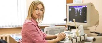
Preparation for ultrasound of the pelvic organs in women
The corpus luteum appears to be a small formation with heterogeneous thick walls and liquid contents. The entry in the ultrasound examination protocol “corpus luteum cyst” is assessed based on the timing of the diagnosis, since detection of a cyst after ovulation indicates normal functioning of the reproductive system, and before ovulation – a pathological formation.
The endometrial layer in the uterine cavity in the first days of the menstrual cycle is defined as a heterogeneous structure of varying thickness (from 0.3 to 0.8 cm). By the time menstrual bleeding ends (4–5 days of the cycle), the thickness of the endometrial layer is only 0.2–0.4 cm and is practically invisible on the echogram. In the early phase of proliferation (day 6–7 of the cycle), a slight thickening of the endometrium up to 0.6–0.9 cm can be noticed, with a simultaneous decrease in echogenicity.
At the same time, its layering is clearly determined in the form of the appearance of a thin echo-negative contour 1 mm thick. By day 10, the thickness of the endometrial layer is about 1 cm. In the secretory phase (days 15–27 of the cycle), as well as during menstrual bleeding, a significant thickening of the endometrium occurs (sometimes up to 1.5 cm), which is noted on the echogram in the form of a thickened reflective surface internal cavity of the uterus.
Important! Detection of the corpus luteum and thickened endometrium at the beginning of the menstrual cycle, in the absence of a fertilized egg in the uterine cavity, can serve as an indirect sign of ectopic pregnancy.
An ultrasound image shows the uterus on the 25th day of the menstrual cycle. The thickness of the endometrial layer is 1.0 cm
Symptoms of uterine fibroids
Most uterine fibroids are asymptomatic, but large tumors may cause the following symptoms:
- prolonged and painful menstrual bleeding;
- bleeding during the intermenstrual period;
- feeling of fullness or heaviness in the lower abdomen;
- frequent urination (if the deformed uterus puts pressure on the bladder);
- constipation (if the tumor puts pressure on the intestines);
- pain in the lower back;
- pain during intercourse:
- increase in abdominal size;
- miscarriages, infertility.
What is not allowed for small uterine fibroids?
Among the drug therapy of traditional medicine, a controversial issue that remains unresolved to this day is the prescription of progesterone drugs such as Duphaston. Some researchers say that the administration of progesterone is pathogenetically justified, as it reduces estrogen levels. affecting the growth of the node. And some, in contrast, point to cases of a jump in the growth of nodes during pregnancy, which itself is a source of progesterone. Also cited as an example is the use of the antiprogesterone drug Mifepristone, which obtained good results of tumor regression. For now this question remains open.
Contraindications for small uterine fibroids:
- The influence of constant stress factors on the body;
- Heavy lifting and significant physical activity;
- Performing intrauterine manipulations, such as abortion or medical termination of pregnancy;
- Physiotherapeutic treatment associated with warming procedures.
Massages and exposure to high temperatures in the form of saunas and baths are prohibited.
Small uterine fibroids: forum
Many people are looking for answers to the question of treating a diagnosis of small uterine fibroids on forums, however, it is worth noting that no Internet page will give you a comprehensive and objective answer like a qualified obstetrician-gynecologist. You should not expose your body to such dangers as incorrect therapy prescribed by strangers, in most cases even without medical education. Only timely qualified diagnosis and treatment is the key to successful fight against such pathological conditions of the woman’s reproductive organ – the uterus.
Source: zaberemenet-pri-miome-matki.ru
What can you do
There is a high probability that you yourself do not know that you have fibroids. They are most often discovered during a routine gynecological examination or during prenatal testing. As long as fibroids do not cause severe pain or a feeling of heaviness in the lower abdomen, do not cause infertility, or do not exceed 10 centimeters in diameter, they do not require treatment.
Visit your doctor once or twice a year so that he or she can monitor the growth of fibroids. These regular checks (with periodic ultrasound examinations) are necessary to detect complications, such as enlarged fibroids with compression of the ureter.
Forecast and consequences
With timely detection and treatment, the pathology does not cause concern and is amenable to conservative therapy. Ignoring symptoms and refusing therapy can lead to:
- purulent processes due to the death of fibroid cells;
- degeneration into cancer;
- anemia;
- torsion of the leg, necrosis;
- infertility.
But more often the prognosis is favorable. After treatment, women do not face any consequences, become pregnant and give birth to healthy children.