Sputum is pathological discharge from the respiratory tract that appears after coughing. Expectoration indicates a dysfunction of the inner lining of the respiratory tract. Sputum analysis is a test widely used to diagnose diseases of the lungs and bronchi. The procedure makes it possible to differentiate pathologies that occur against the background of cough and other typical clinical symptoms. You can obtain sputum for analysis yourself during a cough or using medical manipulation (bronchoscopy).
Why are all these various studies needed?
The main purpose of the described study is to clarify the diagnosis. Under normal circumstances, no sputum is produced. Goblet cells of the ciliated epithelium secrete from 10 to 100 ml of liquid, which is swallowed by a person.
The progression of the pathological process in the bronchi or lungs leads to a change in the activity of the corresponding structures with an increase in cough, shortness of breath, and chest pain. The amount of liquid mucus increases, and bacterial microflora may join. The result is sputum production with coughing.
Based on the expected diagnosis and the results of a visual assessment of the secretion of the bronchial glands, the doctor prescribes the appropriate type of study. The use of different options for sputum analysis makes it possible to assess the physicochemical properties of the fluid, cytological changes (presence of cancer cells), and the presence of bacterial invasion.
Decoding
To correctly diagnose a patient, sputum is assessed according to three different indicators. Macroscopic, bacterioscopic and microscopic analysis is carried out, the results of each study give a clear picture of the human condition. Color, consistency, smell, division into layers and the presence of inclusions are the main indicators of macroscopic analysis of the secretion. For example, clear mucus occurs in people with chronic respiratory diseases.
The rusty hue of the secretion is due to bloody impurities (decomposition of red blood cells), which often indicates the presence of tuberculosis, lobar pneumonia, and cancer. Purulent sputum, which is formed when white blood cells accumulate, is characteristic of an abscess, gangrene or bronchitis. The yellow or green color of the discharge is an indicator of a pathological process in the lungs. The viscous consistency of the secretion may be a consequence of inflammation or taking antibiotics.
Kurshman spirals in sputum, which are white convoluted tubes, indicate the presence of bronchial asthma. The results of microscopic and bacterioscopic analysis provide information about the content of pathogenic microorganisms or bacteria in the mucus. These include: diplobacillus, atypical cells, staphylococci, eosinophils, helminths, streptococci. Serous sputum is released during pulmonary edema; Dietrich's plugs are found in patients suffering from gangrene or bronchiectasis.
Types of sputum tests (studies)
Sputum examination can be carried out either with the naked eye or with the help of specialized equipment.
Depending on the disease that the doctor suspects, the following types of diagnostics may be used:
- General sputum analysis . The doctor evaluates the physical characteristics of the mucus that appears after coughing;
- Cytological (microscopic) examination . To carry out appropriate diagnostics, the doctor needs a microscope. Using image magnification, the liquid is analyzed. The technique allows you to see the presence of pathological cells that appear in certain diseases;
- Chemical Research . Changes occurring in the metabolism of alveolocytes and ciliated epithelium of the bronchi are assessed;
- Bacteriological method or sputum culture . The essence of the study is based on sowing bacteria obtained from the contents of the respiratory tract on a nutrient medium. The growth of colonies confirms the presence of the pathogen in the respiratory tract. An important advantage of culture remains the ability to test the sensitivity of bacteria to specific antimicrobial drugs in the laboratory.
In severe cases, for timely diagnosis of the pathology of the patient’s respiratory system, all three research options are simultaneously prescribed. Based on the results obtained, appropriate therapy is selected.
General sputum analysis
Fact! General or macroscopic analysis allows you to evaluate sputum immediately after it is obtained. This research option has been used by doctors for hundreds of years. Even before the invention of the microscope and modern analyzers, doctors made diagnoses based on the appearance of the expectorated liquid.
Below we will describe the key aspects that the doctor pays attention to during diagnosis.
Quantity
The daily amount of mucus secreted ranges from 50-100 ml to 1.5 liters, depending on the underlying pathology, which disrupts the normal secretion process of goblet cells. Respiratory diseases such as bronchitis or pneumonia are accompanied by the release of up to 200 ml of fluid (daily amount).
A sharp increase in this indicator occurs when pus or blood accumulates with further release through natural pathways. Bronchiectasis, drained abscess, and gangrene of the lung occur with the release of up to 1.5 liters of fluid.
Character
Depending on the nature of the liquid that is expectorated during a cough, pulmonologists distinguish the following types of sputum:
- Mucous . Favorable scenario. Diseases in which it occurs: bronchial asthma, chronic bronchitis, tracheitis;
- Mucopurulent . Additionally, a bacterial infection is added. In addition to coughing and mucus, liquid is released, which is the waste products of microorganisms and bacteria “digested” by immune cells. Diseases – lung abscess, bacterial pneumonia, gangrene;
- Purulent . The reasons for the occurrence are the same as in the previous case. The difference is a higher percentage of pus and tissue breakdown products. The patient's condition worsens;
- Bloody . When individual red blood cells or portions of blood enter the liquid expectorated during a cough, it acquires a characteristic color. The symptom indicates vascular damage. Possible causes are cancer, trauma, pulmonary infarction, actinomycosis.
Assessing the nature of liquid discharge when coughing helps to understand the pathological process that develops in the patient’s respiratory system and select adequate treatment.
Color
The color palette of sputum released during a cough depends on its nature.
Possible combinations:
- Mucous membrane – grayish or transparent;
- Mucopurulent – gray with yellow or purulent patches;
- Purulent - the fluid may be dark yellow, green or brown;
- Bloody - various shades of red. It is important to remember that a “rusty” color indicates the presence of modified red blood cells. If a vessel is damaged, the blood is scarlet or pink (depending on the intensity of blood loss).
Interesting! Anthracosis is an occupational disease of miners caused by inhalation of dust rich in coal. The corresponding pathology is accompanied by the release of mucous sputum, but due to coal impurities it is black in color.
Smell
Sputum in 75% of cases has no characteristic odor. The exception is the release of purulent contents. Dead tissue particles cause a putrid odor. When a lung cyst in which echinococcus (helminth) has developed breaks through, a fruity aroma appears.
Layering
The mucus released when coughing is predominantly homogeneous.
The separation of sputum into layers is characteristic of the following pathologies:
- Lung abscess . In this case, 2 layers are formed - serous and putrefactive;
- Gangrene of the lung . In this case, a third (upper) foamy layer is additionally formed, which is caused by the vital activity of the corresponding microorganisms that produce gas bubbles.
Visual assessment of mucus allows you to quickly establish a diagnosis without performing auxiliary tests.
Impurities
Impurities in sputum include red blood cells, pus, or serous fluid. The presence of the described inclusions allows the doctor to assess the degree of damage to the lung tissue and understand which pathological process is primary for a particular clinical case.
Chemical research
Chemical analysis of the fluid secreted during coughing allows us to determine the severity of the pathological process. Depending on the results of the study, the doctor selects appropriate medications aimed at stabilizing the function of the ciliated epithelium.
Reaction
Normally, the pH of sputum ranges from 7 to 11. The progression of the processes of decomposition of lung tissue leads to the oxidation of the corresponding reaction (indicator below 6). The reason for the change in pH value is based on disturbances in the metabolic processes of salts and minerals.
Protein
Protein is always present in the liquid released when coughing. The norm is up to 0.3%. A slight increase in the corresponding figure to 1-2% may indicate the progression of tuberculosis. A significant increase in the indicator (10-20%) is a sign of the development of lobar pneumonia. Laboratory examination of mucus with protein determination makes it possible to differentiate these pathologies against the background of an analysis of the clinical picture (cough, shortness of breath, chest pain) and the results of other diagnostic procedures.
Bile pigments
Bile pigments, or more precisely, cholesterol microparticles, are secreted with mucus during coughing in the following pathologies:
- abscess;
- formation of echinococcal cyst;
- malignant tumors of the respiratory tract.
Microscopic examination
Microscopic analysis of sputum allows, using an appropriate optical apparatus, to detect the presence of cells or microorganisms that should not normally be present in mucus secreted by coughing.
Epithelial cells
Epithelium in sputum is a normal variant. On microscopic examination, attention is drawn to a sharp increase in the concentration of cells or the formation of epithelial casts. This picture indicates damage to the respiratory tract and inner lining.
Alveolar macrophages
The function of alveolar macrophages is to provide local immune protection. A small number of cells may be present in the mucus. A sharp increase in the concentration of macrophages indicates a chronic inflammatory process (bronchitis, bronchiectasis, asthma, tracheitis).
Leukocytes
The appearance of leukocytes indicates the presence of acute inflammation, which can occur against the background of a bacterial infection. Possible pathologies are abscess, pneumonia, bronchiectasis.
Red blood cells
Blood cells appear in sputum when small or large vessels rupture. The doctor judges the nature of the bleeding by the number of red blood cells. Separately, it is worth highlighting the appearance of modified cells that penetrate through the dilated walls of blood vessels without rupturing the latter. A typical example of the disease is lobar pneumonia.
Tumor cells
Atypical cells in sputum are a sign of a developing cancer process. To clarify the location and type of pathology, additional tests are required.
Fact! With the help of microscopic examination, cell differentiation is also established. The less similar the structures are to the original tissue, the worse the prognosis for the patient.
Elastic fibers
The appearance of elastic fibers in the mucus released when coughing is a sign of serious lung damage with tissue breakdown. Examples of diseases are gangrene, late stage bronchiectasis, tuberculosis and cancer, accompanied by destruction of organ parenchyma.
Detection of Mycobacterium tuberculosis
Microbiological analysis of sputum is one of the important methods for verifying the presence of tuberculosis. The causative agent of the disease is mycobacterium (Koch's bacillus).
Determining the presence of a microorganism is possible using the bacterioscopic method using a microscope. To visualize the pathogen, it is necessary to stain the material under study using the Ziehl-Neelsen method. If Koch's bacillus is detected in the sputum after a cough, the phthisiatrician must indicate BC (+) in the documentation, which indicates the release of the pathogen. Such patients require isolation. CD (-) – the patient does not spread the bacteria.
Fact! Sputum analysis for tuberculosis is also carried out by inoculating the test fluid on a nutrient medium. The advantage of the method is 100% accuracy. If the bacterium is present in the body, it will grow in the laboratory. The main disadvantage of the described diagnostics is the length of time it takes to obtain analysis results (sometimes more than a month).
Bacteriological examination for infectious lung diseases
Bacteriological research for inflammatory lesions of the respiratory tract is usually used to verify community-acquired forms of infection (pneumonia, actinomycosis, etc.).
The analysis is carried out in three stages:
- sputum collection for research;
- sowing the liquid onto a previously prepared nutrient medium;
- reseeding the required colony with the study of the chemical and physical characteristics of the pathogen.
If it is necessary to establish susceptibility to antimicrobial agents, a sensitivity test is additionally performed. Paper mugs treated with antibiotics are placed in a Petri dish where a colony of microorganisms has grown. Those drugs around which the zone of colony destruction is maximum are recommended for use in a particular patient.
Chemical research
Reaction
Freshly secreted sputum has an alkaline or neutral reaction. Decomposed sputum becomes acidic.
Protein
Determination of protein in sputum can be helpful in the differential diagnosis between chronic bronchitis and tuberculosis: with chronic bronchitis, traces of protein are determined in the sputum, while with pulmonary tuberculosis in the sputum the protein content is higher and can be determined quantitatively (up to 100-120 g /l).
Bile pigments
Bile pigments can be detected in sputum in diseases of the respiratory tract and lungs, combined with jaundice, during communication between the liver and lung (when a liver abscess ruptures into the lung). In addition to these conditions, bile pigments can be detected in pneumonia, which is associated with intrapulmonary breakdown of red blood cells and subsequent transformations of hemoglobin.
Diseases for which a doctor may prescribe a general sputum test
Collecting sputum for general analysis can be prescribed for almost any disease that is accompanied by expectoration after coughing. However, appropriate diagnostics are rarely used for seasonal viral infections because they are unnecessary. In these cases, cough and other symptoms regress with plenty of fluids and bed rest.
Pathologies requiring sputum analysis:
- tuberculosis;
- lung abscess;
- malignant neoplasms;
- gangrene of the lung;
- bronchial asthma;
- Chronical bronchitis;
- pneumoconiosis is an occupational disease of the bronchopulmonary system.
Confirmation of the diagnosis is carried out using laboratory, physical, and instrumental methods.
Interpretation of results
In a healthy person, bronchial secretions are swallowed, since their significance is insignificant. If the patient can spit out sputum, then its amount has increased. This indicates a disease of the respiratory system.
Let's look at what results a sputum test can show:
- A viral disease is a transparent, viscous secretion. This discharge is characteristic of an acute inflammatory process.
- An admixture of blood is the most dangerous symptom that characterizes a serious pathology: tuberculosis, cancer, systemic damage to connective tissue. Sometimes small streaks of blood occur with a very strong, dry cough (whooping cough, tracheitis with the flu).
- Allergic sputum has a viscous consistency and an amber tint.
- Purulent sputum usually characterizes a bacterial infection. The discharge is cloudy, yellow-green, sometimes whitish. This symptom occurs in many diseases - bronchitis, pneumonia, sinusitis, lung abscess and others.
- Serous sputum is characteristic of pulmonary edema. It contains an increased amount of liquid component.
- If leukocytes are found in the secretion in numbers exceeding 25 thousand in the field of view, this indicates inflammation, most often of a bacterial nature.
- Microscopic examination reveals a large number of eosinophils. Then they immediately assume a helminthic infestation, which also often causes a cough, or an allergic reaction.
- Specific signs of bronchial asthma are Kurshman spirals and Charcot-Leiden crystals. The first are “casts” of small bronchi, consisting of viscous secretion. Crystals are formed from the secretion of eosinophils and are excreted in the sputum in the form of oblong pyramids.
- Elastic fibers. Their detection is always alarming, as it happens when lung tissue is destroyed (tuberculosis, tumor, abscess pneumonia).
Table for interpretation of the results of microscopic examination of sputum
| Cells | Result |
| Flat - usually indicates improperly collected material when saliva gets into the sputum. The detection of columnar epithelium indicates bronchitis, bronchial asthma or lung cancer. | |
| Alveolar macrophages | The result of a long stay in a dusty room. Sometimes hemosiderin is detected along with them - a breakdown product of hemoglobin (this is a sign of mitral stenosis, pulmonary infarction, stagnation) |
| Leukocytes | If eosinophils predominate - bronchial asthma, pneumonia, tuberculosis If lymphocytes - tuberculosis, whooping cough |
| Red blood cells | A sign of a violation of the integrity of the lung tissue is destructive forms of tuberculosis. tumor |
| Tumor cells | Detection of atypical cells is significant only when there is a large accumulation of them. If there are single ones, the study is repeated. |
| Elastic fibers | Decay of lung tissue due to tuberculosis, tumor, abscess |
Features of sputum analysis for bronchitis
Bronchitis is a respiratory tract disease that usually complicates the course of a viral infection.
Inflammation of the bronchial mucosa is caused by bacteria, viruses or an allergic reaction
Treatment for bronchitis varies greatly depending on the cause, so sputum examination is important to make the correct diagnosis.
The results may be as follows:
- Viral bronchitis
- mucous sputum, without impurities. - Bacterial bronchitis, suspicion of pneumonia
- the appearance of purulent impurities in the mucous discharge. - Bronchiectasis, chronic bronchitis of staphylococcal nature
- completely purulent discharge. - Allergic bronchitis
is a small amount of clear secretion, in which microscopic examination reveals a large number of eosinophils.
When cloudy sputum settles, it usually separates into two layers, which indicates the purulent nature of the inflammation. If the liquid is stratified into three layers, this indicates the presence of a putrefactive process (a sign of incipient gangrene of the lung).
You should not independently draw conclusions about the presence of the disease based on the results of a sputum examination. It is better to entrust this to a doctor, who compares them with clinical manifestations and only after that makes a final diagnosis.
Post Views: 3,501
Sputum analysis is a laboratory test that reveals the nature of the pathological process that occurs in the respiratory organs. Sputum can be heterogeneous in composition; it often contains pus, blood and other inclusions. The analysis also reveals the type of pathogen that provoked the disease and its sensitivity to antibacterial agents.
It is very important to collect sputum correctly to ensure accurate results.
How to prepare for the test in order to pass it correctly?
Preparing a patient to collect sputum for analysis is a responsible process on which the quality of diagnosis may depend. If simple rules are ignored, additional impurities appear in the mucus, preventing the laboratory assistant from establishing the root cause of cough and respiratory pathology in general.
Recommendations:
- Preparing the container . Containers sold in pharmacies remain optimal. If such a bottle is not available, even a half-liter jar or small plastic tank (no more than 1 liter) will do. However, it must be taken into account that such containers are extremely inconvenient and can only be used in atypical circumstances when there is no access to normal containers;
- Two hours before the examination, you need to brush your teeth and rinse your mouth. Removing food particles and saliva improves diagnostic accuracy;
- Consult your doctor . The doctor will explain in detail how to properly collect sputum for analysis.
If a person is giving bronchial mucus for the first time, then it often takes several attempts to complete the procedure correctly.
How to properly collect biomaterial
The correctness of the results and the effectiveness of further treatment depend not only on the correctness of the study, but also on the correct collection of materials for it.
Basic rules for collecting sputum for research
- You need to cough up not saliva and mucus from the nasopharynx, but the contents of the lower respiratory organs.
- To get a productive cough, you should first take 2-3 deep breaths, trying to breathe so that the lungs are completely filled with air and open.
- The patient should hold the container for collecting material as close to his mouth as possible and immediately spit into it what he coughed up.
- If it is not possible to obtain sputum, then the following manipulations are performed: the subject must tap himself on the chest several times, breathe deeply, holding his breath for a few seconds each time. As a last resort, it is suggested to breathe over a container of hot water with the addition of baking soda.
The container for collecting materials for analysis also has special requirements. Dishes do not have to be sterilized. But it must be clean, dry, made of plastic or glass, with a tightly screwed lid.
Important! The material for analysis must be immediately sent to the laboratory - the study must be carried out no later than 2 hours after collection. Until the materials for research are transported to the laboratory, they are stored in the refrigerator in a hermetically sealed container.
How to submit sputum for analysis?
In addition to the preparation nuances described above, the rules for collecting sputum provide for the use of a morning portion of mucus. The reason is the accumulation of secretions from the night, which greatly facilitates expectoration after coughing. You can take bronchial mucus at other times of the day, however, the quantity and quality of the material being studied is reduced.
Algorithm for collecting sputum for general analysis:
- take a deep breath and hold the air for 10 seconds;
- exhale smoothly;
- repeat 2 breaths;
- on the third exhalation, the air must be forcefully pushed out of the chest, after which you need to cough;
- bring the container to your lower lip and spit out the mucus.
This algorithm allows you to collect the required amount of test material (2-5 ml). If difficulties arise, it is recommended to lean forward and lie on your side. To speed up the removal of mucus, you can additionally do moisturizing inhalation with steam or using an expectorant.
Collecting sputum in the described manner does not exclude saliva from entering the test sample during coughing. An alternative to this algorithm of action remains the collection of secretions from the respiratory tract during bronchoscopy. The doctor uses an endoscope to examine the condition of the ciliated epithelium and can take the required amount of fluid for appropriate analysis.
Preparation
The sputum test must be taken correctly, following all the instructions of laboratory technicians and doctors. This is a responsible process that requires careful preparation. If medical prescriptions are ignored, additional impurities may appear in the biomaterial, which, as a result of the study, will interfere and confuse the laboratory technician, which in turn leads to an unreliable diagnosis.
Recommendations for collecting biomaterial for general analysis for pneumonia and VK:
- Preparing a container for biomaterial. It can be purchased at any pharmacy. This is a sterile container with a wide neck and a lid. For sputum, a small 5 ml container is sufficient.
Memo: how to take a sputum test
- Sputum is collected in the morning on an empty stomach. During the night's rest, a sufficient amount of biomaterial accumulates in the mucous epithelium. In some cases, sputum collection can be carried out at any time of the day.
- Before taking the test, you must rinse your mouth thoroughly; brushing your teeth is strictly not recommended. This procedure is performed 2 hours before sputum collection.
- To collect mucus, you need to perform simple manipulations. Take a maximum breath, hold your breath, then exhale lightly. This should be repeated three times, after which you exhale sharply and cough out the air from your lungs. With such an exhalation, the phlegm should leave, so during the last exhalation the mouth is covered with gauze, and the container is held near the lower lip. The emerging biomaterial is spat into the container, after which it is tightly closed with a lid. When a small amount of sputum is discharged, the procedure is repeated. The required volume of biomaterial for research is 5 ml.
If, when following the recommendations described above, difficulties arise with the release of sputum, you need to lie on your side or lean forward. For better sputum removal, doctors recommend doing a moisturizing steam inhalation before collection, or adding an expectorant to the inhaler.
Before the procedure, during the previous day you need to do the following:
- drink a lot of warm water;
- take expectorants;
- rinse your mouth and throat with furatsilin solution;
- In the evening, brush your teeth thoroughly.
Sometimes saliva gets into the container along with sputum, in which case the analysis is repeated or a more reliable method of examination is prescribed - bronchoscopy, in which the biomaterial is removed using a catheter directly from the respiratory tract.
A prolonged cough with sputum production for 3 weeks or more is considered a reason to be tested for tuberculosis (TB), AFB. This is a serious problem, so sputum testing is done only in a hospital setting under the supervision of doctors.
The collection of biomaterial for this study occurs in three stages:
- First. In the morning on an empty stomach.
- Second. 4 hours after the first collection at the medical facility.
- Third. The next day. Control sputum collection.
If the patient is in serious condition and cannot visit the laboratory on his own, a laboratory assistant and a nurse with the necessary equipment are sent to his home.
The rules for preparing for an analysis in a medical institution are identical to the rules for preparing for a general analysis.
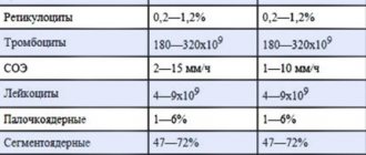
Interpretation of sputum tests, normal indicators, examples
Normally, a pulmonologist or phthisiatrician will interpret the sputum analysis. Below is a table showing the characteristics of mucus released after a cough in the absence of pathology.
After examining the sputum, the laboratory assistant fills out the appropriate form (click to enlarge).
The specified document may look slightly different. It all depends on the specific laboratory. Below are options for study forms with proposed diagnoses.
Interpretation: pink color of mucous sputum in combination with the presence of mycobacterium tuberculosis (MT+) indicates the presence of a corresponding pathology.
Explanation: given the presence of leukocytes, mucopurulent consistency and a large amount of coccal flora, the most likely diagnosis remains chronic bacterial bronchitis.
Explanation: first of all, attention should be paid to a large amount of sputum (50 ml). In combination with the abundance of leukocytes, which cover the entire field of view of the microscope, and the presence of elastic fibers, one can judge the presence of an abscess that has broken into the bronchus.
Bacterioscopic examination
Sputum testing for Mycobacterium tuberculosis (MBT) is performed on a specially stained smear. It has been established that a routine study of a stained smear for MBT gives a positive result only if the MBT content is at least 50,000 in 1 ml of sputum. It is impossible to judge the severity of the process by the number of detected MBTs.
When bacterioscopy of sputum of patients with nonspecific lung diseases can be detected:
- for pneumonia - pneumococci, Frenkel diplococci, Friedlander bacteria, streptococci, staphylococci - 100%;
- with gangrene of the lung - a spindle-shaped rod in combination with Vincent's spirochete - 80%;
- yeast-like fungi, to determine the type of which a sputum culture is necessary - 70%;
- for actinomycosis - actinomycete drusen - 100%.