Among all the various diagnostic medical procedures, ultrasound is the most common. It is this method that reveals all kinds of pathologies in the human body at an early stage. Ultrasound of the pelvic organs in women should be performed regularly. After all, their organs are more susceptible to inflammation, and diagnosis in this way allows you to identify all problems in a timely manner.
The method uses the principle of echo: a sound signal is sent to the body, reflected and returned. All this is done using a special device, which then reads the readings and identifies them. A characteristic feature of the procedure is the complete absence of contraindications to it. Ultrasound of the pelvis in women: how to prepare, what not to eat and which method to choose?
…
What is uterine ultrasound
The principle of ultrasound is based on the ability of tissues to reflect ultrasound waves differently. Sensitive sensors pick up the return signals and send them to the computer. There, the data is processed using a special program and displayed on the monitor in the form of an image. Ultrasound of the uterus is a study that helps assess the size, structure and condition of this organ and others included in the female reproductive system.
Diagnostics can be carried out as a preventative measure or for certain indications:
- cycle disruption or absence of menstruation;
- pain in the uterus or appendages;
- determining the duration of pregnancy;
- before caesarean section;
- complications after childbirth or abortion;
- suspicion of neoplasms (including cancer);
- exclusion or confirmation of pregnancy;
- infertility;
- after menopause;
- bleeding of any intensity;
- assessment of fetal development;
- pain during urination;
- ectopic pregnancy;
- purulent or bloody mucous discharge;
- discomfort during sexual intercourse;
- disruption of the reproductive system;
- uterine fibroids;
- suspicion of inflammatory processes.
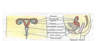
Ultrasound can be performed in several ways. The most common is transabdominal, when the sensor slides across the abdominal cavity. It can be used even in late pregnancy or for virgins.
A more informative method is transvaginal, when the sensor is inserted into the vagina. This helps to more accurately assess the condition of the uterus, its cervix, walls, as well as the fallopian tubes and ovaries. However, this method is not suitable for women from the 2nd trimester, with heavy bleeding and for virgins.
With the intrauterine method, the sensor is inserted directly into the organ. However, it is rarely used, mainly to identify tumors or pathologies of the endometrium. With transrectal ultrasound, the probe is inserted through the anus into the rectum. This allows you to examine not only the reproductive system, but also the bladder.
Indications
Ultrasound scanning, or echography, of the pelvis is carried out to identify various problems in girls and women.
Pelvic ultrasound
Usually a referral is given for an ultrasound of the pelvic organs for the following problems:
- blood or mucus when urinating;
- the appearance of pain in the abdomen, groin and lower back;
- irregular menstrual cycle in girls after 18 years of age;
- severe pain during menstruation;
- gynecological inflammatory diseases;
- in case of difficult childbirth or abortion;
- before or after surgery on the uterus and appendages;
- suspected ectopic pregnancy;
- problems with conception.
If alarming symptoms or one of the above problems appear, the gynecologist or urologist gives a referral for an examination of the ovaries with the uterus and appendages using echography. The same method is used to confirm pregnancy, examine the prostate gland, install an intrauterine device, identify malignant tumors and pathologies of the female organs , and find out the causes of infertility.
Also, women should undergo this diagnostic as a preventive measure once every 1-2 years, and after 40 years - annually, for timely identification of problems.
Preparing for an ultrasound of the uterus
The most common methods are abdominal and vaginal. Preparation before ultrasound of the ovaries and uterus depends on the chosen method of examination, the area being examined and the need to evaluate other organs. For example, with the transabominal method, the accuracy of the data is slightly reduced compared to the vaginal method, since in the second case the sensor is located much closer to the uterus. This increases the information content of the diagnosis.
If it is also necessary to check the genitourinary system, then the transrectal method can be used.
General rules for preparing for the research procedure
There are a number of general rules on how to prepare for an ultrasound of the uterus. The examination is not carried out if there are open wounds or bandages on the abdomen. Diagnosis may be delayed before gastroscopy, colonoscopy or x-ray. Ultrasound of the uterus is often performed when examining the kidneys, so the preparation should be identical. It is not recommended to drink alcohol or smoke before the examination. If you have constipation, you can relieve them with laxatives.
Is it possible to eat before an ultrasound of the uterus: it is better to eat a few hours before the procedure so that it is carried out on an empty stomach. Within 2-3 days, the patient should begin to follow a slag-free diet to eliminate and prevent gas formation. Before the ultrasound, the doctor must be notified in advance about all medications that the patient is taking. You must take cleansing wipes with you to the examination.
Before the examination, the doctor may offer a disposable diaper, but according to the rules of personal hygiene, it is better to take it with you. If the ultrasound is performed transabdominally, then a special gel is applied to the abdominal cavity. After the procedure is completed, the product can be easily removed with wet wipes or a towel. Therefore, you also need to take them with you.
With the transvaginal method (there is a photo in this article), the sensor is inserted into the vagina. For hygiene purposes, a condom is placed on the device. Usually the doctor asks patients to take it with them. To avoid injury to the film, it is better to stock up on several condoms at once.
If churchometry is prescribed, it is performed transvaginally. In this case, you need to follow the appropriate recommendations for preparing for an ultrasound of the uterus and appendages. With the rectal method, in addition to standard recommendations, the intestines must be emptied 6 hours before the procedure. If you plan to examine the fallopian tubes, then the genitals must be washed before the examination.
How to prepare for an abdominal ultrasound
Because an abdominal ultrasound uses a transducer to scan the uterus through the abdomen, proper preparation is important to obtain accurate results. For 4-5 days, all foods and drinks that contribute to gas formation are excluded from the diet:
- legumes;
- sweet;
- flour;
- onion;
- all types of cabbage;
- kvass;
- beer;
- lemonades.
Research process
The event itself is carried out very simply and quite quickly, but requires preliminary preparation, which will be discussed below. There are several types of this diagnosis, but the most common of them is transabdominal .
Preparing for an ultrasound
How to do it:
- The patient covers the couch with a diaper or towel she brought with her.
- Lies on his back and exposes his stomach and groin by raising the top of his clothing and lowering his trousers or skirt.
- The doctor lubricates the device’s sensor and the patient’s stomach with a special gel.
- A session begins, during which the doctor moves a sensor over the patient’s skin and records the necessary data that the device detects.
Any actions during the diagnosis do not cause pain (if there is no acute inflammation of the OMT), and it will take no more than 10-20 minutes. In case of transvaginal:
- The woman is freed from clothes (below the waist) and lies down on the couch.
- Feet are placed on the bottom of the couch, legs bent and spread apart.
- The doctor opens the package with the contraceptive in front of the patient and puts it on the sensor.
- Treats it with a gel that creates good conductivity.
- The sensor is inserted into the vagina.
- The device screen displays the internal organs, and the doctor records them.
If the doctor carefully and slowly inserts the sensor, then there should be no unpleasant sensations.
Transrectal is most often prescribed to men , but can also be performed on women for special indications.
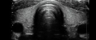
What does a pelvic ultrasound reveal?
It is carried out like this:
- The patient is exposed from the waist down.
- He lies down on the couch, turns over on his left side and presses his knees to his chest.
- The doctor treats the sensor with a solution and inserts it into the rectum.
- The device takes readings.
- At the same time as examining the internal organs, the doctor may take some material for a biopsy.
All ultrasounds of the pelvic organs in women are painless, unless the patient has acute inflammatory processes.
Important ! Transvaginal and transrectal diagnosis is carried out with a special device, which must be covered with a condom.
After the examination, based on the data obtained, the doctor makes a conclusion and makes a diagnosis. It is best to see not just a sonologist, but also a gynecologist and a urologist at the same time, then the doctor will be able to better assess the condition of the OMT and establish an accurate diagnosis.
Useful video
The specialist explains the rules for preparing for the procedure in this video.
A few hours before the examination, Espumisan or activated charcoal is taken. If there are problems with stool (constipation), a microenema is done (for example, “ Microlax ”) or a candle is placed in the anus after eating. In 1-1.5 hours you need to drink a liter of still water or compote. A full bladder helps improve the image and provide more accurate data.
How to prepare for a transvaginal ultrasound
Before undergoing an ultrasound of the uterus using the transvaginal method, preparation is not always required. However, it won’t hurt, then the results will be more accurate. Ultrasound is usually done on an empty stomach, so your last meal should be a few hours before the test. However, this condition is optional. Before the procedure, you need to drink a liter of still water. This will improve signal patency and allow for more detailed visualization of the uterus.
Also, a few days before the procedure, it is advisable to start following a diet, eliminating from the diet everything that causes flatulence. Or, instead, you need to take medications that prevent gas formation.
Why is ultrasound performed only on a full bladder?
An ultrasound scan is done of a full bladder - when it is maximally full, the inside is seen better, since in an empty state it is wrinkled and difficult to see by a specialist.
Before an ultrasound of the bladder, it is necessary to fill it with liquid, then the walls will straighten and stretch without unnecessary folds. When the organ is full, it can bring the intestinal loops closer to the abdominal cavity, to its front part.
Filling of the organ should occur in natural conditions and at a pace, but in some cases the patient may not know about the need for an ultrasound, or if the procedure is emergency and there is no time to prepare according to the rules. In this case, the bladder needs to be filled quickly and efficiently.
To do this, just drink a liter of clean water an hour before the intended procedure. Instead of water, it is allowed to use tea or fruit drink, juice or herbal infusions. Experts do not recommend drinking milk or highly carbonated drinks to ensure the reliability of the examination results.
The bladder of all people is different in volume, and if after an hour the patient does not have the urge to deurinate, then it is worth drinking another half liter of liquid. It is important to prevent overflow; if the urge to urinate appears long before the procedure, you can visit the toilet, but then be sure to replenish the lost volume of water.
As a rule, early urination indicates excellent functioning of the kidneys, which produce urine. Experts advise not to empty your bladder for several hours before the ultrasound, so as not to drink excess water and not put a strain on your kidneys. This is only possible if the patient has a good knowledge of his excretory system.
Preparing for an ultrasound during pregnancy
How to prepare for an ultrasound of the uterus in women while pregnant: it depends on the period. In the 1st trimester (approximately 10-13 weeks), the transvaginal method is used. It helps to more accurately visualize the fetus and the condition of the uterus. Preparation is carried out in several stages:
- For 5 days, all foods and drinks that cause flatulence and fermentation in the intestines are excluded from the diet.
- You need to fill your bladder 30-40 minutes before the examination. To do this, drink 0.5 liters of still water and do not go to the toilet before scanning.
- The examination is usually scheduled for the morning, but glycerin enemas are not recommended for pregnant women. If you try to cleanse your intestines this way, overexertion can cause a miscarriage. It's better to just skip breakfast.
Starting from the second trimester, only transabdominal ultrasound is used, since during this period the uterus is greatly enlarged, the fetus becomes larger and they can be seen even through the abdominal wall and without any preparation. The use of a vaginal sensor can only be to assess the condition of the uterine cervix.
Ultrasound is a painless and safe procedure and is performed very quickly. It allows you to identify many gynecological diseases. Ultrasound examination does not require specific preparation; all the recommendations described above are standard for many types of diagnostics.
How to prepare?
First of all, this is compliance with the necessary diet. Before going for the procedure, you should eliminate foods that can cause gas formation from your diet for 2–3 days. These include dairy and legume dishes, yeast bread and generally all flour products, fresh fruits and vegetables, nuts, coffee, and sparkling water. You can eat lean boiled meat and broths, boiled eggs and fish, dried bread. In addition, before going for an ultrasound, it is advisable to cleanse the intestines with an enema. If the body is prone to gas formation, regardless of the foods consumed, then it is also recommended to take drugs such as espumizan or filtrum.
As for the question of whether you need to drink water before an ultrasound scan of the kidneys, which is often asked by patients, the answer is clear. Yes, it is necessary, even mandatory! During the study, there should be at least 500–700 ml of fluid in the bladder. That is, it should not be overfilled.
Water enlarges organs for the procedure
What you can and shouldn't drink
It is best to drink water before the bladder ultrasound procedure. Also allowed:
- fruit drink;
- tea;
- juice.
Drinks prohibited by doctors are soda and milk, as they contribute to the formation of gases in the intestines, which complicates the examination.
Why is liquid needed?
Often doctors, when referring a patient for an ultrasound, warn that they need to drink 1 liter of water before the procedure. But not everyone explains why this is necessary.
Water is necessary for the bladder to enlarge. This makes the surrounding organs clear on the screen. And the research turns out to be more reliable.
Water is especially needed before diagnosing the condition of the urinary tract. With its help, the presence of stones, tumors, pathological changes in the mucous membrane is better visible, and the condition of the genitourinary organs can be accurately determined.
It is also recommended to drink liquid before examining the uterus and ovaries. As the bladder grows larger, it shifts these organs, making them easier to see on the screen.
During pregnancy, such preparation is necessary in the initial stages for the same reason.
When should you drink?
To keep your bladder full during the procedure, start drinking 1-2 hours before it. First you need to urinate, otherwise it will be impossible to endure the urge for such a time. There is no point in drinking near the office before the procedure itself - the water will not have time to penetrate the bladder.
If you need a little water, for example, during pregnancy, you can drink a half-liter bottle 40 minutes before the ultrasound. After all, the essence of drinking liquid this way is so that it has time to penetrate the bladder and fill it.
Features and advantages of ultrasound diagnostics
This is one of the most accurate and safe diagnostic methods used in gynecology. The principle here is that the internal organs reflect the sound signal directed by the sensors. With the help of modern technology, the reflected impulse is transformed into a symbolic image. It is interpreted by the doctor conducting the diagnosis.
The use of ultrasound makes it possible to study the condition of the necessary organs over time, which helps to make accurate diagnostic conclusions. During an ultrasound, the doctor performing the manipulation assesses the condition of the tissues being examined, comparing the obtained indicators with the standards. For diagnostic procedures, the following data is used:
| Index | Interpretation |
| Form | If the natural contours of an organ are changed, this may indicate structural defects - congenital or acquired. |
| Dimensions | Increased size indicators of the organ indicate inflammation, decreased ones indicate fibrous damage. |
| Wall thickness | An increase in this indicator may be a sign of the presence of malignant tumors and inflammation. |
| Structure | A heterogeneous structure indicates fibrosis. |
| Presence of seals | Swellings and stones are detected. |
| Echogenicity | The density of tissue structures increases in the presence of pathological changes. |
To confirm the ultrasound diagnosis, additional tests are required.
The advantages of OMT ultrasound are that it is highly informative, allows for pain-free manipulation, and the ability to detect pathological changes in the initial stages of the disease. During research activities, the skin is not injured and the administration of pharmaceuticals is not required. An ultrasound scan takes little time and does not create a feeling of discomfort. The entire ultrasound examination process lasts no more than 20 minutes.
Ultrasound can be used to perform diagnostics as many times as needed.
Ultrasound is performed if there are no direct contraindications. There are few of them: an allergy to latex (during transvaginal ultrasound, when a contraceptive is put on the nozzle) or open injuries to the skin.