Ultrasound is a non-traumatic, safe and informative method for diagnosing many diseases. Ultrasound of the esophagus, in comparison with another common research method - gastroscopy - does not cause discomfort in the subject, does not require special preparation, and has no contraindications. Therefore, the method is widely used in gastroenterology, especially in the treatment of children. However, it is not without its drawbacks. Read the article about how ultrasound of the esophagus is performed, when the use of the method is justified, and in which cases the results may be questionable.
Description of the procedure
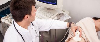
Ultrasound is an informative, harmless method of instrumental examination. This type of examination of the esophagus does not take much time: its duration is about 15 minutes. The main advantage of ultrasound examination is the resolving property of the equipment, which allows one to examine all the features of organs and their sizes. Using ultrasound of the esophagus and stomach, the following examinations are performed:
- study the walls of the stomach and intestines, tissue structure;
- detect motility disorders (movement of food in the tract);
- determine the condition of the vessels;
- lymph nodes are analyzed.
What is the method
Many people have heard about such a method of examining the body as ultrasound of the esophagus, but not everyone can say anything specifically about it. The essence of the method is that it is examined using a high-frequency sound wave propagating throughout the entire space of the internal organ.
Ultrasound is reflected from the boundaries of the organ and displays the resulting image on the monitor. Only a specialist with the required qualifications can decipher the result. He sees what changes and deformations are present or absent in the human body. Nowadays, many medical institutions have 3- or 4D imaging technology. It is capable of displaying a clear picture in color on the monitor. Thus, doctors have the opportunity to accurately assess the patient’s condition and prescribe the necessary treatment regimen.
Indications
To identify some types of diseases, it is impossible to do without ultrasound. An ultrasound examination will determine the stage of the disease. Using this method, you can quickly identify the symptoms and characteristics of the disease, make a diagnosis, and, therefore, prescribe appropriate drug therapy. The organs of the digestive system are checked using an ultrasound examination if there is a suspicion of formations in the esophagus. In such cases, patients may complain of abdominal pain. This method of research helps to make a thorough analysis of inflammatory processes in the colon, to study erosions and ulcerative lesions of the mucous membrane of the gastrointestinal tract. In addition, ultrasound can reveal the location of polyps.
The specialist prescribes the procedure if there are suspicions of the following pathologies:
- neoplasm in the esophagus;
- peptic ulcer of the stomach or duodenum;
- tumor lesions in the stomach;
- hernia development.
Also, ailments that can be diagnosed using the ultrasound diagnostic method include:
- gastroduodenitis;
- polyps;
- any ulcerative lesions;
- cancerous tumors.
Ultrasound of the esophagus should also be done as a preventive diagnosis. In addition, it is sometimes possible for stomach acid to enter this organ. As a rule, this indicates the development of an infectious disease. To determine it, doctors prescribe such a study. Once the disease is diagnosed, doctors will prescribe appropriate therapy.
An organ ultrasound in a small child is primarily necessary to diagnose developmental anomalies . The method is widely used for the initial diagnosis of various diseases in a child, since it is safe for the child’s body.
What to take with you?
Parents must take a referral from the attending physician and an insurance policy (if any) with them to the ultrasound examination. The results of previous ultrasound, computed tomography or magnetic resonance imaging will also be useful.
This will allow the doctor who carries out the diagnosis to make comparisons, as well as to look for a specific pathology in a more targeted manner. When a child is in the hospital, you need to take a medical history with you, in other cases an outpatient card (in written or electronic form).
It is important to consider that the child will be hungry during the study. And immediately after its completion it will be nice to have a snack. Therefore, it is recommended to take with you several sandwiches, biscuits or bananas and warm tea in a thermos. If we are talking about an infant, you should think about whether you will need to breastfeed him or give him ready-made formula.
If the examination is carried out on a baby, then to create more comfortable conditions it is often advised to take with you several favorite toys . They help create a comfortable atmosphere that has a beneficial effect on the child’s psychological mood.
It is advisable to take with you several disposable towels (they may not be available in public medical institutions) and a diaper that needs to be laid on the couch.
Preparation for the procedure
A couple of days before the diagnostic procedure, you should adhere to a strict dietary regimen. Before the study, you should not smoke or drink alcohol. Ultrasound is performed on an empty stomach, with the exception of half a glass of tea and a small cracker. Such preparation will allow doctors to make a more accurate diagnosis. In addition, patients are prohibited from eating the following foods before an ultrasound:
- peas;
- bread made from rye flour;
- cabbage dishes;
- carbonated drinks;
- fresh fruits and vegetables.
Ultrasound of the esophagus. Preparation and recommendations before undergoing an ultrasound examination
You should know that preparation for an ultrasound is necessary in all cases, regardless of which organ is being examined. At its core, the recommendations that a patient must follow before an ultrasound are quite simple. But they should not be neglected, since failure to comply with these instructions may distort the results of the examination. And this will lead to an inaccurate diagnosis.
There are several preparatory stages:
- First of all, two days before the expected time of ultrasound examination of the gastrointestinal tract, the patient must begin to follow a special diet. It lies in the fact that foods that can cause gas and flatulence are excluded from his diet. The list of these food products includes: beans, peas, cabbage, black bread, carbonated and alcoholic drinks, raw vegetables, fruits.
- There is no need to eat before the ultrasound. The time that should be limited for eating is from 10 to 12 hours minimum. The patient should look at what time of day the procedure is scheduled for. If it is early morning hours, then you need to finish eating at 20:00. When an ultrasound is scheduled for the daytime or evening, you will have to be patient and not eat throughout the day. This rule does not apply to children. If the child is older, you can limit his food intake at least 4 hours before the examination time. As for children who are in infancy, they should not be limited in feeding and can come for an ultrasound at any time.
- Smoking is also prohibited before the ultrasound. This is due to the fact that the examination must be carried out on a completely empty stomach. You should also not drink anything. In this case there may be exceptions. They are among those patients who cannot tolerate fasting, and it is they who experience stomach pain. Such people are allowed to drink some warm sweet tea. Its dose should not exceed half a glass. You are also allowed to eat one cookie. It should be dry.
- If an ultrasound examination is done to study the small intestine, then the patient must additionally cleanse it.
How is the procedure performed?
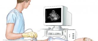
Before the ultrasound begins, the patient must lie down, then a special gel is applied to his stomach. The doctor moves the sensor and examines the organs on the screen, after which the person drinks a small amount of water - this will help the doctor see how it penetrates into the stomach. In this way, the esophagus section can be examined. The diagnostic procedure takes approximately half an hour. During decoding of the results, the echogram is described, pathologies are detected or not, and the person’s health status is determined.
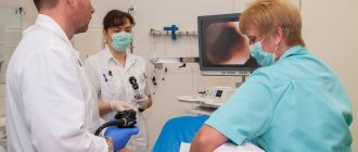
There are several methods for ultrasound examination of an organ. For example, we are talking about the use of fluoroscopy and gastroscopy methods. The latter method involves inserting a special sensor through the pharynx into the esophagus. Thanks to such manipulations, it is possible to determine the disease at the initial stage, when the patient does not experience pain and does not have other unpleasant symptoms. The doctor records changes in the structure of tissues. An ultrasound is performed in the morning, on an empty stomach.
Kinds
Ultrasound includes a number of methods, including:
- Transdermal. During the procedure, a special sensor is located on the surface of the patient's skin on which the gel is applied. The sensor is localized in the cervical region and upper abdominal cavity. Due to this position of the device, different parts of the organ can be visualized. The technique is often used to diagnose diseases in a child.
- Intraesophageal. This method involves inserting a sensor through the lumen in the esophagus, due to which changes in the tissue structures of the organ can be determined. The technique is usually used to diagnose adult patients, since there is a risk of injury to the child. This method is also used to diagnose the heart.
- Using a water-siphon test. This technique allows you to determine the motility of organs. When performing a diagnosis, a person must drink a certain amount of liquid. It, in turn, contrasts in the stomach cavity.
- Endoultrasound. The endoscopic procedure allows you to view organs in detail. EndoUS helps to see pathological changes that are inaccessible to conventional ultrasound. EndoUS is performed using an endoscope with a special optical device and a sensor located at the tip of the device.
The doctor determines the type of diagnostic technique based on the patient’s test results and the characteristics of his anatomy. Thus, you can choose the appropriate method for each case.
Ultrasound of the stomach with water-siphon test for a child
Ultrasound of the stomach with a load on the child is another name for this procedure. With this type of ultrasound, the child is in a sitting or standing position. The doctor collects primary data regarding the pylorus of the stomach using a sensor. Then the patient drinks unsweetened tea and five minutes later a planometry of the pylorus is performed. It is necessary to drink the liquid so that the doctor can calculate the time it takes for the gastric sphincter to return to the state it was before drinking the liquid.
results
It should be noted that ultrasound cannot be a complete replacement for endoscopy or radiography. Nevertheless, this method allows doctors to obtain information regarding the condition of tissues, organ contours, and monitor the development of pathologies. What do ultrasound results show? The method helps to study the peculiarities of organ functioning and reflexivity. The results will provide information regarding the thickness of the gastric walls, the presence of inflammation, and tumors. The technique will make it possible to reliably determine pathologies, blood flow characteristics, and study tissue structures in detail. This diagnostic method will help answer many questions and identify many ailments.
Norms and pathologies
Normally, sections of the esophagus are visible as oval-shaped formations. Its walls are 6 millimeters thick. A glass of water can be evacuated in approximately 20 minutes. The walls must be uniform, the condition of the tissues surrounding the esophagus must also remain normal. In the presence of organ pathologies, reflux is sometimes diagnosed. In rare cases, cysts are found. The formation of hernias and other pathologies is possible.
In a healthy child, the thickness of the wall of the section in the esophagus is several millimeters. In sick babies, polyps, small hernias, disruption of gastric motility, obstruction in the esophagus, and other diseases are detected.
Reviews and features
Most people consider ultrasound an effective technique. At the same time, reviews from the majority of patients who underwent the procedure show that some do not fully understand the features of this diagnostic method. When undergoing an examination for the first time, people are interested in what an ultrasound should show. Those who have completed it claim that the method helps to identify many things. Experts also consider the technique to be effective, since pathological processes can be detected in the initial stages. At the same time, the method is simple and safe for the majority of subjects.