The female body is a unique creation that undergoes many transformations throughout life. Any, even minor, disruption of its activity can lead to irreversible consequences, such as, for example, infertility, which deprives the incomparable joy of motherhood.
To avoid such a sad turn of events, it is recommended to undergo a preventive examination, which necessarily includes an ultrasound of the female organs of the reproductive and genitourinary systems. This will allow many pathologies to be recognized early and appropriate measures taken.
Characteristics of gynecological ultrasound
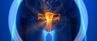
Gynecological ultrasound is a diagnostic method that involves examining the organs of the female reproductive system located in the pelvic area. Its scope also includes studying the condition of the fetus during pregnancy.
The method is based on the ability of ultrasound to penetrate body tissues and cause a certain reaction. The reflected waves are recorded by a device specially designed for this purpose, transforming into a visual image. The latter is displayed on the monitor and allows the doctor to obtain comprehensive information about the object of study.
Ultrasound examination is considered one of the safest methods. The waves produced by the sensor do not cause any harm to the body. This is why the method is so widely used in obstetrics. Another huge advantage of this type of diagnostics is its high information content. Another advantage is painlessness and affordable price.
Ultrasound of the uterus and appendages: rules of preparation, how they are done, norms and deviations
It is recommended that every woman undergo an ultrasound scan of the uterus and appendages at least once every 6 months.
This procedure is a safe, painless and informative diagnostic method that allows you to identify possible pathologies in the pelvic organs. There are no age restrictions or contraindications for the examination. As a result of the diagnosis, the doctor receives data on the condition of the uterus and appendages (ovaries, fallopian tubes), their shape, size and structure.
Based on the results obtained, the specialist can make the correct diagnosis and select an effective treatment regimen.
- Pathologies detected by ultrasound
- about ultrasound examination of the uterus and appendages
- Uterine fibroids. This is a benign formation from the muscular layer of the organ. There are several fibroid nodes, their sizes vary from a few millimeters to 50 cm. Ultrasound determines the number, size, location and growth dynamics of fibroids.
- Hyperplasia and endometrial polyps. Hyperplasia is characterized by diffuse or focal excessive growth of the endometrium. Its thickness in this disease is more than 7 mm in the first phase of the cycle. Polyps form from focal hyperplasia. There may be several of them, the size of the formations can reach several centimeters. Using ultrasound, these conditions are assessed and the necessary treatment is prescribed.
- Cysts and tumors of the ovaries. Cysts can be functional or pathological. The former appear and disappear again during the cycle without any intervention. Pathological cysts require treatment. Ovarian tumors may indicate oncology. Therefore, decoding an ultrasound image of a neoplasm in the ovary is a responsible task that is solved by more than one specialist.
- Apoplexy or rupture of the ovary. This is a dangerous condition that occurs spontaneously, for example as a result of injury. Ultrasound allows you to distinguish apoplexy from ectopic pregnancy or appendicitis, evaluate bleeding, and make the correct diagnosis.
- Tuboovarian formations - hydrosalpings. Appear against the background of inflammation in the pelvic organs. Ultrasound of the appendages and uterus not only reveals this abnormal condition, but also helps eliminate it using hydrotubation of obstructed fallopian tubes under ultrasound guidance.
- Pathologies associated with pregnancy. It could be an ectopic pregnancy, a frozen pregnancy, or a hydatidiform mole. A placental polyp may be detected: if an ultrasound of the uterus and ovaries after a cesarean section, natural birth or abortion reveals a remnant of placental tissue in the organ cavity, its urgent removal is necessary.
Ultrasound of internal female organs: how to prepare?
How to prepare for an ultrasound of the female genital organs? If we are talking about intravaginal ultrasound examination, it is desirable that such diagnostics be carried out on an empty stomach, and the intestines and bladder are empty. Food does not need to be taken eight to twelve hours before diagnosis.
How to prepare for an ultrasound of the female organs? What else do you need to know? Whenever possible, you need to free the intestines as much as possible from gases and feces. This must be done so that the ultrasound can fully pass through and the desired image is displayed on the screen. This means that you need to start preparing three to four days before this ultrasound diagnosis.
Ultrasound of the internal female genital organs: how to prepare to exclude gas formation? On the indicated days, you should remove from the menu those foods that are difficult to digest.
The same applies to fatty foods, as well as those foods that contribute to increased gas formation.
These are fresh vegetables and fruits, which contain a large amount of fiber, legumes, whole milk, carbonated drinks, brown bread, coffee and confectionery products that are high in calories (for example, cakes and pastries).
Get a free doctor's consultation
Ultrasound like a woman: how to prepare if the patient has a constant tendency to gas? In this case, you can drink special medicines, such as Festal, Enzistal, Creon, Panzinorm, Karbolen, Espumisan, fennel or chamomile infusion. If constipation has become a common occurrence for the patient, it is recommended to take laxatives.
It is advisable to carry out a cleansing enema, since the air that enters along with the water accumulates in the lower intestines. Types of ultrasound examination are divided into transvaginal and transabdominal examination. Types of female ultrasound diagnostics are divided only into the variety of accesses.
Ultrasound of internal female organs: how to prepare? Take with you a sterile diaper and a condom (for the sensor).
If the ultrasound examination will be carried out using the transabdominal method (that is, through the anterior wall of the peritoneum), then it is necessary to drink from a liter to one and a half liters of clean water an hour before it to ensure normal filling of the bladder.
Thus, an ultrasound of the female organs (preparation for the procedure) does not pose any difficulties for the woman who will have to undergo it. You just need to follow the recommendations given above, and the procedure will go correctly. If you have any serious diseases or pathologies in the body, you should consult your doctor in advance.
The body of the fair sex is a special mechanism in its structure, characterized by excessive fragility. That is why it requires increased attention and timely care. When situations occur that were previously not characteristic of the nature of a particular organism, the question arises of what to do.
In this case, you need to promptly seek help from a specialized ultrasound diagnostic center and make an appointment. Thanks to the research results, it is possible to completely cure ailments of the reproductive system in women.
What does an ultrasound of the uterus and appendages show?
When conducting a diagnostic study, a specialist may detect the following diseases:
Reliable results of decoding an ultrasound of the uterus and its appendages require a specialist to have excellent knowledge of the anatomy and physiology of the female body, as well as the clinical situation. The results of the study can only be assessed by a person with a medical education. You cannot independently decipher the ultrasound protocol and determine the diagnosis, as some women do.
The appendages are the formations on the side of the uterus. Echo-positive inclusions in their structure contain echo-negative elements of round or oval shape.
Ultrasound examination of the uterus and appendages is performed to diagnose physiological and pathological conditions. The method is indispensable for identifying diseases in girls before sexual activity begins.
Using ultrasound, you can observe the maturation of the follicle and confirm ovulation and intrauterine pregnancy. Ultrasound allows you to identify the threat of spontaneous abortion and prescribe adequate therapy to prevent miscarriage.
Important! During pregnancy, screening ultrasounds are performed to assess fetal development.
Typically, the first ultrasound is performed after the onset of puberty. Diagnostics helps to identify pathologies of uterine development by determining the size of the muscular organ and appendages. The specialist also evaluates their structure, position and functioning.
Anomalies in the development of the uterus often lead to infertility. Ultrasound can reveal the saddle-shaped, bicornuate, horseshoe-shaped nature of the muscular organ, and the presence of a septum. In adolescence, a specialist sometimes diagnoses ductal atresia, complete and incomplete duplication of the uterus.
When performing an ultrasound of the appendages and uterus, the following pathologies are determined:
Source: https://PlastikaPlus.ru/diagnostika/kak-delayut-uzi-matki.html
What does ultrasound show?
Ultrasound in gynecology allows us to identify pathological processes in the organs of the female reproductive system at the earliest stages. Also, using the method, you can learn about the anatomical features of the ovaries, uterus, and appendages. The method is successfully used not only at the diagnostic stage, but also during treatment to evaluate the effectiveness of the therapy used. Among the diseases that can be detected using ultrasound:
- single cysts;
- polycystic disease;
- torsion of the cyst pedicle;
- uterine fibroids;
- endometriosis;
- inflammation of the uterus, appendages;
- pathological accumulation of fluid in the pelvis;
- malignant tumors of the female reproductive system.
It is difficult to assess the role of ultrasound diagnostics in obstetrics. This method is able to detect ectopic pregnancy and the threat of miscarriage in the early stages. A routine ultrasound examination shows how the fetus is developing, whether it is experiencing hypoxia, or whether it has genetic or other pathologies. Ultrasound also allows you to monitor a woman’s condition after childbirth (this is especially true if it was difficult), miscarriages, or abortions.
Ultrasound in gynecology: indications for undergoing
Ultrasound examination (ultrasound) is distinguished by the high accuracy of the results obtained and the absence of harm to the body. The operation of ultrasound is based on the principle of a sound wave, due to which an idea of the current state of the body is formed.
During the examination of the female reproductive system, the uterus, fallopian tubes, and ovaries are subject to diagnosis. In addition, the bladder and rectum are examined. An ultrasound scan determines the presence of a possible pregnancy.
As a rule, a referral for an ultrasound scan is issued in the following cases:
- disturbances or failure of the menstrual cycle;
- presence of changes in menstrual flow;
- increased pain as a result of menstruation;
- suspicions of ovarian dysfunction, the appearance of neoplasms, endometriosis, inflammatory processes;
- the appearance of uncharacteristic discharge with a simultaneous appearance in the lower abdomen;
- tracking existing abnormalities of the genital internal organs;
- identifying the root causes of infertility;
- diagnosing urolithiasis;
- prescribing oral contraceptive methods;
- problems with urination;
- after gynecological surgery;
- prevention and prevention of diseases.
In most cases, ultrasound is used not only to diagnose existing gynecological diseases, but also to monitor the progress of pregnancy, as well as monitor the effectiveness of treatment and the results of its implementation.
| Code | Name of service | Price |
| 18.14 | Ultrasound of the pelvic organs in women (transabdominal) | 1650.00 |
| 18.15 | Ultrasound of the pelvic organs in women (transvaginal) | 1750.00 |
| 18.16 | Ultrasound of the pelvic organs in women (transabdominal and transvaginal) | 2000.00 |
| 18.17 | Ultrasound of the ovaries with follicle monitoring (folliculometry) | 1000.00 |
| 18.18 | Ultrasound of the 1st trimester of pregnancy (up to 13 weeks) | 1800.00 |
| 18.19 | Ultrasound 2nd trimester of pregnancy (14-28 weeks) | 2500.00 |
| 18.20 | Ultrasound of the 3rd trimester of pregnancy (from 29 weeks) | 2700.00 |
| 18.20.1 | Ultrasound Screening 1st trimester of pregnancy | 2200.00 |
| 18.21 | Fetal blood flow Doppler | 1500.00 |
| 18.22 | Ultrasound during multiple pregnancy (twins) in the first trimester | 3100.00 |
| 18.23 | Ultrasound during multiple pregnancy (twins) in the 2nd and 3rd trimester | 4500.00 |
| 18.24 | Determining the sex of the child | 600.00 |
Indications for use
Diagnostics using ultrasound is prescribed for women who suspect the pathologies listed above (malignant and benign tumors, inflammation, etc.). Indications for the study may include:
- pain in the lower abdomen;
- bleeding from the vagina;
- pathological discharge from the genital tract;
- menstrual irregularities;
- discomfort during intercourse or pain after it.
Also, the reason for ordering an examination in many cases is a pregnancy that does not occur for a long time. This diagnostic method helps to determine the causes of a woman’s infertility. Expectant mothers undergo routine ultrasound examinations at 12, 20 and 30 weeks. If there are alarming symptoms, the study is carried out at any stage of pregnancy.
Regardless of the presence or absence of signs of disease, women are recommended to undergo ultrasound during regular medical examinations (once a year) or six months. Many pathologies are asymptomatic, making themselves felt when the disease has progressed very far. This is especially scary when it comes to malignant tumors. Ultrasound diagnostics will allow you to identify the disease “in the bud” and begin treatment in a timely manner, which significantly increases the chances of success.
Standard indicators on ultrasound
To begin with, the transcript of the ultrasound of the female genital organs indicates the size of the uterus. The standard distance between the fundus of the uterus and the internal os of the cervix is 5-8 cm. On average, this distance in a healthy nulliparous woman is 60-71 mm. In female representatives who have given birth, the uterus enlarges slightly; in this case, its size is determined by the number of births.
After studying the ultrasound readings, the doctor can draw a conclusion about the woman’s health
The thickness of the uterus should be 30-40 mm, and the width - 45-60 mm. A few years after reproductive function fades away, the uterus may decrease in size to 40-50 mm. When assessing the health of a woman’s genital organs, it is important to take into account the location of the uterus.
The normal location is in the center of the pelvis with a slight deviation towards the peritoneum. Such a position in the act of research is designated as “anteflexio”. “Retroflexio” is a physiological anomaly in the location of the uterus (the uterus deviates backwards), namely “bending”. The term “lateroflexio” refers to the deviation of the body of the uterus in relation to the center of the body.
When assessing the location of the uterine body, it is necessary to take into account that due to a full bladder, it may deviate slightly from the norm.
Condition of the appendages
The cervix on the echogram is determined as a formation of 2-3 cm, in the shape of a cylinder, the echogenicity is similar to the uterus. Normally, the width of the cervical canal should be 0.3-0.4 cm. The ovaries on an ultrasound image are visualized as an oval-shaped formation located on both sides near the uterus. The ovaries should be 27-37 mm in length, 21-29 mm in width, and 17-21 mm in thickness.
During an ultrasound, the doctor examines the ovaries very carefully
The ovaries can vary in size, since when the follicles grow, the ovaries also enlarge. When a dominant follicle is released, which is determined during the first phase of the cycle and continues to grow vigorously until days 12-14 of the cycle, the others become smaller again, and the ovaries return to normal size.
By the time of ovulation, follicles can grow to 15-29 mm, so they can be easily identified by ultrasound examination. When visually assessed by size, one ovary should be no more than 1/2 the size of the uterus in width. It is impossible to determine normal sized fallopian tubes using ultrasound. At the end of ovulation, the corpus luteum, a temporary hormone-producing gland, begins to form, the main purpose of which is to ensure implantation of the embryo and maintain pregnancy.
The corpus luteum is visualized as a small formation with a heterogeneous thick wall and fluid inside.
The endometrium in the uterus during the first days of the cycle is determined in the form of a heterogeneous structure with different thicknesses (3-8 mm). By the end of menstruation (4-5 days of the cycle), the endometrium is 2-4 mm thick, so it is almost invisible on ultrasound. During the early stage of proliferation (6-7 days of the cycle), a slight increase in the size of the endometrial layer is determined up to 6-9 mm, and in parallel the echogenicity decreases.
The doctor pays special attention to the condition of the endometrium
At the same time, it is easy to determine the emerging thin echo-negative contour up to 0.1 cm in thickness. By day 10, the endometrium increases in thickness to 10 mm. During the secretory phase (15-27 days), and during menstruation, the endometrial layer thickens significantly (in some cases up to 15 mm), this is visualized on an ultrasound image as a thickened reflective surface inside the uterus.
It should be noted that the detected corpus luteum and thickened endometrial layer in the first days of menstruation, if there is no embryonic egg in the uterus, may indirectly indicate an ongoing ectopic pregnancy.
Types of gynecological ultrasound
There are three main classifications of ultrasound of the female genital organs.
The first is based on the specifics of the conduct. From this point of view, ultrasound is divided into:
- Transabdominal is most often practiced. Provides for the passage of waves through the peritoneal wall. The sensor is placed in the lower abdomen. It does not apply if there is damage to this area of the skin (abrasions, scratches, purulent lesions, burns), as well as obesity, which is an obstacle to the passage of waves and can significantly distort the picture.
- Transvaginal involves inserting a sensor with a condom on it into the woman’s vagina. It is considered more informative than transabdominal. It is not carried out for girls who have not yet had sexual experience. It is often used in obstetrics in the early stages of pregnancy, when it is impossible to see the fetus through the abdominal wall. The method requires maximum caution, as it can provoke a miscarriage.
- Transrectal involves inserting a thin sensor into the anus. It is not performed on women who do not have a rectum, or in the presence of inflammatory processes in this area. This type is indicated for virgins when transabdominal ultrasound is for some reason impossible or impractical.
Taking into account the condition of the woman being examined, three more types of ultrasound diagnostics are distinguished:
- done outside of pregnancy, it involves identifying various pathologies of the genital area;
- carried out during the period of bearing a child, provides control over its intrauterine development;
- The purpose of folliculometry is to examine exclusively the ovaries.
The third classification is based on the features of the resulting image. It can be two-dimensional (2D), three-dimensional static (3D), and four-dimensional dynamic (4D).
Reasons for prescribing an ultrasound
Ultrasound examination is a fast, convenient and inexpensive research method that allows you to see the exact structure of the female organs of the reproductive system and the tissues surrounding them, and determine even a slight deviation from the norm or pathology.
The following manifestations may be the reason for referral for an ultrasound examination of the female organs:
- pain in the lower abdomen;
- pregnancy, including in the early stages;
- diagnosis of the causes of infertility;
- diagnosis of urological diseases, identifying the cause of problems with urination;
- menstrual irregularities, too little or too much discharge;
- taking hormonal medications, having an intrauterine device;
- detection of neoplasms in the pelvic organs;
- diagnosis of inflammatory processes in the female reproductive system.
There are three types of gynecological ultrasound: transvaginal, transabdominal and transrectal. How to prepare for a gynecological ultrasound, taking into account the method of conducting it?
Optimal timing of ultrasound
Ultrasound examination in gynecology and obstetrics can be emergency or planned. The first is carried out in case of open bleeding, severe pain, suspicion of an ectopic pregnancy, and in other situations when you cannot hesitate.
The doctor prescribes the optimal timing of routine diagnostics individually. They depend, first of all, on the purpose of the study. Typically, an ultrasound examination is performed in the first half of the menstrual cycle (approximately the third to fifth day after the end of menstruation).
This is explained by the fact that the follicle in the ovary has not yet matured, which can later be easily confused with a cyst. In addition, at this stage, the endometrial layer is thin and cannot cover the neoplasms in the uterus.
There are situations when, on the contrary, it is better to do an ultrasound at the end of the cycle. Among them are suspicions of certain types of fibroids, which are difficult to discern at other times. If the purpose of the study is to assess the “performance” of the ovaries, ultrasound is performed three times – on days 8-10 of the cycle, then on days 14-15, and on days 23-24. The first ultrasound examination during pregnancy can be done as early as the third or fourth week.
Ovarian diseases
Problems of the uterus and ovaries are often ignored by many women. Meanwhile, ovarian cysts can indicate a hormonal imbalance, which in most cases leads to infertility. Such a disease is polycystic ovary syndrome, which is clearly visible on ultrasound and indicates large-scale hormonal disorders. Ultrasound data is performed at the end of the menstrual cycle on the fifth or seventh day. In addition to cysts, examination using ultrasound waves will help identify tumors, both benign and cancerous, as well as inflammation of the ovaries.
How to prepare for the examination?
Preparation for an ultrasound of the female organs largely depends on what technique will be used. All three types (transabdominal examination, transvaginal and transrectal) require a special diet for several days before diagnosis. The goal of nutritional correction is to reduce the risk of flatulence. Gases in the intestines can distort the picture during the study. It is recommended to avoid:
- fat;
- sweet;
- flour;
- cabbage;
- radishes;
- mushrooms;
- legumes;
- sparkling water;
- beer.
To improve the digestive process, you can take enzyme-based medications. These include Creon, Festal. To reduce gas formation, Espumisan and chamomile tea are indicated. Women who suffer from constipation can take a laxative.
Increased attention should be paid to the condition of your intestines before transrectal ultrasound. The last meal should be at least 5-6 hours before the procedure. The night before you need to do a cleansing enema. When using the other two types, it is not necessary to greatly disrupt your diet, but it is better to avoid a large breakfast before the examination.
Transvaginal and transrectal ultrasound are done on a full bladder. This way the picture turns out clearer and more informative. An hour before the procedure, you need to drink about a liter of water and avoid going to the toilet. If an ultrasound is planned with the insertion of a sensor into the vagina, the bladder, on the contrary, should be emptied before the examination.
Techniques
The exact method in which the study will be conducted depends on the indications, contraindications and age characteristics of females. The main types of ultrasound of the genitourinary system that are used for women are transabdominal, transvaginal and transrectal. Despite the common points, these techniques also have some differences that patients should take into account when preparing for the procedure.
Transabdominal ultrasound
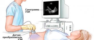
The simplest method used in most cases. Its essence is to study the genitourinary system in women with the usual movement of an ultrasound emitter in the lower abdomen. To improve contact with the skin and minimize friction, the diagnostician applies a special gel to the emitter and then examines the projections of the internal organs of the small pelvis.
Performing a transabdominal pelvic ultrasound
In order for the results to be as accurate as possible, all of the above methods require special preparation, consisting of dietary restrictions and proper filling of the bladder before the procedure. Dietary restrictions look like excluding from the diet foods that increase gas formation in the intestines, since gas bubbles can be mistaken for cysts or tumors.
Normal pelvic ultrasound in women
Prohibited foods include fatty cheeses, meats, fish and broths made from them, raw fruits and vegetables, spicy, salty, fried foods, smoked foods, spices, dairy and bakery products, and sweets.
Alcohol, carbonated drinks and water should be avoided, and for several hours before the procedure you should not smoke, chew gum or suck hard candy. Your menu 3-4 days before the study must be composed of lean meats, poultry, fish and first courses based on them, boiled vegetables, porridges - buckwheat, rice, oatmeal. You can also eat 1 boiled egg and drink 1 glass of kefir or milk per day.
You are allowed to drink weak tea or coffee during preparation for the ultrasound. For a transabdominal ultrasound, it is necessary to ensure that the bladder is full - thanks to this, the intestinal loops will be elevated and the internal organs will be accessible for study. To sufficiently fill the bladder, you can simply refrain from urinating for at least 3 hours or drink at least 1 liter of still water 1–1.5 hours before the ultrasound.
Transvaginal ultrasound
Or, as it is sometimes called, intravaginal ultrasound, despite the slightly more complex access, is also used quite often, like the previous method. During this procedure, a special gynecological sensor with a diameter of about 3 cm is used, which does not cause any pain during examination.
Due to its size, the gynecological sensor allows the procedure to be carried out painlessly
A vaginal examination, like the other two procedures, lasts no more than 10-20 minutes, and only in controversial cases can it take a little longer. Preparation for a gynecological ultrasound through the vagina is simpler than with the previous one - you need to urinate before the examination so that a full bladder does not interfere with the movement of the transducer (sensor). The doctor puts a condom on it for hygienic purposes, the patient lies with her back on the couch, spreads her knees according to the principle of the position on the gynecological chair. Intravaginal ultrasound vaguely resembles an obstetric examination.
Important! If the patient is allergic to latex, then when prescribing intravaginal ultrasound diagnostics, you must tell the doctor about this.
Transrectal ultrasound
In gynecology, the transrectal technique is also used, but much less frequently - mainly in virgins or for some specific indications. To prepare for an ultrasound in this way, it is necessary to clear the rectum of feces, since the procedure is done by inserting a special sensor into it.
The diameter of the rectal ultrasound emitter is even smaller than the vaginal one, so there is no pain or simply unpleasant sensations. When conducting gynecological diagnostics using this technique, a condom is also used, and its lubrication allows the doctor to enter the anus easily and painlessly. There is no need to fill your bladder.
Features of the procedure
Features of ultrasound examination of the pelvic organs also depend on the chosen method. If the most common type is used - transabdominal, the woman lies face up on the couch.
The exposed lower abdomen is lubricated with a special gel, which serves as a conductor for the waves, improving their permeability. The doctor moves the sensor over the treated area, examines the image on the screen, recording all the parameters. After the procedure, the stomach is wiped with a towel or disposable napkin.
During a transvaginal gynecological ultrasound, an oblong sensor is inserted into the vagina and comes close to the uterus. A special condom is first put on it. The woman lies on her back with her legs slightly spread. A gynecological chair is not required.
Transrectal ultrasound involves inserting a thin probe through the anus. In this case, a condom is also used. The patient lies on her side with her back to the doctor. Legs should be bent at the knees. The procedure with any of the chosen methods is absolutely painless. Lasts from five to fifteen minutes.
Transvaginal ultrasound: preparation and research methodology
Transvaginal ultrasound, unlike transabdominal ultrasound, requires less preparation. Preparation for a gynecological ultrasound using this method also involves limiting the consumption of foods that stimulate increased gas formation in the intestines, and warning the doctor about previous studies of the body that were carried out using a contrast agent (barium, sergzin, triotrast, etc.). At the same time, it is better to empty your bladder before a transvaginal gynecological ultrasound.
During a transvaginal examination, an ultrasound probe is placed directly into the vagina. That is why this type of examination of the female reproductive organs is the most accurate. The ultrasound sensor has an oblong shape that follows the anatomical shape of the vaginal cavity. The patient lies with her back on the couch, with her legs slightly spread. Due to the anatomical shape of the sensor, neither a gynecological chair nor special dilators are required.
You should take the following things with you to the sonologist's office for a transvaginal ultrasound:
- A towel, small sheet or disposable diaper. For a gynecological examination, you will be asked to remove clothing from the waist down and lie down on a couch, so it is worth bringing something to put under you.
- Condom.
- Shoe covers, gloves - you can take them just in case, if the doctor has not specified that they will not be required. Usually, free clinics require shoe covers and non-sterile latex gloves.
The doctor places the condom on the sensor before inserting it into the vagina to ensure sterility.
Special condoms for ultrasound are sold in pharmacies.
They are cheaper than regular ones, have a smooth structure, no lubricant and do not have a seed accumulator.
In extreme cases, a special condom can be replaced with a regular one.
Transvaginal ultrasound is not performed for inflammatory processes in the vagina, menstruation, latex allergy and integrity of the hymen.
results
The results of a gynecological ultrasound are usually available immediately after the procedure. They reflect the size of the uterus and ovaries, the thickness of the endometrium, the presence or absence of neoplasms. If a pregnant woman is examined, the size of the fetus, its individual organs, heartbeat, and other important parameters are recorded.
In later stages, special attention is paid to the condition of the placenta and blood flow. To understand whether the fetus receives enough oxygen and nutrients, an ultrasound with Doppler is performed. During the examination and after it, the specialist does not diagnose the patient. It simply captures all the necessary data. The latter are deciphered by the attending physician or obstetrician-gynecologist, if we are talking about a pregnant woman.
Decoding the results
Decoding the results includes:
- location, structure and size of the cervix;
- ovarian structure;
- places where the fallopian tubes exit;
- size and number of mature and emerging follicles;
- the amount of free fluid in the abdominal cavity.
The generally accepted standards for the main research indicators are as follows:
- Uterus. Length - 65-75 mm, width - 55-65 mm, diameter - 39-43 mm.
- Endometrium. The thickness depends on the phase of the cycle: 1 week - 3-8 mm, 2 - 7-16 mm, 3 - 10-18. Before the onset of menstruation it reaches 20 mm.
- Ovaries. Length - 20-25 mm, width - 12-15 mm, thickness 9-12 mm, volume - 1.5-4 cubic meters.
The fallopian tubes are not visible in normal condition; they are visible only during an ectopic pregnancy or the presence of inflammatory diseases. An enlarged uterus in the absence of pregnancy indicates a hydatidiform mole.
Irregular shape and unclear contours of the uterus indicate the presence of oncological processes. A large number of calcifications with compaction of 2 mm or more indicates inflammation of the appendages. The development of cancer is indicated by thickening of the walls of the cervix and/or fallopian tubes.
A vaginal examination alone does not make an accurate diagnosis. An ultrasound is performed to examine the general condition of the organs. If there are disturbances in their work, additional narrow-profile diagnostics are prescribed.
Where is ultrasound performed and what is its cost?
Ultrasound of the pelvic organs is a widely used and accessible procedure. It is done in public medical institutions and private clinics. Budgetary organizations provide this service free of charge if there is a doctor’s referral.
In private centers you will have to pay about 300-500 rubles for a standard transabdominal ultrasound. The procedure with insertion of the sensor through the vagina costs a little more. The most expensive are folliculometry and examination of fallopian tube patency. The first may cost 7 thousand rubles, and the second – 25,000.
Gynecological ultrasound is a simple, painless and very effective procedure. It allows you to identify many diseases and the prerequisites for their occurrence at an early stage. Without it, it is difficult to imagine a successful pregnancy. This diagnostic method is safe, which also sets it apart from many others.
- Ultrasound during pregnancy
- When will an ultrasound show pregnancy?
- Ultrasound of the bladder. Research methods and their features.
- The test is positive, but the ultrasound does not show pregnancy
- Ultrasound of the uterus and appendages. How does it go and what will it show?
Diagnostic methods
Such diagnostics are carried out by three methods:
- Transvaginal. Used to more accurately diagnose pathologies of the genital organs. It is carried out by introducing a specialized device into the patient’s vagina.
- Transabdominal. This method is used to examine a virgin or to detect gross pathology in the pelvic organs. The examination is carried out through the abdominal wall.
- Transrectal. The examination is carried out by inserting a sensor into the anus. The method is as informative as transvaginal examination. Rarely used, only for virgins.