April 18, 2020 mrt Home page » MRI and CT of the pelvic organs Views: 10461
A CT scan of the pelvic organs is a layer-by-layer scan using X-ray radiation of the structure of the organs of this area of the human body.
What does a pelvic CT scan show? Computed tomography is one of the leading methods in diagnosing various organ diseases in this area of the human body. CT allows specialists to study the structure of organs, identify pathologies of bone structures and soft tissues, and diagnose diseases of the lymphatic system.
Unlike a regular X-ray, during a CT examination a person is exposed to less X-ray radiation, which makes the procedure less dangerous for the person.
Indications for prescribing CT:
- injuries of various types in the pelvis and sacral area (fractures, hematomas, etc.)
- pain in the pelvic area
- cancerous tumors in the uterus and its cervix, in the urethra, bladder
- suspicion of a cyst or abscess
- inflammatory processes of the uterus and appendages
- inflammation in women and men of the rectum and bladder
- defects of bones and soft tissues
- need for immediate surgical intervention
- development of tumors, spread of metastases to other structures
- identification of urethral fistula, determination of various changes in the structure of the genital organs.
A CT scan is prescribed to study the condition of the bone structures of the pelvic area and identify pathologies in the lumbosacral region of the spine.
Repeated and additional CT examinations are prescribed to monitor the patient’s condition and the dynamics of the disease after the patient’s operations, radiation or conservative treatment.
When is an MRI of the pelvic organs prescribed?
Scanning based on the generation of a magnetic high-frequency field is based on triggering a response from the smallest particles of living fibers. Under the influence of a magnet, the nuclei of molecules emit a response signature, which is recorded by sensitive sensors of the equipment. After processing the signals, photographic images of the area under study at a given depth and in different projections are sent to the computer monitor.
MRI of the pelvic organs is preferable for use in the following situations and with the symptoms described below:
- pain during urination, a feeling of difficulty, the presence of an obstruction in the urinary canal;
- disruptions in the female cycle, in the absence of menstruation for long periods of time, or vice versa, with too frequent critical days, accompanied by very heavy bleeding;
- pain in the lower back both at rest and during physical activity in the absence of visible causes;
- detection of swelling in the soft tissues of the peritoneal cavity;
- complications after an infection, a protracted inflammatory process that is not amenable to treatment.
MRI of the pelvic organs quickly and efficiently determines the presence of tumor pathologies of various origins. The images of the device show not only the location of the anomaly, but also its contours, the degree of provision with blood channels, the depth of ingrowth into adjacent tissue fibers, the presence of cancerous processes and the level of their migration into adjacent spaces.
In what cases is pelvic CT preferable?
Screening based on the use of X-ray radiation comes first when studying disorders in cavity formations, both empty and filled with liquids. This fact is due to the fact that X-rays penetrate living fibers of different composition and density with varying degrees of penetration. The denser and harder the fabric, the lower the level of permeability for ionization. The softer the fibers, the easier the radiation penetrates through them. The liquid does not block X-rays at all, so such cavities stand out as dark spots in the images.
Bones and cavities appear most clearly in tomograph photographs. Therefore, CT of the pelvic organs better recognizes internal changes occurring in the bladder, urinary canals, and circulatory network. CT scan of the pelvis is used for:
- detection of the localization of solid formations in the urinary system (stones, sand);
- studying the nature of internal “growths”, such as polyposis clusters;
- identifying foci of inflammation before planned surgery;
- in the initial post-traumatic stage to detect internal bleeding, hematomas and accumulation of liquid substances.
Pelvic CT also acts as an alternative diagnostic method when scanning with a nuclear resonance method is impossible due to the presence of embedded metal elements, residual ferromagnetic fragments, electronic stimulators and implants in the body.
When is it prohibited to perform an MRI or CT scan of the pelvic organs?
Like any other diagnostic method, hardware types of screening have both general and individual contraindications. The risk of using tomography should be assessed by the attending physician, as well as the radiologist on duty at the diagnostic center that was selected for scanning.
General prohibitions on instrumental diagnostics include:
- Excess body weight exceeding 120-200 kg. Modern machines are not designed to accommodate overly obese patients.
- Mental or neurological abnormalities accompanied by uncontrolled motor activity. The effectiveness of diagnosis is primarily influenced by such a rule as the immobile state of the patient during the procedure. With involuntary movements, the quality of the images simply disappears.
- Pregnancy in the first stage of development (for nuclear resonance imaging), and for the entire period with a planned CT scan of the pelvic organs. For natural reasons, doctors recommend protecting the developing intrauterine fetus from any external influence, as this can critically harm the child in the future.
Let's consider the prohibitions that apply to each type of diagnostics separately. Thus, MRI of the pelvis is not possible for patients with:
- metal prostheses, implants and electronic devices that do not require temporary removal;
- convulsive syndrome, severe pain, shock or inadequate condition.
CT scan of the pelvis is completely contraindicated throughout pregnancy, as well as in young patients who have not reached the 14-year age threshold. Doctors have long known about the harmful effects of ionizing radiation on actively growing living fibers, so it is worth refraining from exposing intrauterine fetuses and children in the active growth phase to the X-ray distribution zone.
If it is impossible to perform a hardware examination of this department, specialists resort to alternative forms of diagnostics, which may not be as effective, but they cannot be avoided if patients have contraindications to tomography.
Carrying out a CT scan
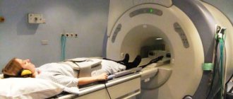
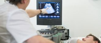
The patient is placed on the tomograph bed. After turning it on, the table slides into the tunnel of the device. A ring equipped with multiple sensors begins to rotate around the patient. They pick up signals reflected from tissues and send them to a computer. The tomograph is equipped with a microphone through which the patient communicates with the diagnostician.
He may ask you to hold your breath for a few seconds. The main rule is that the patient must lie still while the tomograph is operating, otherwise the images will be blurry and the procedure will need to be repeated. During the scan, the person does not feel any discomfort or pain. However, some people may find crackling and buzzing noises annoying. Duration of CT scan is up to 30 minutes.
Carrying out CT with contrast agents
With the use of dyes, the examination time increases to an hour. During this time, the circulatory system is additionally assessed, and, if necessary, pathological foci and tumors are studied in more detail. In general, CT scanning of the pelvic area does not differ from the standard method, only before scanning the patient is injected with an iodine-based substance.
It quickly spreads throughout the vascular system, which helps detect many pathologies at an early stage. In places of malignant tumors and metastases, contrast accumulates, which indicates cancer. The concentration of the dye occurs due to greater blood flow to the affected areas. CT scanning with contrast is prohibited for people whose kidneys cannot quickly remove the substance.
Preparatory measures before the examination
This area of the body is one of the few that must be prepared in advance before the procedure. Since the gastrointestinal tract organs are constantly in dynamics and are also filled with waste products, you should thoroughly cleanse the intestines and calm the stomach before the tomography session. Both before a CT scan of the pelvic organs and before an MR screening, the attending doctor will issue instructions that include a list of preparatory procedures. This list necessarily includes dietary recommendations that must be followed 1-2 weeks before the scan. On the eve of the session, 1-2 cleansing rinses of the gastrointestinal tract are performed.
Both MRI of the pelvic organs and computer screening can be performed using contrast. In this case, be sure to warn your doctor about existing chronic pathologies and the presence of an allergy to a particular drug. Don’t forget about the documents that relate to the disease; they will be needed both during registration and for the diagnostician’s preliminary review of your medical history.
How to prepare for scanning
During a routine examination, before being referred for a CT scan, the patient undergoes ultrasound and MRI diagnostics of the area of interest.
To an appointment with a radiologist, the patient brings the conclusion of consultations with specialized specialists and previous research methods. The referral from the attending physician indicates the diagnosis, goals, and objectives.
If the administration of iodine-containing drugs is required, then the functional abilities of the liver and kidneys are additionally assessed.
Pathology of the hematopoietic system is excluded. 3 days before the tomography, blood is donated to determine creatinine levels. On the day of the test, a test is taken to confirm the absence of an allergy to iodine.
To obtain high-quality tomograms, scanning is performed on a full bladder and cleansed intestines. Preparing the patient for the study on the day of the procedure includes:
- self-administration of a cleansing enema;
- to fill the bladder within two hours before diagnosis, ingest 2 liters of clean water at room temperature;
- in patients with a urinary catheter, the bladder is filled by clamping the tube or retrograde fluid injection.
Diet
Within two days before the diagnosis of OMT, a diet is established for the patient, which allows eliminating excess gas formation and stagnation of feces. To do this, exclude gas-forming and hard-to-digest foods from the diet:
- fresh fruits and vegetables;
- baked goods;
- beans, peas, beans;
- black bread;
- animal meat;
- milk;
- canned food;
- smoked meats, pickles, marinades;
- beer, kvass, sparkling water.
It is recommended to consume liquid, non-rich soups, cereal porridges, compote, and sweet, weak tea. 6 hours before the study, food intake is completely stopped.
How does an MRI of the pelvis differ from a CT scan of the pelvis?
In addition to the above-mentioned distinctive features of these procedures, the diagnostic process itself is very similar. The patient is placed on the conveyor, listens carefully to the instructions of the laboratory assistant, and is fixed in a motionless state. The retractable part of the tomograph smoothly moves inside the device, and screening begins. The examination does not cause painful or uncomfortable sensations, but if complaints arise during the procedure, you can inform the employee about this over the loudspeaker.
The difference between pelvic MRI and similar computer diagnostics is only the duration of screening. Thus, the nuclear resonance method takes approximately 30 minutes, while diagnostics using a computer will take only 15 minutes of your time. If contrast enhancement is planned, then the duration here and there increases by 15-20 minutes.
As a result, the client will have a package of documents in his hands, including photographs of the department under study and a preliminary conclusion. With this documentation, you need to go to the specialist who referred you for examination.
Contrast agents in diagnostics
Contrast enhancement in computed tomography is carried out to detail the detected changes if the tumor nature of the disease is suspected.
The tomography technique with contrast is performed by intravenous and intracavitary administration of drugs:
- For bolus delivery of contrast agents into the bloodstream, a catheter is installed in the vein of the elbow, to which a syringe injector is attached. The flasks of the automatic system are filled with iodine substance in a volume of 90-120 ml and physiological solution 50 ml. The rate of drug administration is determined by the radiologist;
- to contrast the bladder on the day of the study, 1.5 hours before the procedure, the patient takes orally 1200 ml of liquid diluted with 40 ml of a radiopaque iodine-containing substance (Urografin);
- to study the distal parts of the large intestine, the patient drinks 0.5-1 liters of a contrast agent solution on the eve of the procedure and 0.5 liters in the morning on the day of scanning. The rectum is filled with contrast before the examination using an enema.
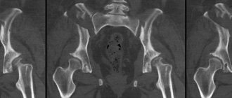
What does a pelvic CT scan show?
The survey provides comprehensive information about the condition of such structures as:
- bone fiber density after trauma;
- pathological accumulation of liquid substances and their chemical composition;
- genetic disorders of the development of the studied region.
A pelvic CT scan is often prescribed when other diagnostic measures are insufficiently informative, for example, when ultrasound diagnostics are ineffective. In addition, tomography allows you to assess the condition of the blood channels and the presence of destructive processes in them.
Which examination will cost more?
Both the first and second methods are expensive procedures, so they are prescribed when absolutely necessary. The cost depends on the coverage area of the beams or field, so bone structures will require less research costs, and internal organs will require more. Hence the final price. A CT scan of the pelvis for studying bone formations will cost less than an MRI of the pelvis for diagnosing diseases of dense organs.
The cost will increase in both cases with the introduction of contrast, since the price of the drug will also be included in the amount. The required volume is calculated individually and depends on the person’s weight category.
What to choose: MRI or CT of the pelvic organs?
The choice of screening method lies entirely with the attending physician, since each of them is used for its own purposes. The department under study has gender characteristics in men and women, so the most appropriate diagnostic method is determined.
MRI or CT of the pelvic organs is equally effective at the preoperative stage, when there is a need to determine the degree of operability of the patient, as well as for monitoring in the postoperative period. Whatever method is chosen by the specialist, the patient can be confident that the diagnosis will be as informative as possible.
Are there any risks for the patient from this study?
This procedure is safe for humans. However, in very rare cases, an allergic reaction to gadolinium is possible. A possible serious complication of the procedure is nephrogenic systemic syndrome.
However, if the kidneys are examined, this risk is completely minimized.
It is best to carry out diagnostics in men using an open-type device - this will be much more reliable and safer.
Comparison of MRI machines. On the left is a closed MRI, on the right is an open type of MRI machine