Ultrasound examination (ultrasound) of the female genital organs is one of the most informative types of diagnostics. Among its advantages are the complete absence of pain or discomfort for the patient, the absence of contraindications (safety), efficiency, instant receipt of data for prescribing treatment or detection of pathologies. Another definite plus is the minimum requirements for preparation for a gynecological ultrasound. A woman is not required to follow strict diets, preliminary manipulations, any training, etc. It is enough just to follow a number of simple rules.
When is it prescribed?
Ultrasound examination is actively used both for the diagnosis of various diseases and for preventive examinations. In the first case, an appointment for an ultrasound is issued if there are complaints:
- for pain in the lower abdomen and groin;
- prolonged tugging or other discomfort;
- frequent urge to urinate;
- disruptions of the menstrual cycle;
- spotting (not during menstruation);
- uncomfortable urination (accompanied by aching or acute paroxysmal pain);
- burning sensation in the urethra;
- too much (scanty) discharge during menstruation;
- the presence of blood or bloody mucus in the urine, etc.
Preventive ultrasound of the female organs is prescribed when planning pregnancy to monitor its course or progress of treatment (confirm recovery); before installing or removing an intrauterine device. An ultrasound examination is mandatory after operations on the organs of the woman’s reproductive system and during the recovery period. In addition, all women are strongly recommended to undergo preventive examinations to identify so-called female diseases at an early stage with a frequency of once every 1–2 years.
In what cases is it prescribed
Ultrasound examinations of organs in the pelvis can be prescribed by a doctor if there are certain indications. These include:
- pathologies of inflammatory etiology, for example endometritis, cystitis, vulvovaginitis, parametritis;
- presence of suspicion of the development of a tumor disease in the pelvic organs (fibroids, neoplasms of various etiologies);
- pregnancy;
- determining the number of available follicles and identifying the process of ovulation in the ovaries;
- clarification of the condition of the cervix (carried out during pregnancy and after childbirth);
- monitoring the status of the installed intrauterine device;
- monitoring the patient’s condition after surgery (especially after artificial termination of pregnancy).
Women in healthy condition should undergo ultrasound diagnostics for preventive purposes at least once every 2 years. Over the age of 40, this procedure is recommended to be performed every year. This will help identify any diseases that occur in a latent form. Preventive ultrasound should be performed in the first phase of the menstrual cycle (5-7 days after the start of menstruation).
During a standard examination, a specialist assesses the condition of certain internal organs, taking into account the following characteristics:
- the location of the uterus relative to the pelvis and nearby internal organs;
- the size of the uterus and the types of its contours;
- structural structure of the muscular and mucous tissues of the uterus;
- the dimensions of the inner surface of the uterus, the structure of its walls;
- the size of the cervix, its location and the integrity of the layers;
- the size of the ovaries and the structural structure of the fallopian tubes;
- the condition of nearby tissues and various internal organs.
What diseases are determined using ultrasound of the female organs?
The list of pathologies that an examination can confirm or refute is very large. A specialist can evaluate several indicators at once: position, size, structure of organs, and compare the obtained indicators with normal ones.
Based on the results of the ultrasound, the doctor can state:
- polycystic disease;
- oncology (indicated by various neoplasms, thickening of organ walls);
- ovarian cysts;
- fibroids;
- endometriosis and much more.
The research data is obtained immediately (no need to wait for processing), but it must be deciphered by a qualified doctor (urologist, oncologist, gynecologist, obstetrician). The patient’s area of responsibility is proper preparation for a gynecological ultrasound, on which the correct diagnosis largely depends.
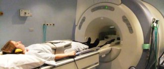
How is a pelvic examination performed using an ultrasound probe?
The patient lies down on a couch or sits in a gynecological chair. If the procedure is performed transabdominally, the subject removes clothing from the abdominal area and a conductive gel is applied to it. The doctor presses the sensor tightly against the body at various angles, moves it across the abdomen, obtaining an image of the internal organs on the monitor.
If we are talking about the transvaginal method, a condom is put on the sensor and hydrogel is applied, after which the doctor inserts it into the vagina. In this case, the sensor looks like a thin long tube, about 3 centimeters in diameter, with a rounded end. The doctor changes the angle of the sensor in the vagina to get a clear image of all the organs being examined. The transrectal procedure occurs similarly.
The duration of the study is from 15 to 25 minutes. Patients note that ultrasound is almost completely painless. In some cases, when the sensor moves into the vagina, unpleasant sensations may occur, and if an inflammatory process occurs there, even pain. They should be reported to your doctor.
How to prepare
To prepare for the procedure, you need to know which method will be used. Today in gynecology, three types of ultrasound examinations are prescribed. As the frequency of use decreases, this series looks like this:
- transabdominal method;
- transvaginal ultrasound;
- transrectal examination.
Preparation for transabdominal gynecological examination
The easiest and fastest option is transabdominal. Almost every woman aged 16 years and older underwent such examination. Its essence is to gradually move the ultrasound sensor along the lower abdomen to study the projections of the internal organs displayed on the screen. For better sliding of the sensor, a special “conductive” gel is applied to the patient’s abdomen. It is non-allergic and does not contain aggressive elements, so it can be used without the slightest risk.
Rules for preparing for this type of gynecological ultrasound:
- 3-4 days before the procedure, it is advisable to start following a simple diet, which involves: Avoiding foods that increase gas formation. This includes fatty meat or fish, rich broths, dairy products, white bread, and sweets. It is undesirable to eat dishes with a pronounced taste - salty, spicy, fried, smoked and spicy.
- Avoid drinking alcoholic beverages and alcohol-containing products.
- Avoiding carbonated drinks.
- It is advisable to include lean soups, lean meats and poultry, buckwheat, rice, and oatmeal in the menu.
- It is not forbidden to eat up to 1 egg per day and drink no more than 1 glass of kefir (milk).
- Ensure that the bladder is full. To do this, you can refrain from going to the toilet (urinating) for 2–3 hours or drink 0.8–1 liters of clean still water 60–90 minutes before the procedure at your usual pace.
Transvaginal method
For transvaginal examination, a special sensor with an elongated end, the diameter of which does not exceed 3 cm, is used. The end of the sensor is inserted into the vagina. The examination itself does not differ in technique from the transabdominal examination (projection of organs on the screen, assessment of their condition in the protocol).
The patient experiences no more pain and discomfort during the transvaginal examination than in a gynecological chair. The entire procedure takes 5-20 minutes (very rarely does the doctor need more time) and is performed on an empty bladder. Preparation for a gynecological ultrasound is simpler here (only diet and quitting smoking for a few hours).
An important point: for hygienic purposes, the ultrasound doctor puts a latex condom on the end of the sensor. If the patient may be allergic to latex, this must be reported in advance.
Transrectal ultrasound
Transrectal ultrasound is performed much less frequently than transabdominal and transvaginal ultrasound, but the preparation rules are similar, only the mandatory emptying of the rectum is added immediately before the procedure.
Preparation for the procedure
Many people are interested in how a pelvic ultrasound is performed on a woman. This procedure can be carried out using the following methods:
- transabdominal (through the anterior wall of the abdominal cavity);
- transvaginal (through the vagina);
- transrectal (through the anus);
- obstetric (in a pregnant woman).
Each research method requires certain preparation, which may differ depending on the research method. However, when performing an ultrasound of the pelvic organs in women, preparation for any method has some similarities:
- Before doing an ultrasound, for several days you should avoid eating foods that can cause increased gas formation. These are legumes, yeast bread, fermented milk products, alcoholic drinks.
- Directly on the day of the examination, in preparation for the procedure, care must be taken to completely cleanse the intestines.
- If a diagnosis using barium or x-ray was carried out a few days before the ultrasound examination, it is recommended not to do a pelvic ultrasound in the near future, since the substance can greatly affect the results.
Ultrasound for expectant mothers
Pregnant women may undergo preventive or therapeutic ultrasounds up to 5–6 times. The mandatory minimum is a three-time study, which is prescribed at 11-14, 19-21 and 30-34 weeks. Ultrasound in the first trimester is the most effective and safe way to detect serious disorders/anomalies in fetal development (including Down syndrome).
In the second trimester, the normal development of vital organs and their systems in the fetus is monitored (sex is also determined here). The latter procedure is aimed at identifying late developmental abnormalities of target organs (heart, kidneys, gastrointestinal tract, central nervous system, etc.). During this study, the doctor can draw conclusions about whether the speed of fetal development is normal.
What is included in the pelvic organs in women?
For women, the most important thing is to maintain the health of the pelvic organs, which are directly related to pregnancy and childbirth.
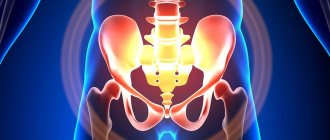
Below we will look at the features of the pelvis in women, their main functions and structure, location and effective methods of research and study. But first, let's look at the definitions. First: the pelvis is the area inside the pelvic bones. All organs located inside the pelvic bones are called pelvic organs. On the side of the pubis, the small pelvis is limited by the symphysis pubis, on both sides by the wings of the ilium, which, unlike the wings of the male pelvis, are located parallel to each other. At the back, the small pelvis is limited by the bones of the sacrum and coccyx. In total, the small pelvis is formed by four bones: two pelvic, one sacral and one coccygeal. The female pelvis is wider in size than the male pelvis, which is due to the female body’s ability to bear children and give birth.
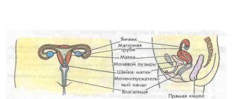
Like the male pelvis, the female pelvis contains the rectum and bladder. This is where the similarities end. The main thing is that here are the internal organs that are directly responsible for childbirth, reproduction and procreation:
Nature arranges it in such a way that a woman’s organs are extremely close to one another. The anatomical structure can be represented as follows: the uterus seems to rest on the bladder. The bladder rests on the vagina. All pelvic organs are equipped with muscles, the elasticity of which allows the organs to seriously stretch.
So, the vagina is essentially a muscular tube, often reaching up to 12 cm in length. During childbirth, the vagina stretches and serves as the birth canal. The vagina is adjacent to the cervix. The uterus is an empty organ consisting of muscle tissue. It is the uterus that is responsible for the process of bearing a child, so the uterus can stretch, take on large sizes, and contract again.
The ovaries are extremely important attributes of the reproductive system in women. The formation of follicles (or eggs) takes place here, and the ovaries are also responsible for the production of female sex hormones.
The fallopian tubes (or fallopian tubes) combine the uterus and peritoneum. It is in the fallopian tube that the male and female reproductive cells find each other and fertilization occurs. And it is through the tube, provided that it functions normally, that the fertilized egg enters the uterus. When the tube functions abnormally, pregnancy begins to develop in the tube cavity, causing it to rupture. This failure is called an ectopic pregnancy, and it is always terminated. Unfortunately, this is often followed by removal of the pipe.
The pelvic organs include the bladder and rectum, which are responsible for removing food debris and the results of its processing from the body. The bladder is also a muscular organ in which urine accumulates; it also has the ability to contract and stretch. The ureter or urinary canal is attached to it. The bladder lies behind the pubis. The rectum is responsible for the excretion of excrement and is a section of the intestine, its lower part.
When we talk about the female pelvic organs, we cannot help but describe its external structure, the external female genitalia: labia majora and minora, pubis, clitoris and vestibule of the vagina.
The pubis covers the symphysis pubis; it consists of soft and fatty tissues. The two folds of skin from the pubis to the perineum are called the labia majora. The labia minora, which look like petals covered with mucous tissue, are located inside the labia majora.
In the area of closure of the labia majora there is a tubercle - the clitoris. This is a very sensitive process, because it is filled with vessels, and, most importantly, nerve endings.
The space from the clitoris to the end of the labia majora is considered the vestibule of the vagina.
Since all the pelvic organs are in close proximity, practically lying on top of each other, inflammation, infection or any kind of disease of one system immediately affects the condition of other organs . Therefore, every woman should carefully monitor her health and regularly visit a gynecologist.