Breast cancer (BC) is a pathology that can lead to death or amputation of the breast in the absence of qualified treatment. All women over 40 years old, as well as representatives of the fair sex who are overweight, take hormone-based drugs, have a genetic predisposition or bad habits, are at risk. Breast cancer is visible very clearly on ultrasound. For this reason, this is the main instrumental study that is prescribed for diagnosing pathology.
Brief introduction to breast cancer sonography
Diagnosis is made using a sonograph with a hand-held probe. Tissue changes are detected by ultrasound through the skin. During the procedure, the parasternal (near the sternum) lymph nodes, the nipple area, and the array of the right and left mammary glands are examined clockwise. The nodular formation and the state of the border tissues are recorded with a freeze frame.
Cancer cells can be spread by lymph. Therefore, it is advisable to study this system. After the mammary gland, the nodes under the armpit, near the collarbones, are scanned. The structures in which metastases will appear are also photographed. The pictures are attached to the ultrasound protocol.
Rules for conducting ultrasound examination
For an accurate diagnosis, the patient needs preparation. It consists of eliminating excess gas formation, this is necessary for accurate diagnosis. Stages of preparation for ultrasound:
- 3 days before the examination, cabbage, legumes, milk, fruits, and baked goods are excluded from the diet;
- a laxative is used a day before the procedure;
- 7-8 hours before the ultrasound you should not eat; diagnosis on a full stomach may be erroneous;
- for better digestion of food, it is recommended to take food enzymes;
- To eliminate gas formation, it is recommended to take adsorbents (activated carbon, Smecta) 2 hours before the ultrasound.
This is interesting: Liver cancer in children: symptoms, treatment and prognosis
Indications and restrictions
According to the schedule, women are advised to be examined 1 or 2 times a year. If there are changes in the mammary gland, symptoms of the disease, or suspicion of breast cancer, the doctor will also refer you for an unscheduled ultrasound scan on the day of the patient’s visit.
Indications for sonography:
- lump in the mammary gland;
- swelling;
- skin redness;
- breast asymmetry;
- changes in nipple sensitivity;
- enlargement of regional lymph nodes;
- other pathological signs.
Ultrasound has time limitations: skin damage and acute inflammation in the breast area. In these cases, the woman is examined later or sent for a tomography.
Symptoms and stages of liver cancer
At first, the disease does not make itself known, is hidden, disguised as colds or food poisoning. The liver remains within the subcostal region and is not detected by palpation, although a feeling of compression in the right side already appears.
The main symptoms at stages I and II of the disease:
- slight episodic pain under the right rib;
- weakness, increased fatigue;
- decreased performance;
- sleep disturbance at night, daytime drowsiness;
- sudden increase in temperature;
- loss of appetite;
- nausea;
- episodic digestive disorders (vomiting, diarrhea);
- dizziness due to anemia, intoxication.
With the development of the oncological process, yellowness of the skin, sclera, and mucous membranes of the eyes appears, and the liver noticeably enlarges. Severe digestive disorders are observed, the patient’s well-being worsens, he loses weight, and his face becomes sharper. The third stage is considered critical, because at this moment the tumor begins to grow throughout the body.
This is interesting: Hepatocellular liver cancer: life prognosis, symptoms and treatment
Signs of cancer at stage IV:
- severe pain;
- exacerbation of chronic diseases;
- obstructive jaundice;
- nasal bleeding;
- dropsy (ascites);
- heat;
- digestive system failures;
- skin itching, rashes.
Ultrasound signs of cancer
The protocol contains a picture of what the change in the mammary gland looks like at the time of the examination. The absence of deviations is indicated in the description of the tissue structures of the breast.
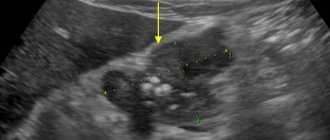
Echo signs of breast cancer:
- high echogenicity (white tumor);
- heterogeneous structure (there is a darkening in the center);
- rigidity of the formation (does not shift, does not compress);
- there is a shadow (dark stripe) behind the tumor;
- increasing the density of border tissue;
- enlargement of regional lymph nodes;
- at stages 3–4 there are metastases.
On a sonograph without additional programs, it is difficult to see whether the lump is benign or oncological. On Doppler ultrasound, there are many new vessels around or inside the changed lesion. Increased blood supply to the tumor and discharge from the nipples are signs of breast cancer. On ultrasound with elastography, the malignant lump is colored blue/purple.
The sonograph will not show incipient tumor foci smaller than 2 mm or whether the changes are malignant. Breast cancer can be detected in later stages by ultrasound. To confirm cancer, a biopsy of the breast tumor is prescribed. Without histological studies, the results of ultrasound or mammography are not accurate diagnostics.
Watch the video of what a tumor looks like on ultrasound:
Difference between cancer and other diseases
The nature of the neoplasm is judged by the presence of signs of cancer, appearance and density of changes, and whether its excessive blood supply is visible.
Echo signs of diseases:
| Pathologies | Differences on ultrasound | |
| Density | Appearance | |
| Malignant tumors | Rigid, does not move when pressed | The edges are uneven, the boundaries are blurred, the area of change is heterogeneous |
| Angiomatous tumor | Soft, hypoechoic formation in the area of the vessel | The contours are clear, pulsates in rhythm with heart contractions |
| Cyst | Soft capsule of normal echogenicity, liquid inside | Round formation with a light gray shell, black cavity |
| Fibroadenoma | Movable, elastic | Smooth outline, clear boundaries, uniform structure |
Increased blood supply to the fibroma or cyst shell is a symptom of the degeneration of benign pathologies into cancer.
Diagnosis of stomach cancer using abdominal ultrasound: is the pathology visible and how accurate is the result?
Stomach cancer is one of the biggest problems in gastroenterology. In the general structure of oncological pathologies, it ranks 4th in frequency (about 1 million diagnosed cases per year), and in overall mortality it is second only to lung cancer. However, it is often diagnosed only at stages 3-4, when there are already local or distant metastases.
Therefore, the issue of using an imaging technique that shows the disease and makes it possible to make an early diagnosis is very relevant in clinical practice. Today we will consider the role and capabilities of the method in this process, whether ultrasound of the abdominal cavity can show oncology, how to prepare in such a way as to maximize the information content of the examination.
Possibilities of the technique
Ultrasound examination is prescribed for almost all patients with suspected benign or malignant disease in the stomach .
High-frequency waves pass well through human tissue and are partially reflected from them (depending on the density indicator).
They are then captured by a sensor located on the front wall of the abdomen, processed by a computer and displayed as an image on the screen.
Ultrasound is a routine test if there are symptoms of dyspepsia, decreased appetite or pain in the upper abdomen. In addition to the stomach, the condition of the pancreas, liver, gallbladder, spleen, lymph nodes and large vessels of the abdominal cavity is examined.
With high-quality preparation of the patient, ultrasound allows one to clearly visualize the walls of the antrum and body of the stomach . The bottom of the organ in most patients is poorly visible due to the presence of gases in it (even a small amount). If the study is carried out by an experienced specialist, he can also approximately estimate the volume of gastric juice that is in the cavity.
Causes
Gastric cancer is a malignant tumor with uncontrolled growth that develops from the cells of the mucous membrane of the organ. Separately, lymphoma is distinguished - a tumor that is formed from lymphatic tissue (there is a lot of it between the layers of the wall). However, during the initial diagnosis before biopsy, they cannot be separated, since symptoms and signs often overlap.
Among the causes of oncological pathology it is necessary to highlight:
- infection with Helicobacter pylori infection;
- chronic inflammatory and erosive processes (gastritis and peptic ulcer);
- long-term presence of Crohn's disease;
- genetic predisposition;
- decreased reactivity of the immune system;
- smoking and alcohol abuse;
- irregular meals;
- exposure to ionizing radiation and environmental pollution.
Classification
A classification of stomach cancer is often used depending on the degree of progression and spread of the cancer process:
- Stage 0 – the process affects only the mucous membrane;
- Stage 1 – growth of pathologically altered cells into the submucosal layer is observed, distant foci cannot be detected;
- Stage 2 - the process spreads to the muscular and serous layers of the wall, several foci were also found in the nearest lymph nodes;
- Stage 3 – the tumor actively metastasizes through the regional lymphatic system (more than 3 confirmed foci), but without involving other organs;
- Stage 4 - when performing an ultrasound, CT or MRI, it was possible to find foci of the process in other organs (liver, spine, brain, spleen, kidneys or others).
Stages of development of stomach cancer
Clinical picture
The symptoms of the disease also differ greatly depending on the stage of the process. But the problem with this oncological pathology is that the clinical picture, even in stage 3, may not differ from gastritis or peptic ulcer. In patients with first detected stomach cancer, the following complaints are most common:
- periodic pain of varying intensity in the upper abdomen (on an “empty” stomach, or after eating);
- feeling of abdominal discomfort or fullness after a small snack
- nausea;
- a sharp change in diet, the appearance of aversion to certain types of dishes;
- decreased appetite and decreased body weight (even with an unchanged diet);
- black feces;
- belching with an unpleasant odor.
Important
If you have the symptoms listed above, you should immediately seek qualified medical help.
How to prepare for an abdominal diagnosis?
Usually, ultrasound of the stomach is performed routinely, both in outpatient and inpatient settings. After ordering a test, the doctor must tell the patient about all the nuances of preparation and diagnostic methods.
The study is carried out on an “empty” stomach, so on the day of the ultrasound the patient should not eat anything.
It is allowed to drink regular table still water an hour before the start of the diagnosis. All prescribed medications are taken as usual in the morning.
//www.youtube.com/watch?v=w-TIB5k03j4
If a patient has problems with increased gas formation (especially in older patients), then a special diet is prescribed a few days in advance , from which all foods and dishes that may contribute to it are removed. Additionally, it is possible to prescribe medications that reduce flatulence (simethicone, absorbents, antispasmodics).
How is the procedure performed?
An ultrasound of the stomach is part of an examination of the abdominal organs and is rarely done separately.
The patient comes to the diagnostic room at the appointed time, takes off his outer clothing, lies down on the couch and exposes the skin of his abdomen. A special gel is applied to it, which increases the permeability for ultrasonic waves, after which the process of examining the patient begins.
For examination, the sensor is placed in a horizontal position in the left upper quadrant of the abdomen, after which its angle of inclination is alternately changed to visualize various parts of the organ. In this case, the left lobe of the liver is used as an acoustic “window”, since a significant part of the stomach is located between it and other anatomical structures (for example, the pancreas).
Reference
Ultrasound examination is absolutely painless and safe. Ultrasound is allowed to be done during pregnancy, in old age and in the presence of any concomitant pathologies.
After the diagnosis is completed, the remaining gel is removed from the skin of the anterior abdominal wall using a disposable towel.
In recent years, endoscopic ultrasound has become more frequent - a diagnostic technique in which a special probe with an ultrasound sensor is inserted through the mouth and esophagus into the stomach cavity (as with FGDS). This allows you to get a more detailed picture of the structure of the walls of the organ (especially in the cardiac region and bottom). The information content of the technique is higher than conventional ultrasound of the stomach through the anterior abdominal wall.
What pathologies will the study show?
Using ultrasound, the following pathological conditions can be determined:
- anatomical abnormalities of the stomach;
- gastritis with hypersecretory state;
- benign and malignant neoplasms of the stomach;
- peptic ulcer;
- metastases of tumors from other localizations;
- intragastric bleeding;
- enlargement of the lower esophageal veins (with increased pressure in the portal vein system of the liver);
- narrowing of the antrum of the stomach;
- the presence of third-party bodies;
- enlargement and inflammation of nearby lymph nodes.
Is cancer visible during the procedure?
Although ultrasound examination of gastric cancer is performed routinely, the informative value of this examination (especially not in specialized oncology clinics) remains low. Whether cancer can be seen on an ultrasound depends on the following factors:
- Sensitivity of information content to the quality of training. In addition, it is not always possible to completely get rid of the presence of gases in the lumen of the stomach (even when using medications).
- Lower quality of visualization of hollow organs. This is the main disadvantage of ultrasound research methods. Therefore, clinical guidelines recommend the use of contrast-enhanced computed tomography if gastric cancer is suspected.
- Depends on the qualifications of the specialist. In most general clinics, gastric examination is carried out by doctors who do not always pay attention to nonspecific signs of the oncological process.
- Use of outdated equipment. In small regional centers, ultrasound is usually performed on devices that are more than 5-7 years old, which reduces its information content.
Whether an ultrasound will show stomach cancer depends on the qualifications of the doctor. A stomach problem can be detected in large oncology clinics, where such patients are diagnosed every day.
note
It is impossible to make a diagnosis of stomach cancer based on ultrasound. It is imperative to supplement the diagnostic program with gastroscopy, biopsy with cytological examination and computed tomography with contrast.
Decoding the results
Ultrasound symptoms, which may indicate oncological pathology, differ at different stages of the process:
- Stage 0 – absence of any ultrasound signs of cancer;
- Stage 1 – an increase in the thickness and layering of the suspicious area of the wall, a moderate decrease in echogenicity;
- Stage 2 – there is a significant change in echogenicity, the surface of the mucous membrane becomes uneven, a defect in the form of a crater or depression may be detected, a sharp change in the diameter of the wall, narrowing of the lumen of the stomach, tuberosity of the external contour, disappearance of the boundary between the different layers (mucosal, muscular and serous);
- Stage 3 - in addition to symptoms of gastric damage, significantly enlarged hypoechoic lymph nodes are detected;
- Stage 4 – additionally, metastases can be detected in neighboring anatomical structures (most often in the liver).
All detected changes are carefully documented in the results, which are given to the patient or attending physician after the end of the study.
Additional Methods
A number of other diagnostic methods are used to diagnose stomach cancer. Their comparative characteristics are given in the table:
| Ultrasound | FGDS with biopsy and cytological examination | CT with contrast | MRI | |
| Characteristic | Non-invasive ultrasound examination | Endoscopic examination with tissue sampling | X-ray examination with intravenous contrast | Using the phenomenon of magnetic resonance |
| Symptoms of stomach cancer | Changes in echogenicity, wall thickness, contour evenness | Violation of the integrity of the mucous membrane, change in its color, proliferation of tissue of various shapes, deformation | “Defect” of contrast accumulation, wall deformation, change in its thickness and shape | Thickening of the wall, changes in the shape of the stomach, changed lymph nodes |
| Information content of primary tumor diagnosis | ++ | ++++ | ++++ | +++ |
| Information content of metastasis detection | ++ | — | +++ | ++++ |
Conclusion
Ultrasound is inferior to other diagnostic methods (FGDS, CT or MRI) in terms of information content for detecting gastric cancer.
Whether an ultrasound can show stomach cancer is determined by the correct preparation of the patient and the qualifications of the specialist. Higher information content of the technique when searching for metastases in regional lymph nodes, or in other organs (liver, spleen, kidneys).
Have you had experience detecting stomach cancer using ultrasound? Under what circumstances did this happen? Share your story with other visitors.
Source: //gastrosvoboda.com/diagnostika/uzi/vidno-li-rak-zheludka.html
Description of malignancy
What is cancer anyway? It is not a crustacean animal that lives in fresh water. A cancerous tumor is more unpleasant in appearance and carries a much greater danger.
A malignant neoplasm is, first of all, an accumulation of atypical cells . To survive, they need nutrients obtained from the body, i.e. they are a kind of parasite. Having created an initial “bridgehead”, the tumor begins to grow and spread to healthy organs. Everything that it encounters on its way (vessels, muscles, nerves) it destroys.
The appearance of the tumor depends on the location and may vary in each specific case. Usually it is a soft knot with a smooth or bumpy surface.
The sizes are also unique. At the initial stages, the diameter is usually 1 cm. However, over time, the neoplasm becomes much larger. At the last stages of development, the diameter can reach 30 cm .
After decay, the neoplasm changes its “usual appearance.” Now the tumor looks like a purulent mass with an unpleasant fetid odor.
Scintigraphy
Another radiological examination method is scintigraphy. The method is based on the selective accumulation of radiopharmaceuticals by body tissues.
To study a cancer patient, he is first injected with a standard dosage of a radiopharmaceutical that can selectively accumulate in tumor cells; the drug molecules labeled with a radioactive isotope accumulate in the tumor, after which the patient is subjected to scintigraphy.
The study allows us to determine not only the main tumor, but also all metastatic foci in the patient’s body, which is important for developing the correct tactics in treating the patient.
Image obtained during scintigraphy of the thyroid gland. The arrows indicate the focus of tumor growth in the left lobe of the thyroid gland. Before the image, the subject was given an intravenous injection of an iodine-containing pharmaceutical, which selectively accumulates in atypical thyroid cells.
Angiography allows you to assess how well the tumor is supplied with blood, which is important information when planning surgical treatment. In some cases, the tumor may be embolized, causing it to regress.
Renal angiography using X-ray contrast agent. In the image, arrows indicate vessels that have arisen as a result of enhanced angiogenesis during the progression of kidney cancer.