An X-ray examination of the thyroid gland is a study that relates to radiological diagnostic methods. Using X-rays, you can examine the physiological changes of the gland, as well as its location. In other words, based on the results of the study, it is possible to determine the presence of pathological formations.
The technique is often used to diagnose thyroid diseases, but its main value is the ability to assess the size of the thyroid gland. An X-ray may show the presence or absence of a substernal goiter, the presence/absence of calcifications, etc.
Where to do research
This procedure can be carried out in government medical institutions and in special diagnostic centers. It is important that the facility has the necessary equipment and a competent radiologist.
Content:
- Who prescribes diagnostics
- Where to do research
- When is radiography indicated?
- Contraindications to X-ray diagnostics
- How to prepare for an X-ray examination
- Decoding the results
- What diseases can be determined using X-ray diagnostics?
Scintigraphy for suspected cancer
The peculiarity of thyroid cancer is that the disease goes away without obvious symptoms. But the appearance of the slightest signs is a reason to consult a doctor. The sooner therapy begins, the greater the chance of a favorable outcome.
One of the most effective ways to identify a disease is radioisotope testing. It is not possible to obtain information about the type of cancer and benignity, but you can determine the size, shape and presence of metastases.
Scintigraphy is used to assess the activity of hormones and the functioning of the gland.
When is radiography indicated?
This study is prescribed extremely rarely, since today there are safer and more accurate methods. An appointment for a study can be obtained if a goiter is visualized, or if there are disorders of the esophagus or trachea.
During the procedure, you can detect: metastases that remain after removal of the cancer; the presence of hyperthyroidism and hypothyroidism; determine the boundary between a node and a diffuse lesion.
It is important to understand that this type of research negatively affects all other vital organs, since the radiation dose increases at least tenfold.
X-ray or ultrasound?
X-rays are prescribed when ultrasound examinations fail to provide an accurate picture for diagnosis. If a person has a very large goiter and it covers the thyroid gland, only with x-rays can its exact size and neoplasms, including cancerous ones, be determined. In addition, unlike X-rays, ultrasonic waves are not able to pass through the calcium salts that cover the thyroid nodes and cysts.
Ultrasound of the thyroid gland
Ultrasound is prescribed to obtain data on changes in the structure of the thyroid gland, the location of the neck organs, and the presence of pathological formations.
How to prepare for an X-ray examination
X-rays of the thyroid gland are usually performed without contrast, therefore, the patient does not need any special preparations. The first thing to do is to completely expose your torso, remove long hair, and remove all jewelry. It is recommended to abstain from iodine-containing foods before undergoing the procedure. The patient is moved to a special couch. An X-ray machine is placed opposite the thyroid gland area. The laboratory technician places a special lead apron on the genitourinary area.
After all the necessary preparatory procedures have been completed, all personnel leave the room (the diagnostician works in a special booth). During the photography period, the patient needs to hold his breath (the laboratory assistant reports this).
In total, the procedure takes up to 10 minutes.
X-ray diagnostics is a painless procedure, but there are some discomforts (occasional holding of breath and being in a static position). Only by strictly following such simple rules can you obtain correct, reliable and clear images.
Indications for diagnosis
Radioactive scanning of the thyroid gland may not be prescribed to all patients and not always, especially in cases of diagnosed goiter.
Main indications:
- abnormal position of the gland, substernal goiter;
- study of the functioning of the thyroid nodules;
- differential study of thyrotokycosis;
- study of metastases of differentiated thyroid cancer;
- some individual indications, for example, studying the patient’s condition after surgery in the presence of a malignant tumor in the gland.
Decoding the results
After the procedure, the following actions are performed:
- After 30 minutes, the diagnostician gives the finished images to either the doctor or the patient;
- the doctor performs a full analysis of the received images;
- a diagnosis is made (if the research data is insufficient, a referral for MRI or CT is issued);
- treatment is selected and an appropriate diet is prescribed.
Trying to treat the disease on your own and prescribing medication for yourself is highly discouraged, so as not to aggravate the situation.
Structure and function of the organ, features of their assessment
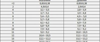
The thyroid gland consists of two lobes (wide and short lower parts, narrow and high - upper parts), which cover the trachea, and an isthmus. It is located under the larynx, its shape resembling a butterfly. The weight and size of the organ is individual: the average weight in an adult varies from 12 to 25 g, its volume in a man is approximately 25 ml, in a woman – about 18 ml.
The thyroid gland is one of the main organs of the endocrine system - its function is to produce biologically active substances necessary for the normal functioning of the human body:
- triiodothyronine (T3);
- thyrocalcitonin;
- tetraiodothyronine (T4).
In a healthy person, these hormones are in an optimal ratio, however, if pathological changes occur in the thyroid gland, this leads to disruption of metabolic processes, increased nervous excitability and interruptions in the functioning of the heart muscle. To assess the condition of the thyroid gland, practicing endocrinologists use physical examination techniques - palpation, ultrasound scanning, computed tomography, MRI.
Identification of morphological changes in the thyroid gland, on the basis of which a diagnosis is made, is carried out during CT scanning. However, this method is based on tissue visualization using x-rays, to which the thyroid gland is very sensitive. That is why it is rarely used to obtain images of an organ. And the MRI method involves exposure to a magnetic field that is absolutely safe for humans.
The choice of method for diagnosing thyroid pathologies remains with a qualified specialist: CT is used to study the physiological state of the organ, MRI is used to assess the chemical structure
What does a fluorography image show?
If you want to know what fluorography is and why it is needed, you should familiarize yourself with the information that will become known as a result of the diagnosis. As a rule, fluorography is used to examine the chest organs, and is more often used to diagnose tuberculosis or neoplasms. It allows you to detect a lot of deviations, while the doctor has the opportunity to assess the condition of the chest structures, and he can prescribe treatment tactics adequate to the disease. You can familiarize yourself with the pathologies that such diagnostics can identify below.
Changes in lung tissue
If you don’t know what fluorography shows, it’s worth finding out that the image will show abnormalities in the lungs. Foci of tissue damage, their location, size and outline. The changes can be sclerotic in nature, when the normal tissue of an organ is replaced by connective tissue, or fibrous, when compactions and scars form on the connective tissue.
The overall picture allows the doctor to make a diagnosis or prescribe additional tests, for example, to determine the nature of the tumor in the lungs.
Inflammatory processes in the lungs
If you don’t know what fluorography is, what it shows and what diseases it will help identify, you should know that any inflammatory processes will be visible on the film. They appear as darker areas, and the darker they will be the more severe the inflammatory process. The procedure will detect:
- inflammation of the lungs, which is called “pneumonia”;
- tuberculosis;
- abscesses;
- etc.
Using fluorographic examination, the disease can be detected at a very early stage.
The order of preparatory activities and the examination
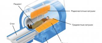
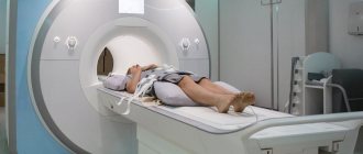
Carrying out an MRI of the thyroid gland does not require the patient to comply with complex preliminary preparation requirements. The only condition is passing general clinical urine and blood tests. However, their results may be required only if it is necessary to use a contrast agent - to identify concomitant diseases and a possible allergic reaction. Before the procedure, the patient should wear comfortable clothing without metal fittings. He is placed in a tomograph cabin that resembles an elevator cabin.
The duration of the diagnosis is approximately 30–35 minutes, during which time the patient must remain motionless. This is important for obtaining a high-quality image. To improve visualization of the organ, specialists use a contrast agent, which is administered intravenously. Thanks to its accumulation in places of pathological tissue changes, the final data of the diagnostic procedure become more accurate.
Using MRI of the thyroid gland, you can identify malignant formations, infectious and inflammatory processes, foreign objects and diagnose autoimmune thyroiditis, Graves' disease, mediastinal tumors
This many-sided goiter of the thyroid gland
How else it will manifest itself in your life depends on the function of the thyroid gland. Many things can happen to it - an increase (hyperthyroidism), a decrease (hypothyroidism), or it will remain unchanged (euthyroidism). The last option is the simplest - goiter with euthyroidism threatens mainly with aesthetic defects. Also
Goiter with hyperthyroidism is also called toxic. Why is this so? Probably because an excess of thyroxine, the thyroid hormone, seems to poison the body, turning from its main “stimulant” into a real poison. Read about diffuse toxic goiter in one of my blog articles. Hypothyroidism, goiter of the thyroid gland is accompanied mainly by iodine deficiency. Most often this is an endemic goiter.
How about endemic?
The word "endemic" means characteristic of a particular area. In the case of thyroid goiter, we are talking about areas where the supply of iodine to the body of the people living there is insufficient. After all, we get iodine mainly from air, water and food. Areas with gray and podzolic soils, as well as mountainous areas, are endemic for the deficiency of this microelement. You understand that in Russia such territories are much vast. As a result of iodine deficiency, endemic goiter develops.
The same condition, which occurs in the population of areas that are quite free in terms of iodine content, is called sporadic goiter. Autoimmune thyroiditis (Hashimoto's disease) leads the list of its causes.
Examination and treatment goals
It is important to assess the size and function of the thyroid gland. As everyone has already understood, in each case it is not just goiter that is treated, but a specific disease. The word “goiter” in the diagnosis simply must be preceded by a couple of clarifying adjectives. They determine the tactics of therapy in specific cases. Large goiters and compression of surrounding tissues may require surgery.
Of course, if you independently detect a visible goiter, it is better to consult an endocrinologist in person. This article is intended only to inform you about the variety of causes and types of thyroid goiter, and not to replace examination and treatment.


