admin Home page » Diagnostics
The thyroid gland is the organ responsible for the production and optimal balance of hormones in the body.
Disturbances in its work seriously affect a person’s well-being, since it is the thyroid gland that regulates metabolic processes.
Today, for a qualitative examination, the MRI method of the thyroid gland is used.
The value of the method
MRI when examining the thyroid gland has the following advantages:
- The accuracy of the results, thanks to which the doctor draws conclusions about the condition of the organ.
- No radiation. During the study, a magnetic field is used. It is considered safe and harmless to humans.
- Receive clear images of the examination results. Thanks to three-dimensional graphics, the thyroid gland is studied in different projections.
- Non-invasive. To carry out the examination, the integrity of the skin is not compromised.
Such advantages make MRI an effective diagnostic method that identifies organ diseases and establishes the causes of unclear pain.
MRI
What the organ looks like, what the study can show
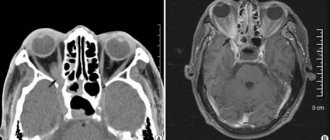
During an X-ray examination with contrast, the thyroid gland is clearly visible against the background of neighboring tissues, since its relative density and blood circulation are higher.
The lobes in the photographs are bean-shaped with an average size (in healthy patients) of 15x25x55. They are adjacent to the anterior surface of the trachea, and their posterior parts are adjacent to the sides of the esophagus. Nerve and vascular bundles and muscles pass nearby. An isthmus is visible between the lobes. The structure of the organ is homogeneous, fine-grained.
For non-tumor lesions in the gland they find:
- micro- or macrofollicular goiter;
- uniform increase in lobes with clear contours;
- the tissue structure remains homogeneous;
- there may be signs of compression of the trachea and esophagus;
- with nodular goiter, the increase is usually insignificant, more often there is one node (solitary), less often there are many of them;
- the contours of a benign node are clear and can be polycyclic (wavy);
- the structures of nodular formations are predominantly homogeneous, there may be areas of low density or deposits of calcium salts;
- large nodes can compress the trachea.
Nodular goiter
With autoimmune thyroiditis, there is a slight enlargement of the gland (first degree), clear boundaries, increased tissue density, heterogeneous structure - small foci of inflammation and strands (sclerosation). A honeycomb pattern is formed.
With papillary cancer, against the background of a slight enlargement of the gland, low-density foci with unclear outlines are found. The nodes are heterogeneous with small calcifications and areas of rarefaction and destruction. There are also signs of tumor growth into neighboring organs - muscles, lymph nodes of the neck, submandibular. As metastases progress, they are detected in the lungs in the form of a chain of nodules along the vessels.
Papillary cancer of the right lobe of the thyroid gland
Follicular cancer usually leads to enlargement of one lobe of the gland. Multiple nodes tend to merge into one with wavy outlines. Their structure is heterogeneous due to the presence of calcium deposits in the center and on the periphery (lime capsule). Often they grow into the sternocleidomastoid muscle, the trachea, and the neoplasm deforms its lumen.
Anaplastic cancer is accompanied by a sharp increase in the size of the thyroid gland; it can move beyond the sternum and mediastinum to the level of the bifurcation of the trachea. The structure of the tumor tissue in most patients is homogeneous, the contours are not clear. The formation spreads to the trachea and greatly narrows its lumen. Metastasizes to the spine, ribs.
A thyroid CT scan is prescribed when changes are detected on ultrasound, the origin of which needs to be clarified. The examination is indicated in case of an abnormal location of the organ, to study the structural features of the surrounding tissues, before surgery in order to determine its volume. CT scanning is contraindicated during pregnancy and intolerance to contrast.
When analyzing the data obtained, typical tissue changes are taken into account, but the final diagnosis can only be established by histology during surgery.
Organ structure and diagnostic methods
The thyroid gland is formed by 2 lobes - the upper and lower, covering the trachea, and the isthmus.
This organ is located under the larynx and resembles a butterfly. Size and weight depending on the gender of an adult are 18-25 cm³ and 15-30 g, respectively.
The gland is an organ of the endocrine system, the function of which is to produce hormones necessary for the normal functioning of the human body.
These include:
- triiodothyronine (T3);
- tetraiodothyronine (T4);
- thyrocalcitonin.
If a person is healthy, then the amount of hormones is at the proper level. When the ratio changes, metabolic processes are disrupted, nervous excitability increases, and interruptions in the functioning of the heart occur.
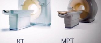
To assess the condition of the gland, the doctor uses the following examination methods: palpation, computed tomography, ultrasound scanning, MRI.
Using CT, morphological changes in the organ are detected, which makes it possible to make a diagnosis. But this method is based on tissue visualization with X-ray irradiation, to which the thyroid gland is extremely sensitive.
Therefore, this method is rarely used. MRI is performed using a magnetic field that is safe for humans.
Serious and not so serious contraindications
Many women are interested in the question of what will happen if they do an MRI during pregnancy. But this particular condition is considered a relative contraindication in the first trimester, since an examination using a magnetic field may not benefit the baby. Especially in situations where the use of contrast is mandatory.
To eliminate the risk of harming the fetus in the form of inhibition of its mental or physical development, it is worth warning the treating specialist about pregnancy in advance, and he will send the expectant mother to be examined using a more gentle ultrasound examination.
But the period of breastfeeding is not a serious prohibition except in cases where the use of a contrast agent is necessary. It’s just that in this case it is necessary to stop lactation for a period of at least two days after the administration of contrast. During this time, potentially dangerous milk is expressed and poured out.
The most important problem for using a magnetic tomograph is the presence of built-in electronic mechanisms or metal elements in the body. The first include pacemakers, built-in hearing prostheses and similar assistants that improve the quality of life. If a person has something similar in any part of the body, then the procedure is abandoned in favor of a safer analogue.
But the presence of knitting needles, pins and other metal components is not always considered a strong contraindication. Here everything is decided by the attending physician, depending on the location of these structures and their composition, as well as the preponderance of benefits from the analysis. Other relative prohibitions include:
- severe general condition;
- childhood;
- mental disorders.
The points listed above are dangerous due to the fact that people at risk are not able to maintain absolute stillness for a long period. After all, it is precisely by maintaining one pose for a long time that you can get high-quality “pictures.” Any careless movement will blur the whole picture, which will become the basis for redoing the analysis.
Just a dozen years ago, fear of closed spaces was also included in the list of contraindications that could not be overcome. To help such patients, exclusively sedative medications were used. Today, technological progress has advanced much further, offering open-type equipment to medical centers. That is, the tomograph has open side panels, maintaining its own functionality, albeit at a lower level.
For what symptoms is examination indicated?
MRI of the thyroid gland is prescribed when the patient has complaints and to clarify the diagnosis. The study is indicated in the following cases:
- nervous system disorder;
- fast fatiguability;
- insomnia;
- sudden change in body weight;
- tachycardia;
- the appearance of a tumor-like formation on the neck;
- enlarged lymph nodes;
- compression in the esophagus and throat;
- pain when swallowing.
This procedure is prescribed if there is a suspicion of a diagnosis of goiter, which is characterized by excessive growth of organ tissue for no apparent reason.
What is found in diseases
Depending on the pathological changes in the thyroid gland, characteristic MRI signs are revealed.
Thyroid cyst
A simple cystic formation has a homogeneous structure; its capsule is most often not visible. There are medica cysts and large ones; they are formed when the node changes. The signal can be low-intensity or amplified, but always the same. Its strength is determined by the protein content of the colloidal liquid. The contours of the cyst are smooth.
Hyperplasia of the thyroid gland
With diffuse goiter, the thyroid gland increases in size, but its tissue does not change. With nodular hyperplasia (proliferation), a homogeneous node and surrounding tissue with a normal structure or minor changes are visible. Large nodes also have uneven density. They can also arise from the merger of several smaller ones. In this case, their outline becomes wavy.
Thyroid adenoma
These formations are low-intensity or similar in density to the surrounding tissues. The structure is usually homogeneous. Their capsule is smooth, the same thickness throughout.
Cancer tumor
There are two options for signal changes during malignant degeneration:
- heterogeneous, of normal intensity with areas of enhancement;
- isointense (equal to the surrounding tissue) with small foci of reduced density.
Stages of thyroid cancer
The boundaries of the tumor are smooth and clear or blurry, lumpy. If there is a transition to adjacent tissues, then the contours are not determined.
After operation
When examining patients, the tomogram reveals:
- low-intensity signal from the scar, as it thickens it intensifies;
- expansion of the space around the esophagus;
- an increase in the diameter of the jugular veins with a weakening of blood flow in them;
- bringing together the nerve and vascular bundles of the neck.
An MRI of the thyroid gland is performed after an initial medical examination, ultrasound and blood tests. It helps to identify nodules and tumor formations, especially with an atypical location of the gland. Contraindicated in the presence of implanted metal structures. To enhance the signal, additional contrast is prescribed. When making a diagnosis, typical MRI signs are taken into account.
Restrictions
Although MRI is considered a safe method of examination, in some cases it is contraindicated. These include:
- First trimester of pregnancy. During this period, the child’s vital organs are formed, so it is extremely undesirable for the mother’s body to be exposed to radiation.
- The presence in the body of foreign objects that support the vital functions of the body. These are implants in the middle ear, pacemakers, etc.
- Psychical deviations. Such patients cannot remain in a lying position for a long time in a capsule that produces unpleasant sounds.
- The patient's body weight is more than 150 kg, because The device has a weight limit.
- Claustrophobia. People with this disorder cannot be kept in a closed cell because they can develop dangerous nervous system complications.
The diagnostician conducting the study pre-examines the patient and can detect reasons that do not allow an MRI to be performed on the day of treatment. This is alcohol or drug intoxication.
The procedure is contraindicated if there is a pacemaker in the human body. During the examination, the operation of the device is disrupted, which will lead to the death of the patient.
Preparation
Before MRI for injuries, an X-ray examination is first prescribed to exclude the presence of metallic foreign objects. For patients with thyroid diseases, ultrasound diagnostics and determination of its function are previously indicated; if contrast is necessary, a kidney examination is performed.
Immediately before the procedure, no special dietary regimen is required, but in order to eliminate the possibility of vomiting (which is rare), the patient is not recommended to eat 2-3 hours before the contrast is administered. A catheter for drug infusion is installed in advance in the vein and left in place, as it may be necessary to add a contrast agent for better visibility.
What is required before the examination
You do not need any special preparation to perform an MRI of the thyroid gland. Unlike an ultrasound or CT scan, the patient does not have to stop taking medications or following a diet.
To make the image as accurate as possible, you need to:
- remove metal objects from clothes, remove jewelry and metal watches;
- put bank cards and phones in a specially designated place, because When such objects enter a magnetic field, they become demagnetized and break, and distort the image;
- remove hearing aids, wigs or dentures.
Sometimes before the procedure the patient is given medications that have a sedative effect.
They are required when examining children or people with hyperexcitability. In addition, some cosmetics contain metal components, so it is better not to use cosmetics before the study.
Advantages and disadvantages
Unlike numerous other techniques, MRI of the thyroid gland sometimes makes it possible to detect the slightest abnormal changes, even tiny tumors. With the help of high-precision imaging, it will be possible to determine the presence of a capsule, as well as clarify whether it has spread to other structures. The latter is especially important if the patient has suspicions about possible malignant tumors.
Content
- Advantages and disadvantages
- When is MRI necessary?
- Serious and not so serious contraindications
- Preparation and carrying out the procedure
Very often, when examining the thyroid gland, an additional study with contrast is required to more accurately confirm the diagnosis. Despite the fact that the cost of manipulation, taking into account the additional stage of contrasting, will be higher, the result will make it possible to verify the diagnosis in detail, establishing the degree of damage, the territory covered by the pathological process, as well as the possible impact on neighboring structures.
If we talk about the advantages of the technique over x-rays, then the relative novelty in the world of medical technologies significantly benefits in several respects. In addition to its ability to capture the bony structure of the area being studied, MRI can visualize muscles and lymph nodes that cannot always be thoroughly examined even with ultrasound technology.
Patients undergoing this examination are also pleased with the fact that there is no radiation exposure. Instead of conventional radiation exposure, the technology is based on the peculiarities of the magnetic field, which was the reason for the name of the method.
The most important advantage is that there is no impact on the thyroid gland itself during scanning. This concerns the counterbalance in favor of magnetic resonance imaging over classical computed tomography.
The latter often requires the use of a contrast agent. It includes iodine-containing components, which in some diseases of the gland themselves pose a serious threat.
For the magnetic analogue, substances of a different type are used, which do not cause any damage to the thyroid gland, which explains the popularity of the technique. This fact, as a rule, also becomes a significant argument in favor of MRI, despite the fact that the examination usually lasts longer than with CT.
The method has one more advantage, namely, when it comes to the inability to recognize with an absolute guarantee whether a patient has a benign or malignant formation, by connecting the contrast stage for clarification, the percentage of error rapidly moves to zero. Moreover, an experienced doctor never makes a verdict about the nature of the tumor only on the basis of examination data. He will definitely take into account the patient’s complaints, his hereditary predisposition, the results of other tests, including a test with oncological markers, and most importantly, the data of histological analysis.
Conduct and results
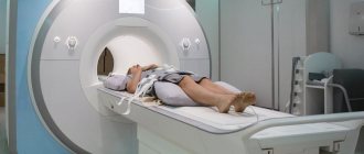
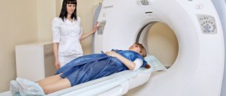
The patient takes off his clothes and puts on a robe, after which he lies down on a movable automatic couch, which slides into the research chamber. The doctor goes into another room where the computer is located.
The person must lie still during the diagnosis, otherwise the results will be inaccurate.
With the help of the device, the magnetic movement of atoms is carried out in the patient’s body and the response to the influence is captured. This radiation is safe for humans.
The MRI procedure lasts from 30 minutes to an hour. The computer saves the received images and then combines them into a three-dimensional image. Sections are made at a distance of 1 mm, which allows the doctor to assess the condition of the thyroid gland.
Many patients are interested in the question of whether it is possible to do an MRI after removal of the thyroid gland. This operation is performed if there is a retrosternal goiter or a malignant neoplasm.
If a person has recovered after surgery, then there are no obstacles to conducting the study.
With contrast
But sometimes after removal of the gland, the patient feels unwell, is unstable, and has a fever. In this case, MRI is prohibited.
When is a soft tissue examination performed?
The thyroid gland is located on the front of a person's neck. It is interconnected with the upper respiratory tract and digestive system. Therefore, when there is inflammation or damage to the trachea, esophagus, or larynx, it is often involved in a dangerous process. MRI of this department is recommended for a comprehensive examination of the following pathologies:
- severe injuries of the cervical spine;
- inflammation of the subclavian nodes;
- identified tumors on the esophagus or pharynx;
- cancer of the salivary glands;
- germination of metastases into large vessels;
- hypoplasia or aplasia of the thyroid gland;
- ulcers or necrosis of glandular tissue.
Examination of the soft tissues and thyroid gland using MRI is recommended if the ultrasound results were controversial and it is difficult to make a diagnosis based on the available data and tests.
Study with contrast
To obtain a clearer image, a contrast agent, gadolinium, is used, which is injected intravenously. It is much safer than iodine-containing contrast agents.
Gadolinium displays a stronger magnetic signal that the machine emits. This substance does not affect human well-being and is safe for him.
It is extremely rare to develop allergic reactions to gadolinium, which are quickly eliminated.
Contrasting is used in the following cases:
- to improve signal sharpness and accuracy;
- if during an MRI of the gland suspicious inclusions of an unclear nature (malignant or benign) are detected;
- to assess the characteristics of the blood supply to the thyroid gland.
MRI with contrast is necessarily prescribed if the patient has previously had problems related to oncology.
The substance is used with caution for liver and kidney diseases. The degree of risk is determined by the doctor, who decides whether the administration of contrast during MRI is permissible.
Contraindications to computed tomography
There are also a number of contraindications associated with the use of iodine-containing substances. The thyroid gland (CT with contrast) is not examined in patients with:
- severe renal or liver failure;
- iodine intolerance;
- hyperthyroidism (additional iodine in any form aggravates the disease);
- diabetes mellitus while taking metformin.
Contrast-enhanced CT scans are not usually performed on children under 12 years of age.
Reverse side
MRI of the thyroid gland has advantages, but also disadvantages - cost. For additional research, an ultrasound is prescribed, which, although less informative, is more affordable.
Magnetic resonance imaging is unable to distinguish between a benign tumor and a malignant neoplasm.
The examination itself can cause discomfort to the patient, because he is in a confined space and at the same time limited in movement. The disadvantages of the procedure include the risk of an allergic reaction to the injected contrast.
When is MRI necessary?
The endocrine system is responsible for the production of hormones, interruptions in the supply of which cause obesity, dry skin and other external manifestations, as well as problems of internal organs.
All this can be prevented if the disease is detected at the beginning of its development. Often what magnetic resonance imaging shows concerns the following abnormalities:
- tumors of various origins;
- autoimmune thyroiditis;
- infectious inflammation;
- nodes;
- foreign bodies;
- mediastinal tumors.
But all of the above is not a definitively complete list of diagnoses that are identified using the results of the examination. In order to send his patient to the diagnostic room, the doctor must have significant reasons for this. Among them are the following complaints and accompanying symptoms in the victim:
- nodular goiter, which grows in the sternum area;
- gland hyperplasia;
- sudden weight gain or loss;
- sleep problems;
- increased fatigue;
- heart failure.
If the patient cannot undergo scintigraphy due to taking thyreostatic drugs, then he will be offered an MRI as a worthy alternative.
Despite the fact that the MRI result can provide quite extensive information about the internal state of the thyroid gland and the surrounding space, an experienced doctor can send the patient to auxiliary tests at his own discretion, such as: ultrasound, CT and various tests to determine the functionality of the thyroid gland.
Alternative replacement
Alternative MRI research methods include:
- computed tomography;
- ultrasonography;
- positron emission tomography;
- multislice computed tomography.
The most common way to diagnose thyroid disease is ultrasound. With its help, the size of the gland, structure, and contours are determined.
A CT scan is prescribed if a detailed examination of an organ is necessary and to clarify the diagnosis.
Differences between CT and MRI
Multislice CT distinguishes a benign tumor from a malignant one. Using positron emission tomography, metabolic processes in tissue cells are studied.
What does a thyroid CT scan show?
Using computer scans, a specialist can judge the shape, size, structure and position of the organ, the condition of the lymph nodes, parathyroid glands, trachea, esophagus, and the presence or absence of pathological changes.
Thyroid tumor on computed tomography
CT scan shows:
- enlargement of the thyroid gland;
- focal changes (cysts, nodes, adenomas, calcifications);
- inflammatory processes;
- neoplasms (benign or malignant will be determined after the material is submitted for analysis);
- tumor invasion of the trachea or neurovascular bundles;
- atrophy or hypoplasia (congenital reduction of the entire organ or underdevelopment of one of the lobes);
- local circulatory disorders (arterial and venous hyperemia);
- hemorrhages into the gland tissue.
Indications for prescribing a CT scan of the thyroid gland
The computed tomography method is in most cases used at the second stage of patient examination. This means that the doctor, after collecting complaints and palpating the thyroid gland, sends the patient for a blood test (determining the level of thyroxine and triiodothyronine, thyrotropin), as well as ultrasound diagnostics. If they do not give an accurate picture or there are doubts about the data obtained, then a CT scan is needed.
The diagnostic procedure is indicated:
- if a tumor is suspected;
- to clarify the spread of the tumor to neighboring tissues, to exclude or confirm near and distant metastasis;
- if signs of compression of surrounding organs by the enlarged thyroid gland are detected;
- before surgery to determine its extent and method;
- in case of abnormal location of the gland - if the lower part is located behind the sternum;
- with congenital developmental disorders;
- if ultrasound reveals a node with reduced echogenicity, unclear contours, heterogeneous structure, or scintigraphy shows an area of low/high accumulation of thyroid-stimulating drug.
Computed tomography and magnetic resonance imaging have approximately the same reliability in detecting thyroid pathology. CT is selected for contraindications to MRI - installed metal prostheses, pacemaker, stent.
Research technique
The procedure is performed on a computed tomograph - a machine that creates virtual “slices” of the object being studied.
- The patient is placed on the machine table, a needle connected to an automatic injector is inserted into the vein.
- Marking tomograms are performed, on which the laboratory assistant marks the study area.
- A contrast agent is injected through a needle into the vein, then scanning begins, taking 10-20 seconds.
The information is transferred to the workstation for review by the doctor.
Where is the diagnosis carried out?
The examination is carried out in any medical organization that has the necessary equipment for computer scanning. The clinic can be either public or private.
For patients registered for monitoring at the oncology clinic, diagnosis is carried out free of charge. Other patients can also undergo testing at a low cost.
To quickly find the nearest clinic, go to the mrt-v-msk main page, mark the desired service and examine the list of medical organizations that opens. All of them are distributed according to ratings and prices for procedures. Compare offers and choose the best ones. Sign up for diagnostics by calling the number indicated at the top of the page and receive 1000 rubles for any type of study.