Oncology
12/18/201701/24/2019 Chernenko A. L. 2302 Views oncology, thyroid gland
Today, thyroid cancer has every chance to occupy one of the leading positions in the list of common cancer diseases. Research methods such as palpation of the neck help to detect the presence of a tumor in 3-5% of patients, and ultrasound diagnoses 50% of tumors. Many diagnoses are benign, but 10-30% are malignant. Therefore, thyroid cancer is considered the most common pathology of the endocrine system.
Considering this trend, one of the leading goals of modern medicine is the development and implementation of innovative methods for non-invasive and cost-effective methods for the early diagnosis of thyroid cancer. Nowadays, tests for thyroid tumor markers are actively used, which can replace a large number of diagnostic procedures and detect the disease in the early stages.
- 1 Names of thyroid tumor markers
- 2 False results
- 3 Indications for tumor marker testing
- 4 Where can I check a tumor marker?
- 5 Preparing for a tumor marker test
- 6 General rules for taking the test
- 7 What is thyroid cancer
- 8 Conclusions
What are tumor markers
The word marker comes from the English verb mark, which translates as “to mark, mark something.” A tumor marker is the general name for a blood or urine test that looks for the presence of traces, that is, “marks” that cancer tumors leave in the body when they originate, form, and grow.
These traces are specific proteins or enzymes and their breakdown products. Such proteins are produced either by the cancer tumor itself or by the immune system as a reaction to cancer processes in the body.
Depending on the location of the tumor, different proteins may be formed. Accordingly, this means that tumor markers show where a cancerous tumor may be located without x-rays. This is why analysis is used as a diagnostic method along with visual ones, such as ultrasound and x-rays.
Types of tumor markers and what they show
According to the standard, there are more than 200 types of tumor markers. The most popular of them are the following:
- PSA (prostate);
- UBC (bladder);
- SA125 (ovaries);
- SA 15–3 (breast);
- SA 19–9 (stomach, intestines, pancreas);
- SA 242 (intestines and pancreas);
- hCG (ovaries, testes, uterus);
- AFP (cancer of the liver, gall bladder, ovaries, etc.);
- B-2-MG (blood and lymph node cancer);
- CEA (carcinoembryonic antigen).
The location of the cancer tumor, indicated by a specific tumor marker, is indicated in parentheses. As you can see, some indicate a specific location, while others have variability in diagnosis. Therefore, a combination of markers is often used. For example, if pancreatic cancer is suspected, SA 242 and SA 19-9 are immediately prescribed, and for ovarian cancer - AFP, SA125 and hCG. But in any case, if there is a deviation from the norm, a comprehensive diagnosis will be required.
When is tumor marker analysis used?
They are prescribed in the following cases:
- if you suspect a tumor that is not visible during visual examination;
- to control relapses;
- at risk of malignancy;
- if metastases are suspected;
- for preventive purposes, for hereditary and other risks;
- as part of a comprehensive diagnosis;
- monitoring the success of treatment; if the outcome is positive, the concentration will begin to decrease.
It is important to understand that one test for thyroid tumor markers, even a cross-sectional one, is not enough to exclude or, conversely, confirm the presence of a tumor.
Indications for delivery
The number of patients facing endocrine system oncology is growing every year. The causes of the disease are chronic stress, autoimmune problems, poor natural resources and other factors surrounding modern man.
Indications for testing for tumor markers of thyroid cancer are the following factors:
- psychoemotional disorders;
- compaction, pathological growth and pain in the cervical lymph nodes;
- feeling of a lump in the throat and discomfort in the neck;
- unexplained hoarseness, loss of voice, hoarseness;
- paroxysmal cough in chronic form;
- surgery to resect thyroid tissue.
Thyroid cancer
This type of cancer is quite rare, mainly affecting women and the elderly. The thyroid gland is an organ that produces many vital hormones. A deficiency or excess of which affects growth, mental development, metabolism and overall well-being. The organ is located on the front of the neck and looks like a butterfly: two lobes are connected by a thin isthmus. Due to its location close to the surface, it is often detected at an early stage during palpation by the patient himself, for example, when washing.
Thyroid cancer has 4 types:
- Papillary is about 75% of all malignant tumors in the thyroid gland. Prone to invasive invasions of neighboring organs and lymph nodes.
- Follicular is the second most common type of thyroid cancer. The main reason is iodine deficiency. With this type of cancer, the tumor usually does not leave the thyroid gland.
- Medullary. It is very dangerous; the tumor with it practically does not accumulate radioactive iodine, which makes treatment with it ineffective.
- Anaplastic. This species is characterized by rapid growth and severe symptoms: hoarseness, coughing up blood, wheezing when breathing, weight loss and difficulty breathing. It is the least common, but the most dangerous.
When thyroglobulin is low
If thyroglobulin is low, the reasons lie in a decrease in the activity of the thyroid gland. This condition is observed after its removal due to tumor diseases.
Also, anomalies in the development of the thyroid gland, as a result of which it is too small in size or there are inactive areas in its structure, autoimmune diseases, chronic stress, diseases of internal organs, insufficient iodine intake from food and other reasons can lead to a decrease in the content of thyroglobulin in the blood. Despite the fact that this condition also requires identification of causes and correction, in general, it is much more favorable than an increase in thyroglobulin levels.
The level of thyroglobulin in the blood decreases when the entire thyroid gland or part of it is removed
Thyroglobulin is a protein from which thyroid hormones are produced. Therefore, its reduced content leads to a decrease in the secretion of T3 and T4, which provokes symptoms of hypothyroidism.
If an examination accidentally reveals a reduced amount of thyroglobulin in the blood, you should definitely consult a doctor to clarify the cause, even if there are no further complaints. The price of frivolity in this matter may be too high, since serious pathologies of the thyroid gland and other organs can be missed.
Signs of thyroid cancer
The following symptoms are considered signs of a tumor:
- hoarse voice;
- sharp fluctuations in weight;
- feeling depressed and loss of physical strength;
- pain when swallowing;
- unexplained increase in body temperature;
- enlarged goiter.
If there is at least one symptom, then you should be concerned, and when there are two of them from this list, then it’s definitely worth getting diagnosed. It is also worth regularly undergoing diagnostics for those who have a history of thyroid tumors in their immediate family.
Diagnostic methods
The most informative diagnostic methods will be the following:
- Ultrasound of the thyroid gland;
- morphological examination (biopsy of a tumor fragment for cytological examination);
- blood test for thyroid tumor markers.
But none of them is used separately to make a diagnosis. If a tumor is suspected, analysis for tumor markers requires an ultrasound examination to detect that very tumor and its exact location. If detected, a morphological examination will be required to determine the degree of malignancy. And only then is a diagnosis made.
A uniform increase in size indicates pathological processes in the gland. Normally, the thyroid gland in women is no more than 19 cm³ and 25 cm³ in men. A cancerous tumor is characterized by an uneven growth, which is visible on ultrasound.
Early diagnosis of the disease can save the patient’s life, because at stages 1 and 2 cancer can be successfully treated, and cases of healing at stages 3 are rare. At stage 4, doctors can do little except prolong life and alleviate suffering.
Tumor markers informative about thyroid cancer
So what tumor marker shows thyroid cancer? These tumor markers are hormones that the gland itself produces if it has a tumor or other degenerative changes. These are hormones such as calcitonin and thyroglobulin. Additionally, the level of carcinoembryonic antigen, or CEA for short, indicates tumor growth.
The thyroid tumor marker calcitonin is produced by the C cells of the thyroid gland. It is indicative in diagnosing medullary cancer.
Thyroglobulin is produced by epithelial cells. A blood test for thyroglobulin is indicative only for papillary and follicular types of cancer. If there is a deviation from the norm, tests for the level of triiodothyronine and thyroxine (T3 and T4) will be indicative.
Each has its own specificity:
- The level of carcinoembryonic antigen increases in various locations of the cancer tumor, including in the thyroid gland.
- Thyroglobulin levels usually increase with relapses of thyroid cancer.
- The hormone calcitonin increases in medullary thyroid cancer.
Decoding the results
Having received the results of the analysis, the oncologist can conclude about the presence or absence of a malignant process in the body, as well as the need for additional examination. An increase in the concentration of tumor markers in the patient’s blood serum may indicate:
- thyroid cancer
- the presence of metastases or recurrence of cancer if the gland has already been removed;
- autoimmune thyroiditis - damage to gland tissue by one’s own immune cells;
- hyperplasia of secretory cells.
Preparing for analysis
For the most reliable result, before taking the test for thyroid tumor markers, it is recommended to fulfill the following requirements:
- The test is taken in the morning on an empty stomach. It is recommended to stop eating at least 8 hours before the test, but you can drink some water.
- Stop taking any medications, dietary supplements, or alcohol within 48 hours.
- It is better not to eat spicy, salty or smoked foods within 24 hours.
- Do not overexert yourself and do not be nervous if possible; stress causes hormonal disruptions.
- Stop taking hormonal medications for a week.
If any requirements cannot be met, you should inform the laboratory assistant about this, he will make a note. For example, failure to regularly take any medication poses a risk to your life.
5 minutes before donating blood to check calcitonin levels, the patient is administered pentagastrin for stimulation.
Where can I get tested?
Tumor markers for thyroid cancer can be taken at public cancer centers or private laboratories. According to statistics, many people prefer to go to independent diagnostic institutions, where, for a certain cost, they will be given a result in a short period of time. But this does not eliminate the need for the study to be deciphered by a specialist.
So, where can you get tested for tumor markers in Moscow and St. Petersburg?
- Medical, Moscow, st. Tverskaya, 24/2. Cost: TG - 760 rubles, CEA - 700 rubles, calcitonin - 950 rubles.
- Clinic "Family Doctor", Moscow, st. Baumanskaya, 58/25. Cost: TG - 720 rubles, CEA - 650 rubles, calcitonin - 820 rubles.
- Therapeutic and diagnostic, St. Petersburg, st. Ryleeva, 3. Cost: TG - 770 rubles, CEA - 750 rubles, calcitonin - 790 rubles.
In the regions of Russia, you can get tested for tumor markers in the Invitro network of laboratories. Let's consider their availability and research prices using the example of some cities.
- Veliky Novgorod, st. Svobody, 23. Cost: TG - 550 rubles, CEA - 545 rubles, calcitonin - 720 rubles.
- Barnaul, st. Popova, 27. Cost: TG - 520 rubles, CEA - 495 rubles, calcitonin - 750 rubles.
Standards for women and men
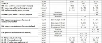
The amount of thyroglobulin in the blood of a healthy person usually does not exceed 10 ng/ml, but a slight increase is not scary, since the norm is slightly higher. The levels of some hormones, such as calcitonin, may differ between men and women.
| Tumor marker | Normal thyroid gland in men | Normal thyroid gland in women |
| Thyroglobulin | From 2 mg/ml, but not more than 20 mg/ml; according to other sources, the norm is up to 60 mg/ml | |
| Calcitonin | 0.68–33 mg/ml, preferably closer to 8 mg/ml | 0.07—12.97 mg/ml |
| Carcinoembryonic antigen (CEA) | No more than 5 ng/ml | |
The standards are not uniform; they vary from laboratory to laboratory. But even if you receive an analysis with deviations from the norm, you should not panic.
Statistics show that people often go to laboratories, bypassing the doctor, focusing on the norms, but this is wrong. After all, as an isolated diagnostic method it is useless.
But if this happens, then the analysis should be assessed by an oncologist, who will make a diagnosis or refute it, referring for additional examination. It is worth remembering that in the presence of a tumor, the indicators are likely to be significantly overestimated, by 10 or even 20 times. A slight increase indicates a malfunction of the thyroid gland.
About 10% of healthy people have elevated levels. During pregnancy, indicators can increase significantly and this is normal, as various hormonal changes occur. After breastfeeding ends, hormones should return to normal. With age and with inflammatory processes in the body, indicators also increase.
Can a tumor marker show a false result?
The presence of a high concentration of tumor markers does not mean that the patient has a thyroid pathology. Increased protein synthesis is influenced by some changes in the human body. Tumor markers can give an erroneous result in the following cases:
- pregnancy;
- hormonal disbalance;
- presence of bad habits;
- inflammatory processes in the liver, liver cirrhosis;
- ulcerative colitis;
- tuberculosis;
- pneumonia;
- hemorrhoids in the acute stage.
Incorrect results may be detected in the presence of benign tumors or cysts; such pathologies respond well to gentle treatment and do not require urgent intervention.
About 10% of the healthy population has increased rates. During pregnancy and breastfeeding, indicators can increase greatly due to hormonal changes in the body. After breastfeeding ends, the indicators should return to normal. With age and in the presence of inflammatory processes in the human body, data may also increase.


