Features of the structure of the lungs
The lungs themselves are organs that ensure gas exchange between the blood and the environment. The grooves divide the lungs into several lobes. The right lung consists of three, and the left lung consists of two lobes. The shares, in turn, consist of segments. They are truncated cones, the apex of which is directed towards the pulmonary roots. The latter are depressions on both lungs from the mediastinum, through which the pulmonary arteries enter the lungs and the pulmonary veins exit. Of these, each segment includes a branch of the pulmonary arteries and segmental bronchi, into which the two main bronchi are divided.
Pulmonary arteries - branches of the pulmonary trunk extending from the right ventricle
They occupy a central position in the segment, and veins pass between them, inside connective tissue partitions. The number of segments in the lobes of the lungs varies. There are 10 of them on the right:
- Upper lobe – 3 segments.
- The average share is 2 segments.
- Lower lobe – 5 segments.
On the left, both lobes have 4 segments, 8 in total.
Upper Lobe - upper lobe; Middle Lobe - middle beat; Lower Lobe - lower lobe
Segments of the right lung: posterior section
This area is located dorsal to the apical one. There are 5 boundaries in a segment. Two of them are projected between the apical, superior and posterior on the medial surface. Three boundaries are on the costal surface. The bridge that forms the anterior and posterior segments of the lung has a vertical orientation. To the vein, artery and bronchus of the posterior element, it is carried out from the medial side in the dissection of the pleura of the portal surface or from the initial section of the horizontal groove. Between the vein and artery there is a segmental bronchus. The blood channel of the posterior element connects to the vessel of the anterior one. Together they enter the pulmonary vein. Between the II and IV costal plates, the posterior segment is projected onto the surface of the sternum.
What are segments?
Inside, the segment consists of lobules, which measure approximately 20 by 15 millimeters, and have their bases facing the outside of the segment. The segmental bronchus is divided into terminal bronchioles, and enters each of the numerous apices. The lobules themselves consist of the main functional unit of the lungs - the acini. It is they who ensure gas exchange between the blood that flows through their capillaries and the air in their cavity.
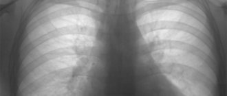
On an x-ray, the doctor can see the lobes and segments. For easier analysis of the images, the image of the lungs is divided into three conventional parts, drawing horizontal boundaries.
Conventional division of the lungs into three zones
Classification
According to its etiological factor, segmental pneumonia in children and adults is:
- primary - this means that the pathology acts as an independent disease and is caused by one of the above microorganisms;
- secondary – characterized by the fact that it develops against the background of other pathologies, i.e. it acts as a complication of them.
The primary inflammatory process in a particular segment of the lungs is divided into:
- typical – caused by the influence of bacteria from the category of the most common pathogens;
- atypical - in such cases, inflammation is caused by rare varieties of the pathogen;
- viral – caused by an increase in the number of viruses;
- fungal – the root cause most often is aspergillus or candida;
- drug;
- toxic.
In addition, there are several routes of infection through which pathogenic microorganisms enter the human body:
- bronchogenic – the causative agent of the disease enters through inhaled air;
- hematogenous - with blood flow;
- metastatic - from another source of inflammation in the body.
Division of the disease depending on the place where the infection occurred:
- community-acquired or home segmental pneumonia;
- nosocomial or nosocomial pneumonia;
- inflammation associated with medical care, in particular the procedure of artificial ventilation of the lungs.
Classification of pneumonia
According to the severity of the disease, there are three degrees:
- light;
- moderate;
- heavy.
Depending on the number of affected segments, the disease is divided:
- segmental pneumonia itself - in this case only one segment of the left or right lung is affected;
- polysegmental pneumonia - this type is spoken of when several segments are simultaneously involved in the pathology;
- subsegmental pneumonia is inflammation of part of the pulmonary segment, i.e. the entire upper, middle or lower lobe.
In addition, according to the localization of the inflammatory focus, left-sided and right-sided segmental pneumonia are distinguished.
Topography of a normal lung
Topographically, the lungs are distinguished by zones of the apexes, which are located above the shadow of the clavicles. Below the collarbone, the upper part of the lungs begins, the lower border of which is the anterior segments of the second ribs. From the second to fourth costal segments there are the middle sections, and down from them there are the lower sections. Thus, there are three landmarks on the radiograph - the collarbones, and the anterior ends of the second and fourth pairs of ribs.
If we draw vertical lines through the point where the clavicle intersects with the outer contour of the ribs and the middle of the clavicle, then the pulmonary field will be divided into internal, external and medial zones.
Since the segments are layered on top of each other, their detailed study is carried out in a lateral projection image.
The right lung is represented by ten segments. The 1st segment of the apex is located in the dome. The posterior C2 of the upper lobe begins from its posterior surface, and C3 begins from the anterior outer surface.
C4 of the middle lobe is located outside, located between the horizontal fissure and the lower parts of the oblique. Ahead is C5.
If you draw an imaginary line from the accessory interlobar fissure back, it will become the lower border of the 6th segment of the lower lobe. Segments C7 to C10 are located at its base. The most medial is the 7th, it overlaps the 8th and 9th, lateral. At the rear is C10.
On the left their location is slightly different. C1-C3 are united into a large posterior apical segment. Below, in place of the middle lobe, there is a lingular segment, which is divided into C4 and C5.
X-ray anatomy of the chest (lung segments are indicated by numbers)
Lateral division
This segment is projected from the side of the medial part only as a narrow strip lying above the interlobar oblique groove. The bronchus has a posterior orientation. In this regard, the segment is located on the back part in the middle lobe. It is visible from the surface of the ribs. There are five boundaries in the department. Two of them lie along the medial surface, separating the anterior and lateral, lateral and medial segments of the lung. The first boundary lies in accordance with the terminal portion of the oblique groove. The other three are located on the costal surface of the organ. They separate the medial and lateral segments of the middle lung.
The first boundary runs vertically. It runs from the center of the horizontal furrow to the edge of the oblique. The second border runs between the anterior and lateral segments. It corresponds to the location of the horizontal groove. The third border is in contact with the posterior and anterior segments in the lower lobe. The vein, artery and bronchus are deep. The approach to them is only possible below the gate along an oblique furrow. The lateral segment is located in the area between the IV-VI ribs.
Indications for the study
A plain radiograph of the chest organs is a routine examination method. Moreover, fluorography, which is a modification of this study, should be performed on all healthy people approximately once a year.
When a patient is admitted to the hospital, doctors in most cases order an x-ray, since it is imperative to make sure that there are no pathological changes in the lung fields, which may be signs of the initial stages of the disease. After all, some pathologies can be identified using this method even before a person has complaints.
In order for an x-ray to be prescribed, the following symptoms must be present:
- Cough.
- Complaints of shortness of breath.
- Complaints about lack of air.
- Whistles when breathing.
- Wheezing when breathing.
- Changes in respiratory movements of the chest.
- Chest pain, especially when breathing.
- Swelling in the legs.
- Mantoux reaction different from normal.
Diagnostics
In order for a clinician to suspect the occurrence of segmental pneumonia, it is necessary to carry out not only a series of laboratory and instrumental examinations, but also manipulations performed directly by a pulmonologist.
Thus, the first stage of establishing the correct diagnosis includes:
- familiarization with the medical history - to identify a chronic disease that could lead to the development of segmental inflammation in the lungs;
- collection and analysis of the patient’s life history;
- a thorough physical examination, aimed at listening to the patient using a phonendoscope and measuring temperature;
- a detailed survey of the patient is necessary to establish the first time of appearance and severity of symptoms, which, in turn, will indicate the severity of the disease.
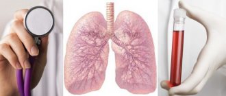
Laboratory and instrumental diagnostic measures include:
- general clinical blood test;
- blood biochemistry;
- analysis of sputum produced when coughing;
- bacterial blood culture;
- chest x-ray;
- CT and MRI.
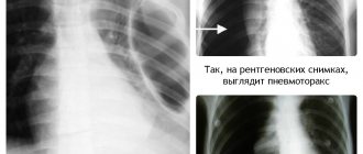
X-ray of the lungs with segmental pneumonia
All of the above procedures allow not only to make the correct diagnosis, but also to differentiate segmental pneumonia from:
- pulmonary tuberculosis;
- primary or metastatic cancer of the left or right lung;
- pulmonary infarction;
- bronchiolitis obliterans;
- foreign object in the bronchi;
- focal, interstitial and lobar forms of pneumonia.
Lung image analysis
Thus, X-rays can be analyzed in stages, which allows doctors not to miss changes that are subtle at first glance. However, it must be remembered that this is a conditional division, and the radiological zones are not equivalent to the pulmonary segments. First you need to evaluate their symmetry and the presence of obvious defects. They can be represented by elements of darkening or clearing, as well as changes in the shape and size of the lungs, as well as a violation of their contours.
Since the lungs are filled with air, which transmits x-rays well, on x-rays they look like light tissue with high transparency.
Their structure is called the pulmonary pattern. It is formed by small branches of the pulmonary arteries and veins, as well as small bronchi.
Since from the roots to the periphery the vessels and bronchi are divided into smaller branches, which are less visible on x-rays, the intensity of the pattern from the center to the periphery decreases. It becomes paler and almost indistinguishable at the outer edges of the lungs. It also becomes depleted in the upper sections, becoming thickest towards the bottom.
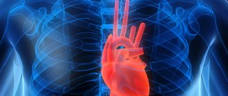
Blood supply
The smallest “details” of the respiratory organ are the alveoli. Alveolar sacs are vesicles covered with a thin network of capillaries through which our lungs breathe. It is in these pulmonary “atoms” that all gas exchange occurs. The lung segments contain several alveolar ducts. In total, there are 300 million alveoli in each lung. They are supplied with air by arterial capillaries. Carbon dioxide is taken up by the venous vessels.
The pulmonary arteries operate on a small scale. That is, they nourish the lung tissue and make up the pulmonary circulation. The arteries are divided into lobar and then segmental, and each feeds its own “section” of the lung. But bronchial vessels, which belong to the systemic circulation, also pass here. The pulmonary veins of the right and left lung enter the flow of the left atrium. Each segment of the lung has its own grade 3 bronchus.
On the mediastinal surface of the lung there is a “gate” hilum pulmonis - depressions through which the main veins, lymphatic vessels, bronchi and arteries pass to the lungs. This place of “intersection” of the main vessels is called the root of the lungs.
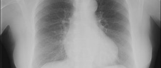
Pathology detected on images
Almost all diseases that can occur in the lungs change the density of their tissue and its airiness. On an x-ray, this appears as areas of darkening or clearing. For example, segmental pneumonia leads to tissue infiltration by leukocytes and macrophages, dilation of blood vessels, and, as a result, edema. As a result, the area becomes denser, transmits x-rays less well, and a darkened zone is visible in the image.
The oval indicates the darkening area
You need to carefully examine the root area and pulmonary pattern. Their strengthening indicates either the early stages of the inflammatory process, or an obstacle to the outflow of blood, for example, thrombosis, edema in heart failure. Knowledge of the segmental structure can help in differential diagnosis. Thus, tuberculosis most often affects the apical segments, since they have poor oxygenation, which allows mycobacteria to easily grow and multiply. But pneumonia often develops in the lower and middle sections.
Surgery: indications
If the functions of any area are impaired, its resection (removal) is carried out. This need may arise in the following cases:
- Destruction of tissue due to inflammation caused by infection (tuberculosis, most often).
- Degeneration of the lung in the process of tumor growth (malignant and benign).
- Acquired or congenital formation of a hollow area.
- Purulent tissue decay against the background of a number of pathologies.
- Injuries.
Treatment
The goal of therapy for lingual pneumonia is as follows:
- stopping the source of infection;
- eliminating breathing problems;
- resorption of areas of inflammation;
- After identifying the nature of the disease, treatment is prescribed:
- drugs to fight viruses or bacteria;
- mucolytics;
- expectorants;
- anticoagulants;
- bronchodilators;
- antispasmodics;
- immunomodulators;
- vitamin complexes.
Sometimes antibiotics are recommended for viral etiologies of the disease. This is done to avoid the addition of a bacterial infection.
The patient is recommended to eat a balanced diet; his menu must include foods high in fiber, protein and vitamins. We must not forget about the drinking regime - in case of pneumonia, drinking plenty of fluids is recommended. In addition, physiotherapy and inhalations are indicated.
Treatment of the disease is complicated by the anatomical features of the left bronchus - getting the drug to the site of inflammation is difficult.
As for the treatment of lingual pneumonia in children, it is similar to the treatment for adults, but the dosage of drugs will be different. Also prescribed:
- chest massage course;
- rubbing;
- tapping on palms.
Indications for hospitalization of the patient:
- acute form of lingual pneumonia;
- development of respiratory failure;
- childhood or old age of the patient;
- critical temperature indicators;
- dehydration;
- presence of purulent sputum.
The duration of therapy depends on many nuances - the extent of inflammation, concomitant chronic pathologies, and the timeliness of started therapy. However, usually the disease begins to recede after 5 days from the start of taking medications. As for complete recovery, it occurs in 14-20 days. Treatment of patients with HIV will take longer.
Clinical picture
Since left-sided pneumonia is difficult to treat, the urgency of seeing a doctor plays a vital role . The disease begins abruptly. This is a characteristic feature of this form of the disease. The clinical picture is as follows:
- a sharp rise in temperature to critical levels - 40ºC;
- heavy night sweats;
- cough;
- fever;
- dyspnea;
- general weakness.
Also, a bluish tint of the nasolabial triangle and nails is considered a specific sign of lingual inflammation.
The cough with lingual pneumonia is sometimes dry, although more often with expectoration of sputum. In adults, the pathology is often asymptomatic , and this poses a threat, since the lack of therapy can lead to unwanted complications.
In children, lingual pneumonia can be expressed by shortness of breath, prolonged hiccups and chest pain.
With viral etiology the following is observed:
- paroxysmal cough without sputum production;
- weakness;
- migraine;
- breathing disorders.
Characteristics of the disease and sources of its occurrence
As the disease progresses, scarring of the lung air sacs is observed, making normal gas exchange difficult. The alveoli (air sacs) gradually lose their elasticity, lose the ability to fully contract and extract oxygen out. Breathing becomes constricted, partial oxygen starvation is observed.
Connective tissue gradually replaces lung tissue. But connective tissue is denser in structure and does not allow air to pass through. As a result, there is an increase in lung volume due to connective tissue.
In the early stages of the disease, it is still possible to overcome its manifestations by relying on therapeutic treatment and eliminating the underlying causes of the disease itself. This disease is considered progressive, and in its final stages it is characterized as fatal.
The root cause of this disease does not depend on age. This diagnosis is made to both children and adults.
Changes in the lungs that are fibrotic in nature most often occur against the background of:
- past infectious disease,
- tendency to allergies,
- the use of radiotherapy,
- prolonged contact with dust.
Factors that cause the development of linear fibrosis can be triggered by external pathogens. The degree of the disease directly depends on the state of the environment. The worse the environment, the greater the likelihood of linear pulmonary fibrosis.
Smoking, of course, is one of the factors that inevitably provokes the destruction of lung tissue and the destructive functioning of the alveoli. The phenomenon of pulmonary fibrosis is 80% more common in regular smokers.
Medicine has not fully identified all the reasons for the occurrence of this disease, but in addition to the above factors, we can add the following:
- interaction with toxic secretions,
- hereditary predisposition,
- low ecological level of the environment,
- age barrier (after 45 years).
Among the assumptions of doctors is the hypothesis that lack of sleep may be a factor provoking the disease. An insufficiently restored body constantly feels a lack of oxygen. The active functioning of the lungs is constantly deteriorating, and as a result, the onset of the disease.
One of the most common causes is diseases of the connective tissues of the internal systems of the body, for example, scleroderma, rheumatoid arthritis. Infectious diseases and associated inflammatory processes can lead to linear pulmonary fibrosis. This division includes advanced forms of tuberculosis and pneumonia.
Fibrosis often occurs due to complications of many ailments. Most specialists try to eliminate the root causes of the disease first.
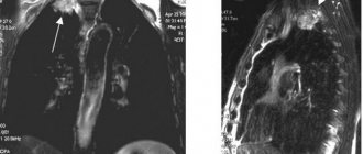
Segmental structure of the lungs on x-ray and CT
The lungs are a paired organ consisting of tubular systems. They are formed by segmental bronchi, their branches, pulmonary, blood, and lymphatic vessels. The growth of tubular formations is parallel to each other. They form bundles of bronchi, veins, and arteries. The image shows that each lobe of the organ consists of small sections that determine the segmental structure of the lungs.
Description and classification of bronchopulmonary segments
The bronchopulmonary segment is a functional part of the main respiratory organ. In medicine, there are several versions of the classification of lobar areas. Specialists of different profiles (radiologists, thoracic surgeons, pathologists) divide the lobes of the lung into an average of 4-12 segments. In relation to the official classification, in accordance with the anatomical nomenclature, it is customary to distinguish 10 segments of the organ.
All sectors figuratively resemble pyramids or irregular cones. They are located in a horizontal plane, with their base towards the outer surface of the lung, and their apex towards the hilum (the entry point of nerves, main bronchi, blood vessels). The sections differ in pigmentation, so their boundaries are visually visible.
Segmental constitution of the right lung
The number of segment plots depends on the shared structure.
The upper lobe of the right lung includes three lobes:
- S1 – located under the pleural vault, protruding in the upper aperture of the chest (the opening formed by the sternum, ribs, thoracic vertebra);
- S2 – lies posteriorly on the border with 2-4 ribs;
- S3 – partially intervening with the vena cava coming from the head and the right atrium, the base abuts the anterior chest wall.
The middle lobe is divided into 2 segments. S4 – moves forward. S5 – touches the sternum and anterior chest wall, fully communicates with the diaphragm and heart.
The lower lobe is formed by 5 sectors:
- S6 – basal section, lies near the spinal column in the area of the wedge-shaped lobar apex;
- S7 – contacts the mediastinum and diaphragm;
- S8 – the lateral part is in contact with the chest wall, the entire segment lies on the surface of the diaphragm;
- S9 – looks like a wedge between other areas, the base touches the diaphragm, the side – the area of the chest near the armpits, anatomically located between the 7th and 9th ribs;
- S10 - lies along the paravertebral line, is located further away from all other segments, penetrates into the depths of the organ, into the sinus of the pleura (the depression formed by the ribs and the diaphragm).
Segmental structure of the left lung
The segments of the left lung are different from the right. This is due to the different structure of the lobes and the organ as a whole. The left lung is 10% smaller in volume. At the same time, it is longer and narrower. The dome of the organ is lowered. The width is smaller due to the heart located on the left side of the chest.
Dividing the upper lobe into segments:
- S1+2 – the base touches 3-5 ribs, the inner part is adjacent to the subclavian artery and the arch of the main blood vessel (aorta), can be in the form of one or two segments;
- S3 is the largest section of the upper lobe, located in the area of ribs 1-4, touching the pulmonary trunk;
- S4 – in front of the chest is located between 3-5 ribs, in the axillary region – between 4-6 ribs;
- S5 – located under S4, but does not touch the diaphragm.
S4 and S5 are lingular segments that topographically correspond to the middle lobe of the right lung. From the inside, they touch the left ventricle of the heart, pass between the pericardial sac and the chest wall into the pleural sinus.
Segmental structure of the lower lobe of the lung
- S6 – located paravertebrally;
- S7 – in most cases includes the bronchus (trunk and beginning of the bronchus of the underlying segment);
- S8 – participates in the formation of the diaphragmatic, costal and inner surface of the left lung;
- S9 – located at the level of 7-9 ribs in the axillary region.
- S10 is a large section located posteriorly in the area of ribs 7-10, touching the esophagus, descending line of the aorta, diaphragm, the segment is unstable.
What do the segments look like on an x-ray?
Since the structural unit of the lung (acinus) is not determined on x-ray, lobar segments are assessed to identify pathological processes. In the photographs they give a distinct shadow with the exact localization of altered or inflamed tissues (parenchyma).
To determine the boundaries of areas, diagnosticians use special markers. First, the lobes are identified, and then the segments of the lungs are identified on the radiograph. All sections of the organ are conventionally separated by an interlobar oblique strip or fissure.
To separate the upper lobe, focus on the following indicators:
- in the rear view of the chest, the line begins from the process of the 3rd thoracic vertebra;
- at the level of the 4th rib it passes into the horizontal plane;
- then rushes to the highest midpoint of the diaphragm;
- in the lateral projection, the horizontal gap starts from the 3rd thoracic vertebra;
- goes through the root of the lung;
- ends at the diaphragm (midpoint).
The line in the right lung separating the middle and upper lobes runs along the 4th rib to the root of the organ. If you look at the picture from the side, it starts from the root, runs horizontally and leads to the sternum.
In the diagram, the slots are indicated by a straight line or a dotted line. Knowledge of the topography of the segments and the ability to correctly decipher images determines how accurately the diagnosis will be made and successful treatment will be carried out.
When examining X-ray films, it is necessary to be able to distinguish pathological processes from the abnormal structure of the chest organs, individual human anatomy, and congenital defects.
How are segments determined on computed tomography?
The tomography method is fundamentally different from x-rays. Lung segments on CT and their structure can be viewed layer by layer in several projections.
On cross-sections of a CT scan, the layers of the pleura, connective tissue layers between parts of the lung, and fissures are not visible. Their location can be guessed from the vascular pattern. In the area of the pleura, arteries into veins are not visualized, therefore, in places where there should be interlobar fissures, an area without vessels is determined. High-resolution tomography, in which the pattern thickness can be reduced to 1.5 mm, allows you to see the layers of the pulmonary membrane.
In the frontal projection, the main interlobar line departs from the chest and goes to the mediastinum. It ends on the back at the level of the 3rd thoracic vertebra. Passing through the organ, it affects the root and a third of the diaphragm. If you make a thin axial cut, the main gap between the lobes will look like an even horizontal line of white color.
If there is an additional interlobar fissure in the image, this is the right lung. In the area of the white zone without blood vessels, there are ring-shaped stripes of low density with blurred contours. This is due to the fact that the right lung is larger than the left. This sign is also characteristic of thickening of the pleural film between the lobes and indicates an inflammatory process.
The localization of bronchopulmonary segments is distinguished by the direction of blood vessels and bronchi of different sizes. Each segmental section faces the root with its apex, and its base faces the muscular septum and chest wall. In the root region, arterial and venous vessels, bronchi are clearly visible in transverse and longitudinal projection. At the base of each section, the vessels decrease in size.
Differences in segmental anatomy of the lungs in children
The peak of segmental formation of the respiratory organ occurs in the first 7 years of a child’s life. The size of the structural units of the parenchyma (alveoli) in children of the first year of life is half that of children 12 years old. In terms of their structure, the bronchi penetrating the segments are not yet fully formed.
There is a denser layer between the segments themselves, which clearly delimits them. The structure of the interlobar pleura is loose and easily amenable to morphological changes.
On x-rays and CT scans, the lines between the segments are unclear. In infants under 2 years of age, they resemble notches on the surface of the organ. Groups of lymph nodes flow into the main slits, which is due to the close location of the lung root.
Externally, the boundaries of the lobes are determined by passing furrows. In children, to distinguish segments, the arrangement of the bronchial tree and branches extending from it is used.
Each segment is independently supplied with blood, innervated and ventilated. This fact helps to highlight individual areas with their projection on the chest. This is important during lung operations and detection of focal inflammation.
Treatment of linear fibrosis
The above material provides a complete description of the diagnosis of linear pulmonary fibrosis, what does this give the patient? The answer is the correct choice of treatment method.
It is no longer possible to get rid of the connective tissue that has formed as a result of the disease. Therefore, the treatment of this disease includes measures that stop the process of creating new connective tissues.
First of all, it is necessary to exclude the possibility of inhaling harmful substances that stimulate the development of the disease. If the patient smokes, then getting rid of this addiction is simply necessary. It is much more difficult to protect yourself from infectious diseases, but it is still necessary to take all preventive measures.
Conservative therapy is effective only in the early stages of the disease. Drug treatment can eliminate the symptoms of the disease and improve the patient’s quality of life. To achieve the greatest effectiveness in the early stages, it is advisable to use therapeutic breathing exercises, oxygen therapy and a moderate diet in combination with medications.
With such a complex effect, it is possible to exclude further possible complications. Expectorants and medications that thin sputum are widely used. The use of steroid drugs in a short time interval can help relieve the patient from the unpleasant sensations of the disease.
For typical heart failure, the patient must be prescribed a number of cardiac glycosides. To strengthen defenses and restore immunity, it is advisable to take a full vitamin course.
At an early stage of the disease, doctors often prescribe a cycle of therapeutic chest massage, which alleviates coughing attacks and minimizes the manifestation of other symptoms of the disease. As an additional therapeutic effect, therapeutic massage strengthens the pectoral muscles and restores pulmonary processes, which is important for the elderly and children.
As an additional therapy, you can independently perform therapeutic breathing exercises, which leads to oxygen saturation of the circulatory system and strengthening of the respiratory system. With proper training, breathing exercises help in the removal of mucus, improve lung ventilation and prevent congestion in the lung tissues.
Patients with linear fibrosis must be observed by a doctor. Life expectancy with this diagnosis directly depends on the stage of development of the disease, as well as the timeliness of therapeutic treatment.
It is very important not only to minimize the manifestations of pulmonary fibrosis, but also to eliminate the disease that provoked this disease.
How to keep your lungs healthy?
The greatest harm to the entire respiratory system is caused by an unhealthy lifestyle, poor nutrition and smoking.
Even if a person lives in a stuffy city and his lungs are constantly “attacked” by construction dust, this is not the worst thing. You can clear your lungs of dust by traveling to clean forests in the summer. The worst thing is cigarette smoke. It is the toxic mixtures inhaled when smoking, tar and carbon monoxide that are scary. Therefore, you need to quit smoking without regrets.
C1. Apical C2. Posterior C3. Front
C1-2. Apical posterior C3. Anterior C4. Upper reed C5. Lower reed
C4. Lateral C5. Medial
C6. Apical C7. Medial basal C8. Anterior basal C9. Lateral basal C10. Posterior basal
C6. Apical C7. C8 is missing. Anterior basal C9. Lateral basal C10. Posterior basal