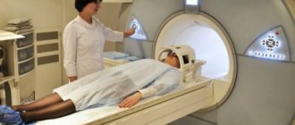
Magnetic resonance imaging of the brain makes it possible to see images of brain sections in different planes and with high resolution. Nowadays, MRI is considered the most accurate method for diagnosing diseases of the nervous system.
MRI belongs to the category of absolutely safe examinations and can be prescribed to patients of any age, including young children. Due to its harmlessness, MRI examinations can be performed regularly, which is necessary for some diseases. High quality brain images are achieved by analyzing the results of the interaction of the magnetic field and radio waves with the human body.
What does a head MRI show?
The capabilities of tomography make it possible to study the structure and functioning of the brain in a short period of time and identify the origin and development of various pathologies.
Diseases detected by MRI scans include:
- congenital anatomical anomalies and defects;
- disturbances in the blood supply system;
- inflammatory processes;
- post-traumatic consequences;
- processes leading to dementia;
- tumor or cystic formations.
Clear and detailed images on tomographic images will give an answer to how functional and productive the human nerve center is.
Inflammatory diseases of the brain
MRI is most often used to diagnose inflammatory brain diseases such as encephalitis and abscess.
With encephalitis, MRI shows foci of inflammation in the brain, swelling of the frontal and temporal regions, as well as microhemorrhages. Encephalitis can be diagnosed using MRI only on days 3-5 of the disease. MRI diagnostics are also performed to exclude other diseases with similar symptoms.
MRI for brain abscess shows the location and size of the abscess. The tomograph clearly visualizes cerebral edema and deformation of liquor-containing formations - this is considered concomitant signs of a brain abscess.
How to do an MRI of the brain
MRI of the head in adults is not only informative, but also a safe research method that does not require special preparatory procedures before it is performed.
Before the tomography, the attending physician examines the patient and talks in detail about the intricacies of the upcoming procedure.
The patient must inform the doctor about the individual characteristics of the body, which may affect the choice of magnetic tomography as a diagnostic method:
- tendency to be allergic to any drugs (with intravenous contrast);
- chronic diseases in the acute stage;
- the presence of prostheses, implants, clips and other metal objects in the body;
- intolerance to confined spaces;
- inability to maintain body immobility due to pain or mental characteristics;
- fact of pregnancy (indicating the period) or breastfeeding.
The quality and detail of the images taken depends on the power of the tomograph, expressed in magnetic field strength. The higher this value, the more informative the screening result.
In Krasnodar, a medical specialist specializes in magnetic resonance imaging. The clinic has a PHILIPS INTERA tomograph (installed in 2019, high-field, closed type) with a voltage of 1.5 Tesla. The specified technical parameters allow for quick and effective diagnosis of children over 5 years of age and adults.
Modern tomograph Philips Intera 1.5 T
Why and how is MRI of the thoracic spine performed?
MRI of the thoracic spine is prescribed for people with back pain, suspected malignancy, or to clarify the diagnosis. How long the examination lasts depends on the size of the area of interest. So, with a normal study of the thoracic spine, an MRI lasts 10–20 minutes, but if additional enhancement of the picture is needed, the procedure will take longer.
Modern high-field tomographs have a higher acquisition speed, therefore, when using them, MRI takes less time.
Magnetic resonance imaging of the thoracic spine is prescribed if the patient has:
- injuries, fractures, dislocations in the thoracic region;
- degenerative diseases (osteoarthrosis, spondylosis, spondyloarthrosis);
- changes in intervertebral discs (hernias, protrusions);
- narrowing of the spinal cord canal;
- benign tumors (hemangiomas, fibromas);
- malignant tumors and metastases from other organs;
- changes in the vertebrae due to infectious diseases (osteomyelitis, tuberculosis, syphilis);
- osteochondrosis of the thoracic intervertebral discs and joints;
- osteoporosis;
- diseases of the spinal cord (stroke, inflammatory processes).
In order to improve visualization of the thoracic vertebrae and obtain a three-dimensional image, it is often necessary to inject a contrast agent. It is necessary when studying the vessels supplying the spine and surrounding tissues. An MRI of the thoracic spine with contrast takes 10–15 minutes longer because it requires taking pictures first without the drug and then after it is injected.
There is no special preparation for an MRI of the thoracic spine, but if it is performed with contrast, you must not eat four hours before the examination, as the drug can cause nausea or vomiting.
Typically, patients are asked to remove all metal objects and jewelry, are helped to lie down on a retractable table, and are offered headphones (the machine makes noise while the examination is ongoing). Then the scanning begins. If necessary, the medical staff gives an intravenous injection of a contrast agent, and the body is scanned again.
The question often arises: “How is MRI of the thoracic spine performed in children?” The only difference is general anesthesia, and it is not indicated for all children. Teens and school-age children who understand lying still can have an MRI without sedation. Small and hyperactive children are put under anesthesia and only then do they begin an MRI of the thoracic spine. General anesthesia does not affect the duration of the MRI, but you will have to stay in the hospital and wait until the child fully wakes up and the anesthesiologist allows you to take him home.
MRI of the thoracic spine is a safe, painless and informative examination that lasts 15–40 minutes, depending on the conditions.
How does the procedure work?
Immediately before the scan, the patient is asked to free himself from removable metal objects (watches, jewelry, hairpins, etc.). Electronic devices must also be left in the doctor's office.
The process of preparing for the examination includes the following steps:
- The patient lies down on a couch that is placed inside the tomograph. Doctors monitor the general condition of the patient, the correct position of the body on the table and install a gradient coil on the scanned area, which is necessary to convert magnetic pulses into an image on a computer monitor.
- If contrast is used, an allergy test for the injected drug is first performed. The coloring agent is injected into the cubital vein. Sometimes so-called dynamic contrast is used, in which the drug is administered gradually through a catheter. With this method, you can see how quickly the substance is distributed throughout the tissues, which helps in diagnosing demyelinating diseases and cancer.
- The radiologist records the activity of the tomograph on the computer. In the medical field, doctors are always present on shift. This means they can begin to interpret images in real time as they appear on the computer screen, significantly reducing wait times for conclusions.
The operation of the tomograph is associated with some sound signals, reminiscent of knocking or clicking. To neutralize noise, patients are given headphones so that the screening time can be spent comfortably with pleasant music.
The duration of an MRI of the brain, depending on the scope of the tasks, takes about 20 minutes, and with the introduction of contrast it takes 15 minutes more.
What can affect the duration of an MRI?
The duration of the examination depends on the power of the equipment. It can be low-, medium- and high-field. The choice of device depends on the disease. Low-field tomographs are mainly used to assess changes after therapy, when the size and location of the lesion have already been determined. To identify tumors or prepare for surgery, the examination is carried out on high-field ones.
Tomographs are also divided into open and tunnel (closed). The latest devices are more powerful and have additional functions. On low-field (open) images the image clarity is much lower. Such devices are also less powerful. Also, the duration of the scan depends on the type of examination:
- Diffusion-weighted helps to detect earlier circulatory disorders in the brain.
- Diffusion tensor allows you to examine the vascular system without contrast. This information is very important before the planned operation.
- Perfusion involves intravenous contrast. It is estimated how much dye reaches the lesion and the speed of fluid flow.
- Spectography is performed on third-generation devices. With their help, the chemical formula of the brain is determined.
The duration of the examination depends on the type of tomography. Diffusion-weighted - 45 minutes, with perfusion for 15 minutes. longer. Since tractography produces a large number of images, it takes much more time to process them. Modern tomographs have ultra-fast spin echo, so a regular scan is completed in a quarter of an hour.
What does a brain MRI show?
Magnetic tomography is one of the most highly accurate visual diagnostic methods. Using magnetic scanning, you can obtain detailed and detailed images of the organ in three projections. Even small structures such as the pituitary gland are clearly visible in the images taken. Magnetic resonance scanning makes it possible to see the structural features of the brain, which means carefully studying the structure of the nervous substance of the cerebral cortex for abnormalities, to see hemorrhages and inflammation of the meninges.
MRI for traumatic brain injuries
An MRI of the brain in case of a skull injury will allow you to determine the presence of damage and determine its type:
- brain contusion;
- diffuse axonal injury (DAI);
- intracranial hemorrhage;
- traumatic subarachnoid hemorrhage.
Brain contusion
A brain contusion is characterized by damage to the nervous tissue and the presence of a focus of necrosis, which is visible on MRI. Foci are of the following types:
- cytotoxic edema (type I contusion);
- infiltrative-gliotic changes and pinpoint petechial blood loss (type II contusion);
- a combination of edema and a hemorrhagic component in the form of intracerebral hematomas (type III contusion);
- intracerebral hematomas (type IV contusion).
All types of bruises are equally well diagnosed on MRI due to clear visualization of the lesions.
Diffuse axonal injury
DAP on MRI is visualized by an increase in brain volume, which occurs due to diffuse edema. Finely focal hemorrhages in the corpus callosum, brainstem and periventricular structures may also indicate DAP.
Intracranial hemorrhage
Intracranial hemorrhage contributes to the formation of a subdural, epidural or intracerebral hematoma. MRI helps determine the type of hematoma, its size and location. The subdural hematoma in the image has a crescent shape, the epidural hematoma is shaped like a biconcave lens, and the intracerebral hematoma is round in shape with an uneven contour.
Traumatic subarachnoid hemorrhage
Traumatic subarachnoid hemorrhage (TSH) is diagnosed using MRI several days after its occurrence. MRI allows you to determine the extent and location of hemorrhage.
Indications for MRI of the brain in adults
Typically, a referral for an MRI examination is issued by the attending physician. The following symptoms and conditions may serve as medical indications for scanning:
- suspicion of the presence of a benign or malignant tumor;
- injuries;
- persistent pain, migraine attacks;
- dizziness and loss of consciousness of unknown etiology;
- constant ringing in the ears;
- convulsions, epileptic attacks;
- sharp deterioration of memory, hearing, vision, speech disorders.
MRI of the brain is also used for dynamic monitoring of the patient’s condition after neurosurgery and to identify congenital developmental anomalies.
Epilepsy
If epilepsy is suspected, MRI helps determine changes in the structure of the brain, as well as the source of the pathology. For example, in young children, the cause of epilepsy may be malformations of the brain, hemorrhages, or the consequences of intrauterine infections. In adults, epileptic seizures can be caused by:
- brain tumors;
- strokes;
- traumatic brain injuries;
- infectious and inflammatory diseases and complications after them;
- cortical dysgenesis;
- hippocampal sclerosis.
All of the above changes are diagnosed using MRI and allow you to confirm or refute epilepsy in the patient.
What diseases will an MRI of the brain show?
Magnetic resonance scanning is used to identify the following brain pathologies:
- tumors of the brain and meninges;
- demyelinating and degenerative diseases (Parkinson's disease, Alzheimer's disease, multiple sclerosis);
- cysts, including pituitary cysts;
- empty sella syndrome;
- acute pathologies of cerebral vessels (hemorrhagic and ischemic strokes, thrombosis, aneurysms);
- complicated inflammatory processes (meningitis, sinusitis and others).
Degenerative brain changes on MRI scans
The final diagnosis is established by the doctor who gave the referral for the study. Usually a neurologist deals with brain diseases, but if a tumor is suspected, a referral can be made by an oncologist.
The radiologist conducting the study does not make a final diagnosis, but analyzes the images obtained and describes the detected abnormalities and formations. After the examination, the images are sent to the attending physician, who confirms or removes the initial diagnosis.
MRI shows stroke
MRI shows what changes in the structure of the brain the stroke caused, what the severity of these changes is, and what type of stroke (ischemic or hemorrhagic). The examination also allows you to accurately determine which part of the brain was affected by a stroke.
With an ischemic stroke in the early period, the image shows a zone of the brain in which a circulatory deficit has formed; in the late period, areas of brain necrosis are visible. If there is a lack of cerebral circulation due to the formation of a blood clot and blockage of blood vessels, the cause and source of hemorrhage can be determined.
In a hemorrhagic stroke, an MRI shows an area of increased tissue density in the brain - this indicates a hemorrhage that occurred as a result of an acute circulatory disorder.
MRI of the brain - contraindications
Despite all the advantages of MRI examination, this type of diagnosis is not indicated for everyone. There are two categories of reasons that prevent an MRI scan.
Absolute contraindications
Brain tomography is not allowed if:
- the presence of metal objects in the body (clips, prostheses made of ferromagnetic metal, bullet fragments, etc.);
- weight category from 130 kg and waist circumference over 150 cm;
- implanted pacemaker, neurostimulator, cochlear implant;
- in the first trimester of pregnancy;
- children under five years of age.
Undesirable moments
There are a number of situations that create problems for prescribing MRI diagnostics or complicate its implementation:
- complicated cardiovascular diseases;
- mental disorders in the acute stage;
- claustrophobia;
- tattoos with metal in dye;
The presence of implants made of non-ferromagnetic materials does not interfere with magnetic resonance examination, provided that documents for the prosthesis are available, three months have passed since its installation and the implant is outside the area of interest of medical specialists.
Preparation for the procedure
To perform magnetic tomography in normal mode, no special preparation is required. However, to make the procedure easier, there are these recommendations before MRI:
- leave metal jewelry at home - you will be asked to remove it before the procedure; also, do not take your watch with you; if possible, remove dentures (if they are removable);
- 1-2 hours before the tomography, try to empty your bladder and large intestine;
- To avoid the desire to go to the toilet during the procedure, do not drink a lot of water or eat 3-4 hours before the examination;
- do not smoke an hour before the test and do not drink alcohol the night before - toxic substances change the tone of the blood vessels in the brain, and the test result will be distorted.
During the entire scanning period (MRI time of the brain is from 15 to 60 minutes), you cannot move. This is easy to explain to an adult. But this is difficult to convey to young children. If diagnostics are prescribed for a child, try to get him or her ready for the procedure. MRI does not cause discomfort or pain - explain this to your child. Before the procedure, take a walk around the courtyard and clinic, let your child get used to the environment.
When is contrast needed during MRI of the brain?
A contrast agent is used for magnetic resonance imaging to improve visualization of soft tissues. The contrast is unevenly distributed in healthy and diseased tissues, helping to identify abnormalities and determine the boundaries of the development of the pathological process.
A contrast agent is used when it is necessary to diagnose brain pathologies such as:
- neoplasms;
Tumors have a developed vascular network. Distributing throughout the tissues, the contrast agent brightly colors the pathologically changed areas. Using contrast, you can analyze the size of the tumor and its exact location. Contrast also helps determine the malignancy of the tumor. Benign tumors have clear boundaries and regular shapes, while malignant tumors grow into healthy tissue and have uneven boundaries and shapes. These features are clearly visible on MRI scans obtained using contrast.
- demyelinating diseases.
Diseases such as Alzheimer's and Parkinson's disease manifest themselves in changes and degeneration of the nerve substance. Contrast is used to diagnose the extent of the disease.
In both cases, the use of a contrast agent makes it possible to clearly differentiate healthy tissues from altered ones, and to see the extent and extent of the pathological process.
How is the examination carried out?
Carrying out any type of MRI of the brain consists of several stages. Immediately upon arrival at the diagnostic center, the patient meets with a doctor and a nurse, who tell him about the features of the procedure. The next steps look something like this:
- The patient removes jewelry, watches, jewelry and all items that contain metal. After putting on a special robe, you can go into the room with the tomograph.
- The person is laid on the table. If necessary, a contrast agent is injected through the catheter.
- The scan is done after the table moves into the tomograph tunnel. The patient will hear a low background noise. To protect against it, you can wear headphones or take earplugs. For 15-30 minutes (with contrast MRI 30-60 minutes) you must remain motionless.
- As soon as the procedure is completed, the table will move back. The patient cannot leave immediately. The doctor needs to monitor his condition after contrast is administered.
After all necessary actions have been completed, the patient can be free.
Knowing how brain imaging occurs will help you tune in and prepare for the procedure in advance.
How to prepare for a head MRI
Preparing for any type of brain MRI, be it angiography or fMRI, is important. It depends on it how accurate the results will be. What can and cannot be done before the procedure?
- It is not recommended to drink a lot of liquid.
- Immediately before the tomography, you need to go to the toilet.
- The day before the procedure, you should not drink alcohol or smoke.
- No need to take jewelry or watches with you.
- A person preparing for an MRI should not be nervous. To calm down, you can ask a relative or close friend to go together.
Can I eat and drink before an MRI? There are no special rules regarding whether you can eat it or not.
How long does the procedure take?
A typical MRI of the brain, including preparation, lasts from 15 to 30 minutes. The duration of the procedure using a contrast agent lasts much longer (from half an hour to an hour). During this time, the specialist will have time to identify all developing pathologies and disorders.
results
https://youtu.be/A-ixmtsb7Q0
The MRI results are assessed by a specialist. The conclusion of a study of the brain of a healthy person looks something like this:
- The substance of the brain and its cerebral hemispheres is not changed.
- The spaces with cerebrospinal fluid are not displaced, their structure is not disturbed.
- Convexital spaces are unchanged, the ventricles are of normal size.
- The basal cisterns have not changed.
- The vault and base of the skull are in normal condition.
- The cerebellum and brain stem are not changed.
- There are no pathologies observed in the structures of the eye orbits.
- No changes are observed at the location of the mastoid processes and maxillary sinuses.
The patient receives a copy of the protocol on a disk or any other medium.
How is data decrypted?
Deciphering an MRI is essentially a comparison of the resulting images with images of healthy tissue. They differ from damaged ones in shade.
When a contrast agent is injected, damaged areas and blood vessels stand out strongly against the background of others. Thanks to this, you can see the development of diseases, tumors or blood clots.
Examination process and types of devices
There are two types of MRI machines: open and closed. The magnets used in the procedure, as well as the process itself, are divided into types.
Decoding MRI of the brain
For diagnosticians at DiMagnit MC, 15-20 minutes are enough to analyze the received images.
The center’s specialist doctor will inform the patient about the results obtained and advise how to proceed further. This consultation is free.
Upon completion of the diagnosis, the patient is given a disk with the image and a conclusion, which are included in the cost of the study. If necessary, the survey results can be duplicated onto the visitor’s flash card for an additional fee.
Interpretation of MRI scans
What determines the duration of the procedure?
How long an MRI takes depends on several parameters: the area that will be examined (one organ will take approximately 20-25 minutes, a general examination will take at least an hour), the power and type of diagnostic device (a more powerful device will scan faster and better) , the patient's condition.
The diagnostic examination consists of three stages: preparation for the procedure, examination and decoding and recording of the results.
to contents ^
The principle of operation of magnetic resonance imaging
The operation of the tomograph is based on the phenomenon of nuclear magnetic resonance. This method of obtaining information consists of measuring the intensity of the response of the nuclei of a hydrogen atom to the influence of a powerful magnetic field. The fact is that the concentration of this chemical element in different tissues differs.
Thanks to modern technologies, it has become possible to obtain high-resolution images and display them on the screen in 3D mode. Scanning of the required area is carried out in several projections with a high slice frequency. Magnetic resonance imaging may be performed using contrast. Preparations containing gadolinium (Omniscan, etc.) are used as a dye.
The contrast extremely rarely causes allergic reactions and is excreted by the kidneys without causing harm to the body. When administered intravenously, gadolinium is distributed throughout the circulatory system and “highlights” areas of increased blood supply on the tomograph screen. This examination is indispensable in the diagnosis of tumors because they have a developed network of blood vessels.
Contrast is widely used in oncology to detect pathological tumors
Contraindications
At first glance, MRI diagnostics are considered absolutely safe, but is it possible to do it indiscriminately for all patients? No, MRI is done taking into account existing absolute and relative contraindications. The doctor cannot allow the patient to undergo the procedure in the following cases:
- artificial heart valves;
- stents in coronary vessels;
- pacemakers, artificial heart pacemakers;
- insulin pumps;
- cochlear implant;
- auditory ossicular prostheses;
- electrostimulation of the vagus nerve;
- intraocular lenses;
- metal clips after operations on the brain and blood vessels;
- metal implants;
- carrying a child in the first trimester.
Relative contraindications to MRI: general serious condition of the patient, epilepsy, schizophrenia (only when accompanied), the patient’s inability to remain still due to pain or neurological diseases, fear of closed spaces.
Dentures are generally not a contraindication, but their presence should be reported to the doctor, as some may distort the image on MRI images.
The essence of manipulation
Magnetic resonance imaging of the brain is a modern diagnostic that provides maximum useful information about the state of brain tissue without interfering with its structures. This MRI of the head is performed on a tomograph. This is a special device that creates a strong magnetic field that maintains a stable state of magnetism in the patient's body.
Radio signals emitted by the patient's body are transmitted to computer equipment, and a specialist can view a high-quality, detailed image of the organ being examined. Modern tomographs make it possible to obtain an image in any plane, while the patient is in one position. During an MRI of the brain, the bones of the skull will not spoil the image, since they are not illuminated or overlapped.
During the manipulation, the radiologist can independently adjust the desired frequency on the MRI unit to detail the objects of study. Therefore, an MRI of the head allows you to see nasal formations, deformations in the oral cavity, as well as problems in the ear canals.
Indications
Patients are often interested in why an MRI of the brain is done and what conditions and diseases such an examination can detect? In fact, brain examination allows us to identify many serious diseases and disorders.
MRI of the head and neck may be prescribed for the following conditions:
- frequent headaches and dizziness;
- hearing loss, sensation of sound in one or both ears in the absence of an external sound source;
- pre-fainting states;
- first signs of stroke;
- numbness and weakness in the arms and legs;
- rhythmic, rapid contractions of the muscles of the trunk or limbs;
- decreased memory and concentration, speech impairment, obvious decrease in intelligence;
- heart rhythm disturbance;
- persistent hypertension;
- impaired coordination of movements and fainting;
- visual impairment;
- vascular dystonia;
- chronic fatigue syndrome;
- mental disorders.
MRIs of the head are also performed to examine the eyes, ears, auditory and optic nerves, diagnose diseases of the pituitary gland, and detect the causes of swelling, abscesses or infections. If patients have a hereditary predisposition to stroke or heart attack, they should have a head MRI every 2 years.
Indications for MRI using contrast:
- Suspicion of an infectious, inflammatory process or carcinoma. Damaged tissues, as a rule, accumulate contrast agent well.
- Malignant tumors. With contrast enhancement, the boundaries of the tumor and its structure are better visible.
- After brain surgery. MRI with contrast helps to assess the quality of tumor removal and exclude the appearance of recurrent formations.
- Search for metastases in the brain. Contrast enhancement increases the likelihood of detecting the spread of tumor cells from the site of origin (primary tumor) to other parts and organs of the patient’s body.
- Assessment of the activity of the multiple sclerosis process. This helps determine treatment methods.
What MRI of the brain shows cannot be expressed in a few words. In the process, a neoplasm, an inflammatory process, selective damage to the myelin sheath, cerebrovascular accident (stroke), pathology of the structure of cerebral vessels, and gerontological manifestations can be detected. Most brain diseases are diagnosed using MRI. It is also performed to clarify research results obtained during other examinations or to monitor the course of diseases over time.
Often MRI of the head is performed for preventive purposes, even if there are no complaints at all
Types and modes of MRI
When understanding the question of what an MRI of the brain is, the patient may be faced with a certain classification. To increase efficiency, certain magnetic tomography modes have been developed:
- A standard study allows you to identify benign and malignant space-occupying formations, as well as areas with the most pronounced decrease in blood flow.
- Magnetic resonance angiography allows you to evaluate the anatomy of blood vessels and the functional characteristics of blood flow.
- Tractography produces diffusion-weighted images. Thanks to this, it is possible to examine the tracts of the white matter of the brain, recognize their direction, identify displacement or deformation, and assess their integrity.
- Perfusion MRI can evaluate blood flow in even the smallest blood vessels. Also, this method shows not only the amount of blood, but also the time during which it passes.
- Functional MRI is performed to measure changes in blood flow caused by neuronal activity. The process determines the activation of a certain area of the brain against the background of its normal functioning under the influence of physical factors or pathological conditions.
In addition, there are 2 main types of brain MRI - with and without contrast enhancement. The first type, which provides a complete diagnosis, is considered priority. MRI without contrast is appropriate only if the MRI procedure of the brain is performed for preventive purposes or if there are contraindications to its administration.
MRI can be used to evaluate patients with Alzheimer's disease and developing dementia
Alternatives
- Multislice computed tomography is a method based on exposure to x-rays. It has proven itself well in diagnosing pathologies and diseases of bone tissue.
- Positron emission tomography (PET) is used to diagnose cancer, neoplasms of various types and locations. Doctors recommend performing PET scans together with computed tomography to obtain a more detailed picture of the course of the pathological process.
- Ultrasound diagnostics of the brain (neurosonography) has proven itself only in the study of infants with loose fontanelles. Not effective in adults.