04/12/2020 274 Biopsy
Author: Tatyana
Kidney biopsy is a diagnostic method aimed at identifying inflammation and diseases of the organ. The procedure involves tissue collection and further histological analysis, including for the development of cancer cells.
- 2
Types of kidney biopsy2.1
Percutaneous puncture
- 2.2
Open - 2.3
Laparoscopic - 2.4
Endoscopic - 2.5
Transjugular
How to properly prepare for the procedure
How is a kidney biopsy done?
results
Possible risks, consequences and complications
Pros and cons of kidney biopsy
How much does the procedure cost?
Photo gallery
Video
Comments and Reviews
Indications and contraindications
Indications:
- complications after donor kidney transplantation;
- rapidly progressing renal failure and glomerulonephritis;
- nephrotic syndrome of unknown etiology (and suspicion of it);
- persistent isolated proteinuria and hematuria (protein and blood in urine analysis);
- nephrogenic hypertension;
- pyeelectasia;
- urinary tract infections;
- if it is necessary to make a diagnosis: chronic nephritis or chronic pyelonephritis;
- suspicion of cancer;
- with elevated levels of creatinine, uric acid and urea.
Diagnostics should absolutely not be carried out if:
- the patient has only one functioning kidney;
- poor blood clotting;
- aneurysm and thrombosis of renal vessels;
- kidney tuberculosis;
- suspicion of glomerulosclerosis;
- purulent paranephritis;
- pyonephrosis and hydronephrosis;
- increased pressure in the veins of the systemic circulation.
Diagnosis is not recommended if:
- renal failure;
- kidney mobility;
- severe diastolic hypertension;
- multiple myeloma;
- severe hypertension;
- nephroptosis;
- vascular atherosclerosis in the last stage.
In what situations is research not carried out?
There are absolute and relative (in which the study is undesirable) contraindications to kidney biopsy.
Absolute contraindications
In case of absolute contraindications, this diagnostic procedure is not carried out in principle, since the risk to the life and health of the subject is too great. These include:
- having only one kidney;
- low blood clotting rates;
- vascular pathologies;
- polycystic kidney disease;
- hydronephrosis;
Hydronephrosis (pathological dilatation of the renal pelvis due to obstruction of urine outflow) is an absolute contraindication to biopsy
- some types of tumors;
- infectious diseases in acute form.
Relative contraindications
Relative contraindications should be understood as those conditions in which the use of this method is undesirable. Its use is possible only if without it there is a direct threat to the patient’s life. In addition, if there are relative contraindications, constant medical supervision is necessary both during and after the procedure. Such conditions include:
- arterial hypertension (high blood pressure);
- chronic renal failure in the stage of decompensation;
- nephroprotosis (prolapse of the kidney);
Nephroptosis of the right kidney is a deviation from the norm, in which the kidney in a vertical position of the body drops down to a height of more than 2 cm
- multiple myeloma;
- severe forms of vascular atherosclerosis;
- pregnancy.
Types of kidney biopsy
The most common methods for diagnosing kidney pathologies in modern medicine are:
- percutaneous puncture;
- open;
- laparoscopic;
- endoscopic;
- transjugular.
Percutaneous puncture
Peculiarities:
- percutaneous (transcutaneous) kidney biopsy is performed with a long needle under the control of an X-ray machine;
- if difficulties in the procedure were previously identified, then contrast is injected into the organ to facilitate tissue capture;
- The entire operation is performed under local anesthesia.
Open
Specifics of the study:
- an open kidney biopsy is most often performed in the case of tumors (benign and malignant) and suspicions of them;
- diagnosis is also indicated for arterial hypertension, if it is difficult to determine the cause of the disorders by other means;
- a parenchyma sample is captured from several areas of the organ and analyzed under a microscope for changes in the structure of tissue cells;
- the operation is carried out after a comprehensive examination: ultrasound, arteriography and x-ray of the kidneys;
- Before the examination, the patient is given general anesthesia.
Laparoscopic
Features of the procedure:
- instruments are inserted into the abdominal cavity directly near the kidneys;
- control of the capture of organ tissue is carried out using a video camera;
- No tissue incisions are provided.
Endoscopic
Features of endoscopic biopsy:
- instruments are inserted into the kidneys through the urinary tract and bladder;
- this type of examination is prescribed to patients after transplantation, children, pregnant women and the elderly;
- Since no incisions or punctures are made to the patient, the operation has the least impact on the body.
Transjugular
The procedure involves inserting a catheter into the jugular and then into the renal vein.
This technique is used in the following cases:
- blood clotting disorder;
- severe obesity;
- severe respiratory diseases;
- inability to use general anesthesia.
The essence and purpose of the examination method
Kidney biopsy is a surgical diagnostic procedure, the purpose of which is to remove part of the affected kidney tissue for a number of laboratory tests: histology and cytology. Thanks to the analyses, they determine not only the morphology of the changed tissues, but also possible inflammatory and cicatricial foci, the nature of the tumors, and blood circulation features. A biopsy is also performed after nephrotransplantation to identify the cause of renal dysfunction.
A biopsy is performed to evaluate structural changes in the kidney tissue and helps determine the diagnosis. Unfortunately, there are no alternative minimally invasive techniques for assessing structural changes in renal tissue.
How to properly prepare for the procedure
Preparation includes the following:
- Donating blood for a coagulation test. The platelet count, clotting time, and coagulogram are examined.
- Carrying out a test for the functional ability of the kidneys (together and separately): echography, CT, urography.
- Determination of blood group and Rh factor.
- Stop taking painkillers and blood thinners.
- Do not eat 8 hours or drink 2-3 hours before surgery.
You can learn about modern medical techniques for kidney biopsy from the video from the Niioncologii channel.
When is a biopsy ordered?
The procedure for collecting biological material is carried out if there are certain indications. A biopsy is prescribed in the following cases:
- Established complex infectious pathologies.
- Long-term chronic diseases, the cause of which has not been established.
- Suspicion of necrosis.
- of protein in the blood or urine .
- Clarification of data obtained using CT or ultrasound.
- Increased urea in the blood.
- Development of glomerulonephritis in order to identify the degree of organ damage.
- Suspicion of cancer .
- Determining the severity of kidney damage or deformation.
- Drawing up a further forecast.
- Determining the need for transplantation.
A kidney biopsy may also be ordered to determine the effectiveness of treatment. The procedure allows you to identify many diseases, reveal the extent of the spread of the pathological process and is one of the most informative diagnostic methods.
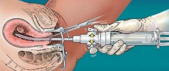
How is a kidney biopsy done?
The patient and doctor perform the following actions:
- The patient undresses and lies down on the operating table.
- The doctor determines and treats the puncture site (incision).
- The anesthesiologist injects local anesthesia into the indicated area. Sometimes general anesthesia is used.
- If the patient is conscious, he is asked to hold his breath and not move.
- The doctor, under ultrasound guidance, inserts a biopsy needle and collects tissue. Sometimes it is necessary to insert the needle 3-4 times to collect the required quantity and quality of material.
The operation lasts no more than an hour, on average 30–40 minutes.
Carrying out the procedure
Initially, the nephrologist asks about diseases, finds out if there are allergies, and about previous operations. Many patients do not know what a kidney biopsy is. Therefore, before the examination, the doctor describes the procedure and the risks associated with it. The patient voices questions, then signs consent for surgery.
Preparatory stage
Preparation begins 2 weeks in advance. In 10-14 days, the use of non-steroidal anti-inflammatory drugs and blood thinners is canceled. You should not eat before the test; at least 8 hours must pass between your last meal and the biopsy. Mild tranquilizers may be prescribed to susceptible people.
A few days before the puncture, the patient’s blood will be taken for general and biochemical analysis, urine will be given, a coagulogram will be done, as well as fluorography, kidney ultrasound and ECG. For diseases of other organs, a consultation with a specialist is prescribed.
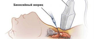
Description
The study can be carried out in a hospital, treatment room or operating room. A kidney puncture lasts an average of 30 minutes, and local anesthesia is most often used. If the patient is emotionally unstable, a drug is administered that puts him into semi-sleep. At the same time, the person is conscious. General anesthesia is done in exceptional cases.
Every patient should know how a kidney biopsy is performed. The person is asked to lie with his back up, his face down. A pillow or cushion is placed under the abdomen or chest. They allow you to raise the organ closer to the back. Pulse and blood pressure are monitored throughout the study.
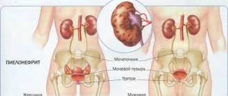
The location of the kidney can be determined under the 12th rib along the posterior axillary line using ultrasound or another method. The skin at the puncture site is lubricated with an antiseptic, and a painkiller is administered.
Then an incision 2-3 mm long is made, through which the instrument is inserted along the trajectory determined earlier. The incision and puncture usually do not hurt. Once the needle is under the skin, the patient is asked to inhale and not breathe for 30-45 seconds. This allows the organs to be fixed in one position.
The needle penetrates the organ 0.1-0.2 cm. The material is collected automatically. Afterwards the instrument is removed. An antiseptic is re-applied to the skin and a bandage is placed on top.
Recovery period and complications
After the procedure, it is recommended to rest for 10-12 hours. During these hours, pulse and blood pressure are measured, and doctors look to see if there is blood in the urine.
You can eat immediately after the procedure, but you need to drink more liquid. The back sometimes hurts slightly; analgesics are prescribed if necessary. If there are no complications and the indicators are normal, the patient is discharged on the same day.
After the study, you should not lift weights for 2 weeks, and you should limit physical activity for several days.
Possible consequences after the procedure:
- Obstruction of the urinary tract.
- Renal rupture or various bleeding in the calyx area or in the pelvis.
- Hematoma of subcapsular or perinephric tissue.
- Damage to other organs, blood vessels, as well as infections, inflammation and purulent paranephritis.
Advice! If some complications occur, you need to visit the hospital. These include increased body temperature, excessive weakness, inability to empty the bladder, dizziness, pain in the lumbar region, fainting, and the presence of blood in the urine on the second and subsequent days after the study.
Evaluation of results
The collected material is examined for 1-2 days. Sometimes the results are ready only after 1.5-2 weeks. Cases are considered normal if there are no manifestations of tumors, infections, inflammations and scars. The presence of the latter often indicates glomerulonephritis and other pathologies.
An abnormal result indicates a change in renal structure, infection, systemic connective tissue disorders, or lack of blood flow. If a transplanted kidney is examined, a negative result indicates its rejection.
results
A negative kidney biopsy for:
- infections;
- scar changes;
- neoplasms;
- atypical cells;
- foreign inclusions;
- inflammatory processes.
If tissue examination shows the presence of at least one of the listed points, the attending physician is obliged to prescribe treatment.
If it is necessary to examine the tissue for hidden infections, the result will be ready in two weeks. In other cases, it can be received within four days.
The results of a biopsy for glomerulonephritis determine the need for organ transplantation.
What to do after the biopsy?
After taking tissue samples, the patient is transported on a gurney to the ward. Bed rest for 6–10 hours is required to reduce the risk of bleeding. Doctors continue to monitor blood pressure and pulse. A urine test is taken to identify blood fractions.
If there is no fever and blood pressure is normal, there are no signs of hematuria, the patient is sent home the same day. If alarming symptoms appear, the time spent in hospital is extended and therapy is prescribed.
Rehabilitation
The patient is recovering at home. Doctors' recommendations for rehabilitation:
- staying in bed for two days;
- increasing the volume of fluid consumed;
- exclusion of physical labor, sports and heavy lifting for the first two weeks;
- You cannot take a bath or shower for three days; the puncture site must be kept dry and clean.
Rehabilitation after a biopsy does not impose dietary restrictions unless the diet is prescribed by a doctor as a method of complex therapy.
Detection of blood in the urine on the second day after the procedure or later, problems with urination, and an increase in temperature are signals to visit a doctor.
Possible risks, consequences and complications
There is a risk when performing any surgical intervention, however, the high probability of returning the full functionality of the organ justifies it. In addition, complications are extremely rare and can be successfully resolved in most cases.
Possible consequences of the procedure:
- bleeding that stops without intervention (1 in 10);
- blood loss requiring transfusion (1 in 50);
- bleeding that can only be stopped by surgery (1 in 1500);
- bleeding requiring kidney removal (1 in 3000).
Complications that are extremely rare:
- puncture of a large blood vessel, which may require a blood transfusion, surgery, embolism, or renal angiography;
- rupture of the lower pole of the kidney;
- suppuration of fatty tissue around the kidney;
- massive bleeding under the capsule and paraneral tissue, resulting in the possible formation of a pararenal hematoma;
- damage to muscle tissue (including neighboring organs), bleeding and pain associated with this;
- if air enters the pleural area, pneumothorax may occur;
- organ infection;
- formation of arteriovenous renal fistula;
- death due to blood loss.
Symptoms of the development of these complications may include:
- blood in urine;
- elevated temperature;
- pain in the lower back and abdomen.
If the above symptoms are detected within 24 hours after surgery, you should immediately consult a doctor.
Preparatory stage of biopsy
To obtain correct biopsy results, there are certain requirements for the patient at the stage of preparation for the study. In addition, the patient must pass some tests, on the basis of which the doctor will decide whether a biopsy is permissible for this person.
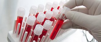
Lab tests
Before performing a biopsy, the following tests are performed:
- general blood analysis;
- general urine analysis;
- blood test for prothrombin time.
With the help of these examinations, the presence of acute infectious and inflammatory processes in the body is determined, which serves as an absolute contraindication to the procedure. The quality of blood clotting is also examined to avoid the possibility of bleeding during the study.
Rules of patient behavior before the procedure
Before conducting the study, the following requirements are imposed on the patient:
- 2 weeks before the biopsy, the subject must stop using medications that have antiplatelet properties (blood thinners), including Aspirin and Ibuprofen. If it is necessary to take certain medications, the patient must notify the attending physician.
Acetylsalicylic acid and drugs containing it prevent blood clotting, so I do not recommend using them 2 weeks before the biopsy
- The last meal should be the day before the procedure no later than 18:00. On the day of the biopsy, immediately before it, you should not drink.
- You should also refrain from drinking alcoholic beverages the day before the test.
Photo gallery
Percutaneous kidney biopsy
Transjugular kidney biopsy
Laparoscopic kidney biopsy
Endoscopic kidney biopsy
Recovery period
After the biopsy is taken, the patient is transferred to the recovery room, where he recovers under the constant supervision of medical staff.
The task of qualified health workers is to monitor the functioning of the body:
- regular measurement of pressure and temperature;
- Constantly checking your breathing and heart rate;
- laboratory testing of blood and urine.
If pain begins to appear, it is relieved with mild analgesics. The patient can remain under such observation for up to 8 hours, after which, if vital signs are stable, he is sent home with a recommendation to refrain from excessive physical activity and not lift heavy objects for 2 weeks.
Video
To learn how the kidney biopsy procedure is performed, watch the video provided by the Hamo Askaryan channel.
Do you have any questions? Specialists and readers of the HROMOSOMA website will help you ask a question
Was this article helpful?
Thank you for your opinion!
The article was useful. Please share the information with your friends.
Yes (100.00%)
No
X
Please write what is wrong and leave recommendations on the article
Cancel reply
Rate the benefit of the article: Rate the author ( 1 vote(s), average: 5.00 out of 5)
Discuss the article:
What is a biopsy, and what are the features of its implementation for kidney cancer?
A kidney biopsy is a diagnostic procedure in which microscopic pieces of tissue (biopsy) are taken from a suspicious area for further examination under a microscope. When taking biopsy material, the doctor performing the manipulations can use various instruments: a scalpel, an endoscope, a thick needle. The choice of instruments is directly dependent on where exactly the biopsy will be taken from.
How is a kidney biopsy performed and what complications can arise?
Most often, biopsy is used in oncological practice and has several purposes:
- confirmation of the malignant nature of the tumor structure as part of differential diagnosis;
- monitoring the effectiveness of therapeutic antitumor measures during the elimination of malignant neoplasms.
But if a patient is suspected of having kidney cancer, a biopsy may not be justified, as it may do more harm than good.
Doctors prefer not to use this method to confirm the malignancy of a neoplasm developing in the renal parenchyma for 2 reasons:
- There is a high probability of spread of mutated cellular structures beyond the kidney. This threat is associated with a violation of the integrity of the renal capsule (fatty tissue) during a biopsy, since it is a natural barrier that holds the cancerous tumor at the site of primary localization. After puncture of the organic fence of the main organ of the urinary system, abnormal cells have a real chance to spread into tissue structures located in the immediate vicinity.
- High rate of false negative results. More than 20% of such procedures do not show the presence of a malignant process in the kidney tissue, i.e., they miss existing kidney cancer. Obtaining incorrect results that mislead onconephrologists and contribute to medical errors often leads to the death of the patient.
But still, in some cases, a biopsy to confirm the diagnosis of cancer is justified. This diagnostic technique is practiced when a specialist suspects the presence of a small malignant neoplasm in the renal parenchyma, the structure and appearance of which is difficult to discern in images taken using MRI or CT.
Where is the procedure performed, what is its cost and patient reviews?
A kidney biopsy can be performed in highly qualified clinics and specialized, private or public, medical centers, which employ personnel with appropriate qualifications. The average cost of the percutaneous type of procedure varies from 2.5 to 24 thousand rubles. Feedback from specialists about this complex study is quite high, because it is the most informative way to identify the disease and assess the nature of its course.
Despite the fact that the price of a kidney biopsy is quite high, and serious complications may arise during the procedure, such diagnostics should not be abandoned under any circumstances. Only by studying a biopsy can a correct diagnosis be made and the most effective course of treatment prescribed in each specific case.