Deciphering a biochemical blood test allows us to identify whether there are any problems in the patient’s body. Biochemical blood test is a frequently used method for studying diseases of internal organs. Find the answer Are you having a problem? Enter “Symptom” or “Name of the disease” into the form, press Enter and you will find out all the treatment for this problem or disease.
The site provides reference information. Adequate diagnosis and treatment of the disease is possible under the supervision of a conscientious doctor. Any medications have contraindications. Consultation with a specialist is required, as well as detailed study of the instructions! Here you can make an appointment with a doctor.
Any deviation from reference values means that a person has a disease.
Rules for preparing for biochemical analysis
To obtain an accurate result, you must follow the algorithm for collecting blood for biochemical testing:
- The collection is carried out on an empty stomach, the last meal no earlier than 12 hours ago. You can drink water, but only still water. The use of chewing gum, sweets and lollipops is excluded.
- 2 days before the test you should not drink alcoholic beverages.
- The day before, exclude fatty, spicy, fried foods from your diet.
- 3 days before the test, avoid excessive physical activity.
- On the day of the study, you should not be exposed to any emotional stress or even minimal physical activity.
- It is customary to take the test in the morning, from 7 to 11 am.
- It is recommended to stop taking any medications 2 days before the test. They may interfere with obtaining correct analysis results.
- You should not smoke 1 hour before the test.
- Before the analysis, it is recommended to rest for 10-15 minutes. Arriving at the laboratory, you need to sit and relax for some time.
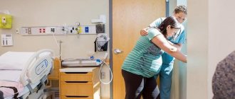
There is a certain procedure that a nurse must follow when drawing blood from a vein for a biochemical study.
This sequence includes:
- Preparing the patient for analysis;
- Blood sampling technique.
Various samples
When collecting urine for bacteriological examination, it is assumed to collect morning urine, its average portion. Toilet of the external genitalia is performed first. The collection is carried out in a sterile container holding 10-15 ml. The test must be delivered to the laboratory within no more than two hours.
If you have to examine a swab from the throat and nose, then in the morning it is forbidden to brush your teeth or rinse your mouth and nose with disinfectant solutions. It is forbidden to consume food and water. If it is necessary to perform bacterial culture of stool, then stool is collected from the morning stool using a sterile spatula into a sterile container; the volume of analysis should be 15-30 grams. It is important that no urine gets into the sample.
The test must be delivered within five hours. It is prohibited to store the test overnight or freeze it. Collecting stool using an enema and laxative should also be excluded.
If you need to examine sputum, then collection is performed in the morning on an empty stomach in a sterile container during a coughing attack along with mucus. Before collection, it is permissible to brush your teeth, but only boiled water should be used for rinsing. The analysis is delivered to the laboratory within an hour.
Breast milk is collected after preliminary hygiene procedures. The area near the nipples should be treated with a swab, which is moistened with 70 percent ethyl alcohol. The first 15 ml cannot be used for donation. 5 ml is decanted from the average portion. Delivery of the analysis must be completed within two hours.
If an analysis of genital discharge is performed, then for women there is a ban on testing during menstruation; at least a week must pass after it. If an antibiotic course was previously administered, a month should pass upon completion. Toilet of the external genitalia is performed first. It is advisable not to urinate for two hours. Men are advised to extend the time they do not go to the toilet to five hours.
Standards for biochemical analysis - explanation
| Index | Normal in adults | In children under 14 years of age | |
| in men | among women | ||
| Total protein (tp) | 60 – 85 g/l | 60 – 85 g/l | 45 – 75 g/l |
| Albumin (albu) | 35 – 50 g/l | 35 – 50 g/l | 40 – 55 g/l |
| Total bilirubin (tbil) | 8.5 – 20.5 µmol/l | 8.5 – 20.5 µmol/l | up to 250 µmol/l (newborns) |
| Indirect bilirubin (dbil) | 1 – 8 µmol/l | 1 – 8 µmol/l | up to 210 µmol/l |
| Direct bilirubin (idbil) | 1 – 20 µmol/l | 1 – 20 µmol/l | up to 40 µmol/l |
| Aspartate aminotransferase (alt) | up to 37 units/l | up to 31 units/l | up to 30 units/l |
| Alanine aminotransferase (ast) | up to 45 units/l | up to 35 units/l | up to 35 units/l |
| gamma glutamine transferase (ggt) | up to 55 units/l | up to 40 units/l | up to 45 units/l |
| Alkaline phosphatase (alp) | 30 – 130 units/l | 30 – 110 units/l | Up to 350 units/l |
| Triglycerides (trig) | 0.4 – 1.8 mmol/l | 0.4 – 1.8 mmol/l | 0.5 – 2 mmol/l |
| Cholesterol (chol) | 3.5 – 5.5 mmol/l | 3.5 – 5.5 mmol/l | 3.5 – 7.5 mmol/l |
| VP lipoproteins (hdl) | 1.7 – 3.5 mmol/l | 1.7 – 3.5 mmol/l | 1.7 – 4.5 mmol/l |
| Fibrinogen (fg) | 2 – 4 g/l | Up to 6 g/l (during pregnancy) | 1.2 – 3 g/l |
| Amylase (amyl) | 25 – 125 units/l | 25 – 125 units/l | 25 – 125 units/l |
| Uric acid | 210 – 420 µml/l | 150 – 350 µml/l | 150 – 350 µml/l |
| Creatinine (crea) | 62 – 120 µml/l | 55 – 95 µml/l | 50 – 100 µml/l |
| Urea (urea) | 2.8 – 7.2 mmol/l | 2.8 – 7.2 mmol/l | 1.8 – 6.2 mmol/l |
| C-reactive protein (crp) | up to 0.5 mg/l | up to 0.5 mg/l | up to 0.5 mg/l |
| Antistreptolysin O (also, aslo) | up to 200 units/l | up to 200 units/l | up to 200 units/l |
| Glucose (glu) | 3.8 – 6.3 mmol/l | 3.8 – 6.3 mmol/l | 3.8 – 5.3 mmol/l |
Preparing the patient for the study
Here are the rules a nurse should follow before drawing blood to study its biochemical composition:
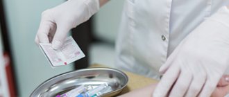
- Tell the patient the goals and progress of the study, and obtain his consent to the procedure.
- Write the patient's name and surname on the blood tube, prepare the necessary equipment, and demonstrate it to the patient.
- Help the person find a comfortable position in the chair.
- Wash your hands, dry them, put on sterile medical gloves.
- Place the patient's hand on the oilcloth cushion.
General blood test algorithm
1. Equipment:
sterile scarifier, packing with sterile Panchenkov capillaries (20 pcs. in 1 package), 70 0 alcohol, tablet with test tubes, tablet for blood dosing, Sali capillary (0.02 ml.), rubber bulb, container with disinfectant solution , disposable (non-sterile) gloves, gown, apron, cap, mask, plastic glasses, cotton wool, 2 sterile glass slides (package - 10 pcs.), test tubes: No. 1 for red blood cells - 4 ml - 3.5% sodium chloride solution , No. 2 for hemoglobin - 5 ml of transforming solution, No. 3 for leukocytes - 0.4 ml-3% acetic acid solution, 3 4 for ESR - 3.8% solution of sterile sodium oxylate 0.025 ml.
2.Preparation for the procedure:
1. blood is drawn from patients on an empty stomach;
2. greet the patient and introduce himself;
3. explain to the patient the purpose and course of the upcoming procedure and obtain his consent;
4. invite the patient to sit comfortably;
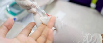
5. wash and dry your hands, put on gloves;
6. treat the patient’s 4th (ring) finger with 70 0 alcohol;
7. Fix the patient’s finger with your left hand, and take the scarifier in your right hand.
3.Performing the manipulation:
1. puncture to a depth of 3-4 mm;
2. Use a dry sterile cotton swab to remove the first drop of blood;
3. Use a sterile Panchenko capillary to collect 0.025 ml of sterile sodium citrate, wash it and pour it into a test tube with sodium citrate;
4. With the same capillary, half a capillary of blood is collected and poured into one dosing cell, from which 0.02 ml of blood is collected using a Sali capillary with a pear, the remaining blood is removed from the pipette nozzle and poured into test tube No. 1 (for red blood cells);
5. 0.02 ml of blood is taken from the dosing cell and poured into test tube No. 2 (for hemoglobin);
6. 0.02 ml of blood is dipped into tube No. 3 (for leukocytes);
7. 1 drop of blood is lowered onto a glass slide using a Panchenko capillary and a thin smear is prepared (at an angle of 45 0 with a special grinding glass);
8. Panchenko’s capillary is lowered into test tube No. 4 (for ESR);
9. treat the patient’s finger with sterile cotton wool soaked in alcohol. The patient is released.
4.Complete the procedure:
1. Sign the blood smear, air dry it, and place it in a tripod for fixation. Pack a rack with test tubes, glasses and a rack for blood dosing in a container for transportation.
2. In the laboratory:
a) counting red blood cells in the Gorev chamber;
b) measurement of hemoglobin on FEC;
c) counting leukocytes in the Gorev chamber;
d) installation of the Pachenkov capillary in a Panchenkov stand with blood drawn up to the “0” mark for 1 hour - determination of ESR;
e) fixation, staining of a blood smear and counting of cellular elements (leukoformula) under a microscope (immersion, magnification 7*90)
3. Disinfection mode:
a) disposal of the scarifier for 1 hour in a 0.05% anolyte solution;
used cotton wool and rags – 1 hour in a 0.05% anolyte solution;
residual blood and reagents with blood - for 1 hour in a ratio of 5 6 1 with bleach.
b) “0” – disinfection of test tubes, capillaries, bulbs, stands in a 0.03% anolyte solution.
c) disinfection and pre-sterilization cleaning are combined - 90 minutes in a 0.03% anolyte solution. Rinsing and sterilization of capillaries and glasses - in packaging in a container by autoclaving (1 hour at 132 0 C, 1.2 atm.) The glasses are boiled in a washing solution with hydrogen peroxide for 1 hour, dried and sterilized by autoclaving.
All materials presented on the site are solely for the purpose of information for readers and do not pursue commercial purposes or copyright infringement. Studall.Org (0.004 sec.)
source
Blood sampling technique
After all of the above steps, the nurse can begin drawing blood.
This is what the analysis technique looks like:
- Place a rubber band in the middle third of the shoulder and tell the patient to clench his fist tightly. The procedure of clenching and unclenching your fist needs to be done several times. During this time, the nurse finds a vein from which blood will be drawn.
- Wipe the vein in the elbow area with napkins moistened with 70% alcohol.
- Perform venipuncture.
- Check that the needle is in the patient’s vein and pull the plunger towards you.
- Pull the plunger towards you until a sufficient amount of blood has been collected.
- Before removing the needle from the vein, you need to untie the tourniquet.
- Press the blood collection site with a sterile cloth soaked in 70% alcohol, then remove the needle.
- Tell the patient to clasp the elbow while holding the alcohol pad.
- Remove the needle from the used syringe and throw it into the disinfectant solution.
- Mix the contents of the test tube by inverting it.
- Place the syringe in a container for disinfection.
After performing these steps, the nurse should help the patient get out of the chair. She needs to put the test tube in a container, take off her medical gloves, and throw them in the trash. you need to deliver the box with the serum to the laboratory.
Blood culture for blood culture - algorithm
So, the material for bacteriological culture is blood in an amount of 5–10 ml obtained from the ulnar vein.
The skin area is treated first with a cotton ball soaked in 70% ethyl alcohol, and then with another cotton ball soaked in a 1–2% iodine solution until the skin is completely dry.
The algorithm here can be the same as when collecting blood with vacuum syringes, with the difference that instead of a test tube, a blood culture bottle is inserted into the adapter.
At the site of venipuncture, the iodine is wiped off with alcohol and the area is sealed with a plaster. The plastic cap of this bottle is broken off and the inner surface of the cap is disinfected with 70% ethyl alcohol.
At a temperature of 37°C, blood should be stored for no more than 2 hours!
It is best to culture blood on a nutrient medium immediately after collecting it.
The ratio of blood and nutrient medium is 1:10, mix carefully.
To obtain the most reliable result, the analysis should be carried out at least twice from different hands with a time interval of 30 minutes.
Advantages of blood culture using this method
The advantage of the method described above is that it has an adequate cost, high information content, ease of implementation, and most importantly – accessibility!
Currently, there are two ways to culture blood on a nutrient medium and determine sensitivity to antibiotics:
- flora culture to identify sensitivity to a wide range of antibiotics;
- flora culture to identify sensitivity to the main spectrum of antibiotics.
Indications for the procedure
A biochemical blood test can be prescribed for preventive purposes to determine the correct functioning of internal organs.
Some donate blood once a year for biochemistry to make sure that there are no pathologies in the body.
But more often, this blood test is prescribed to the patient by a doctor to diagnose the disease. Analysis is one of the main methods for correct diagnosis.
Analysis is prescribed in the case of:
- The patient has inherited diseases;
- Detection of diseases of the cardiovascular system;
- Disorders of the pancreas, intestines, stomach;
- Leukemia;
- Causes of infectious processes in the body;
- Diabetes mellitus;
- Deviations in the gynecological area;
- Diseases of the musculoskeletal system (arthrosis, arthritis, osteoporosis);
- Causes of acute intoxication;
- Pathologies of the organs of vision;
- Skin diseases.
Decoding
Blood culture results are divided into the following three groups:
- no bacterial growth
- net growth
- mixed growth
No bacterial growth
Lack of bacterial growth is a normal result of blood culture, which is obtained if the patient's blood is sterile. Before interpreting the result in this way, it is important to consider the possibility that it is a false negative, i.e. the patient has bacteremia but it was not detected by this test.
Decoding the main indicators of the study
Biochemical research includes many indicators.
Often the doctor prescribes only a few indicators for testing that can confirm the expected diagnosis.
A standard study consists of the following points:
- Glucose. The indicator is the main one for identifying the presence of diabetes mellitus (an increase in the parameter indicates this). A decrease in the parameter includes diseases of the liver and endocrine system.
- Total protein. The indicator is prescribed for studies on the presence of infections in the body, diseases of the gastrointestinal tract and genitourinary system, oncology (the indicator is reduced). Its increase may indicate injuries, burns, overheating of a person, or rheumatism.
- Total bilirubin. An increase in its level includes the development of cirrhosis, anemia, hepatitis, and cholelithiasis. Total bilirubin consists of direct and indirect. The direct one is associated with the outflow of bile, and the indirect one is associated with liver diseases.
- CRP or C-reactive protein. The main indicator of the presence of an inflammatory process in the body.
- Urea. An increase in urea levels indicates poor kidney function, gastrointestinal diseases, heart failure, and neoplasms.
- Creatinine. An increase in creatinine indicates pathologies of the kidneys or thyroid gland. Often this indicator is prescribed together with a urea test.
- Total cholesterol. An increase in the indicator indicates suspicion of atherosclerosis of the liver or thyroid gland.
- Amylase. The analysis is studied for diseases of the pancreas, gall bladder and kidneys. The rate is usually higher than normal in people with diabetes, pancreatitis or cholecystitis.
- ALT (Alanine aminotransferase). An increase in this indicator may indicate hepatitis, cirrhosis, liver cancer, and blood diseases.
- AST (Aspartate aminotransferase). An indicator for checking the condition of the heart, liver, muscle disorders.
- Lipase. An increase in lipase indicates the presence of pancreatic diseases.
Study of vaginal microbiocenosis with determination of sensitivity to antibiotics
Bacteriological examination of material obtained from the vagina, which allows you to assess the quantitative composition of the microflora, the ratio of microorganisms, identify a decrease in the number of lactobacilli, an increase in the growth of facultative microorganisms or the appearance of atypical microorganisms and determine their sensitivity to antibacterial drugs.
Synonyms Russian
Culture for vaginal dysbiosis with sensitivity to a/b, culture of a vaginal smear on a tank. biota and sensitivity to a/b.
English synonyms
Vaginal Culture with Antibiotic susceptibility testing.
Research method
Microbiological method.
What biomaterial can be used for research?
Urogenital smear.
How to properly prepare for research?
- Women are recommended to take a urogenital smear before menstruation or 2-3 days after it ends.
General information about the study
The normal microflora of the vagina, due to the stability of its quantitative and species composition, prevents the colonization of the vagina by pathogenic microorganisms and suppresses the excessive proliferation of opportunistic pathogenic microorganisms (OPM), which are included in small quantities in the normal microcenosis. The vaginal microecosystem consists of permanently inhabiting (obligate) and transient (occasional) microorganisms. The main representatives of the normal vaginal microflora are:
- gram-positive obligate anaerobic and microaerobic bacteria (lactobacteria, bifidobacteria, peptostreptococci, clostridia, propionobacteria, mobiluncus),
- gram-negative obligate anaerobic bacteria (bacterioides, prevotella, porphyromonas, fusobacteria, veillonella),
- facultative anaerobic microorganisms (gardnerella, corynebacteria, mycoplasma, staphylo- and streptococci, enterobacteria, yeast, fungi of the genus Candida).
At different periods of a woman’s life, depending on the activity of the reproductive function and hormonal balance, the vaginal microcenosis has certain characteristics. Before the first menstruation and the formation of menstrual function against the background of non-functioning ovaries, the microflora of the vagina is dominated by gram-positive cocci - epidermal and other coagulase-negative staphylococci, micrococci, non-hemolytic streptococci. Non-pathogenic Neisseria and Corynebacteria are less common, and Escherichia and Enterococcus are even less common. As puberty progresses, the number of lactobacilli increases, and in sexually mature girls the microflora is almost entirely represented by lactobacilli.
In healthy women of reproductive age, the total number of microorganisms in vaginal discharge is 107-109 CFU/ml (colony forming units per milliliter) and consists of more than 40 different species. The predominant species are Doderlein bacilli – Lactobacillus spp. (95-98%) - a large group of bacteria, mainly microaerophiles. Despite the diversity of the species composition of lactobacilli isolated from the vagina of healthy women (more than 10 species), it is not possible to identify a single species that would be present in all women. Most often it is possible to isolate the following lactobacilli: L. acidophilus, L. brevis, L. jensenii, L. casei, L. leishmanii, L. Plantarum. Among transient vaginal microorganisms, the most common are coagulase-negative staphylococci, primarily S. epidermidis, Corynebacterium spp., Streptococcus spp., Bacteroides, Prevotella spp., Mycoplasma hominis, which are usually present in moderate quantities (up to 104 CFU/g). Micrococcus spp., Propionibacterium spp., Veillonella spp., Eubacterium spp. are found just as often, but in smaller numbers. Among the relatively rare microorganisms (less than 10% of those examined) are Clostridium spp., Bifidobacterium spp., Actinomyces spp., Fusobacterium spp., Ureaplasma urealyticum, Staphylococcus aureus, Neisseria spp., E. coli and other coliform bacteria, Mycoplasma fermentans , Gardnerella vaginalis, Candida spp.
A decrease in the number of lactobacilli and excessive growth of opportunistic microorganisms leads to dysbiotic disorders, which can clinically manifest as inflammation of the vaginal walls - vaginitis, accompanied by severe itching, burning, and abnormal discharge. Pathological changes in the vaginal microcenosis can occur during treatment with antibiotics (local or systemic), cytostatics, hormones, radiation therapy, especially against the background of endocrinopathies (primarily diabetes), with anemia, congenital malformations of the genital organs, with the use of contraceptives and disorders in the immune system.
A study of vaginal microbiocenosis (bacteria culture) helps to diagnose dysbiosis (bacterial vaginosis), identify the causative agent of nonspecific bacterial vaginitis, fungal infection, pelvic inflammatory disease or sexually transmitted infections.
This research method is based on the ability of microorganisms to multiply on artificial nutrient media, which makes it possible to identify the types of bacteria and fungi living on the mucous membrane, determine their concentration and sensitivity to antibacterial drugs. When a probable causative agent of an infectious-inflammatory process is identified, it is incubated with antibiotics or diffusion disks impregnated with a drug are used - a drug to which the isolated microorganism is sensitive - this prevents the growth of bacteria.
The bacteriological method is indispensable for infections caused by opportunistic microorganisms (urogenital mycoplasmas, yeast fungi, Entrobacteriaceae, Streptococcus spp., Staphylococcus spp., etc.), since only with its help can the amount of the pathogen be assessed. Determining sensitivity to antibacterial drugs allows you to avoid the prescription of useless but unsafe drugs and select adequate therapy.
What is the research used for?
- For the diagnosis of dysbiotic disorders of the vaginal microflora;
- to identify the microorganism that caused the development of the infectious and inflammatory process of the vagina and pelvic organs;
- to determine drugs to which the causative agent of the infectious-inflammatory process is sensitive (to select effective antibacterial therapy);
- for the diagnosis of nonspecific bacterial vaginitis/vulvovaginitis, bacterial vaginosis, vulvovaginal candidiasis.
When is the study scheduled?
- With clinical signs of inflammatory diseases of the vagina and pelvic organs (itching, burning, leucorrhoea);
- when infectious and inflammatory changes are detected by microscopy of a vaginal smear;
- if treatment for vaginitis is ineffective;
- when selecting antibacterial drugs for the treatment of inflammatory diseases.
What do the results mean?
The result is interpreted by the attending physician taking into account the patient’s complaints, medical history, clinical manifestations of the disease and the exclusion of sexually transmitted infections.
Sensitivity to antibiotics is determined by identifying diagnostically significant growth of opportunistic biota.
Normally, lactobacilli predominate in the culture; opportunistic microorganisms are absent or detected in small quantities - less than 104.
With bacterial vaginosis, the growth of lactobacilli is sharply reduced or absent, and the number of opportunistic microorganisms is increased. When cultured, anaerobic microorganisms, Gardnerella vaginalis, Mycoplasma hominis, Mobiluncus spp., Bacteroides spp., peptostreptococci, can be isolated.
With vulvovaginal candidiasis, against the background of a decrease in the number of lactobacilli, an increase in Candida spp is observed. more than 104. With this pathology, microscopy of the material should reveal pseudomycelium of the fungus.
Nonspecific bacterial vulvovaginitis is characterized by the growth of one or more opportunistic microorganisms in a diagnostically significant titer with a decrease in the number or absence of lactobacilli in the culture.
Important Notes
- It is recommended to combine the study with microscopy of genital discharge, Gram-stained. If there are clinical signs of infectious and inflammatory diseases of the genital organs, it is necessary first of all to exclude sexually transmitted infections.
- It should be remembered that all opportunistic microorganisms can also be found in healthy women and exhibit their pathogenic properties only at significant concentrations. The results of the study should be interpreted taking into account the symptoms of the disease and the patient’s complaints.
Also recommended
- Microscopic examination of discharge from the genitourinary organs of women (microflora), 3 localizations
- Analysis of vaginal microbiocenosis. 16 indicators, DNA quantitative [real-time PCR]
- Analysis of vaginal microbiocenosis. 8 indicators, DNA quantitative [real-time PCR]
- Culture for Gardnerella vaginalis with determination of titer and sensitivity to antimicrobial drugs
- Neisseria gonorrhoeae, DNA [real-time PCR]
- Trichomonas vaginalis, DNA [real-time PCR]
- Ureaplasma species, DNA quantitative [real-time PCR]
- Culture for Ureaplasma species with determination of sensitivity to antibiotics (with a titer of 1x10^4 and above)
- Chlamydia trachomatis, DNA [real-time PCR]
- Culture for Chlamydia trachomatis with determination of sensitivity to antibiotics
Who orders the study?
Obstetrician-gynecologist.
Literature
- Daniels R. Delmar's Guide to Laboratory and Diagnostics Tests // Cengage Learning – 2009 – 1003 p.
- Larsen B., Monif G. Understanding the bacterial flora of the female genital tract. // Clin Infect Dis. – 2001 – 32 (4) – p.69-77.
- Infections in obstetrics and gynecology / Ed. O. V. Makarova, V. A. Aleshkina, T. N. Savchenko. – M.: MEDpress-inform, 2007. – 464 p.
Recommendations and diet before the procedure
Biochemical analysis is a common method of blood testing; by correctly interpreting the results, you can make an accurate diagnosis and identify serious diseases. For example, blood biochemistry diagnoses diabetes mellitus, benign and malignant tumors, hepatitis and pathologies in the urinary system.
The accuracy of the result is determined by the preparation and behavior of the patient before the biochemical study:
- It is forbidden to eat in the morning before taking blood; you can only drink clean water (no tea, coffee, sweet drinks). The last meal should be before 20.00, so that there is a break of at least 10 hours between dinner and the procedure.
- The test result can be affected by overexertion, strong physical activity or drinking alcohol, so the day before the test you should avoid stress and alcohol consumption.
- If it was not possible to donate blood on an empty stomach in the morning, then the study can be carried out during the day (after a 5-hour fast).
Collection, storage and transportation of blood for bacteriological research:
Blood is drawn from the ulnar vein.
Treatment of the venipuncture site:
– wipe the surface with a swab soaked in a 5% iodine solution in a circular motion from the center to the periphery;
– the treated area is dried and then the skin is thoroughly cleansed with 70% ethanol.
The treated area is dried again and then:
1. Pierce a vein with a sterile needle and syringe at the sampling site and take 10 ml of blood for blood culture. Open the bottle with the medium over the flame of the alcohol lamp and carefully add blood from the syringe (so as not to wet the stopper), mix the contents of the bottle.
2. For serological diagnosis, blood is taken in a volume of 2-5 ml into a sterile tube.
– the collected blood is brought into containers with aerobic or anaerobic conditions;
– if the patient has a fever of unknown origin, then blood is taken in stages:
– first stage – take 2 samples from different blood vessels;
- second stage - after 24 or 36 hours, two more samples are taken at the peak of temperature;
– taking blood from an indwelling intravenous or intra-arterial catheter is allowed only in cases of suspected catheter-associated infection. If the patient is already being treated with antibiotics, then 6 samples are taken from him within 48 hours.
Pathologies that cause increased bilirubin levels
Bilirubin appears after the breakdown of red blood cells, so it is regularly formed in the body. First, indirect bilirubin is formed in the bloodstream and tissues: it is insoluble in water and has increased toxicity. Normally, indirect bilirubin travels through the blood to the liver, which converts it into the direct form.
Impaired release of the direct form of bilirubin from the liver indicates:
- Infectious diseases;
- Acute viral hepatitis;
- Infectious mononucleosis;
- Damage to cytomegalovirus;
- Syphilis (secondary or tertiary form);
- Presence of parasites;
- Toxic hepatitis;
- Fatty hepatosis;
- Liver cancer;
- Impaired functioning of the biliary tract (cholecystitis; cholangitis).
Impaired release of indirect bilirubin indicates:
- Hypothyroidism in newborns;
- Hereditary pathologies;
- Obstruction of bile ducts by stones;
- Neoplasms of the pancreas;
- Biliary cirrhosis;
- Sclerosing cholangitis.
Diseases in which the amount of ALT and AST increases
A study of the amount of ALT and AST indicates the condition of the liver, which is why these tests are called liver tests. Screening the amount of ALT and AST helps determine the function of this organ and see the development of disorders in its work.
The level of aspartate aminotransferase (AST) increases in the following pathologies:
- Heart attack;
- An attack of angina;
- Pulmonary embolism;
- Malignant neoplasms in the liver;
- Bruises or muscle injuries;
- Acute form of rheumatic carditis;
- Fatty hepatosis;
- Myopathies;
- Hepatitis caused by toxins, viruses or alcoholic beverages;
- Pancreatitis in the acute stage;
- Cholestasis (stagnation) of bile;
- Gangrene.
The amount of alanine aminotransferase (ALT) in the blood increases in the following diseases:
- Fatty hepatosis;
- Obstructive jaundice;
- Poisoning;
- Hepatitis;
- Burns and injuries;
- Cirrhosis or cancer;
- Myocarditis or myodystrophy;
- Heart attack;
- State of shock;
- Myositis;
- Blood diseases.
Pathologies that cause increased levels of amylase and lipase
Amylase is an enzyme produced by the pancreas and affects the breakdown of carbohydrates into glucose. Amylase is actively involved in the digestive process, so it is found in large quantities in the gland itself.
There are 3 types of enzyme - alpha, beta and gamma amylase. More often, the activity of alpha-amylase, which is divided into S and P types, is studied in the laboratory. About 55% of S-type amylase is found in the bloodstream. If its amount increases, then this process is called hyperamylasemia.
Diseases in which blood amylase increases:
- Mumps caused by a viral infection;
- Pancreatitis in the acute stage (the maximum level increases 4 hours after the first attack, and decreases to normal levels 4-5 days after the onset of the disease);
- Acute and chronic pancreatitis (usually enzyme activity increases many times);
- Alcohol abuse;
- Malignant or benign neoplasm in the pancreas;
- Ectopic pregnancy.
Lipase is a digestive enzyme whose main function is considered to be the process of breaking down fats. Lipase works through the action of bile acids and colipase (a coenzyme). Lipase, which is formed in the pancreas, is important for diagnosis; therefore, the activity of the enzyme is determined to identify disturbances in the functioning of this organ.
An increase in lipase activity in the bloodstream occurs when:
- Cholestasis;
- Acute pancreatitis;
- Cholecystitis;
- Pancreatic tumors;
- Ulcers;
- Gout or excessive weight gain;
- Drug abuse, especially painkillers and narcotic drugs;
- Bruises, fractures and other injuries;
- Kidney failure.
When diagnosing pancreatic diseases, lipase is considered a more different test than amylase activity. Increased lipase levels remain normal in mumps, tubal pregnancy, acute appendicitis, and liver disease.
If there is a suspicion of pancreatitis, doctors advise determining the activity of amylase and lipase at the same time. The increase in blood lipase activity in acute pancreatitis can be from 3 to 40 times higher relative to the physiological state.
To determine the acute form of alcoholic pancreatitis, the ratio of the amount of amylase and lipase is used. When this proportion is greater than 3, the cause of pancreatitis is alcohol intoxication.
When acute pancreatitis occurs, serum lipase activity increases earlier and more strongly than amylase activity. The increase in amylase levels in the bloodstream begins only 6 hours after the first attack of pancreatitis and reaches its peak after 13-25 hours. Enzyme activity remains elevated for 7-11 days.
Norm and interpretation of results
What does a blood culture show? These studies are classified according to criteria such as growth, pure and mixed growth of bacteria.
Data are good if there is no growth. This indicates that the person's blood is pure. Pure growth refers to the growth of a specific type of microorganism. This is observed in sepsis.
In case of combined growth, it is worth interpreting the presence of more than two types of bacteria.
Source
Blood culture is a bacteriological research method used to reliably determine the presence of bacteria in the blood.
Sowing is used to identify various microorganisms in it and has to be done for many diseases. Blood cultures are performed for isolation purposes to identify pathogenic bacteria in bacteremia.
Bacteremia is the penetration of bacteria into the blood during the generalization of infection in the human body, blood poisoning (sepsis). This type of analysis is carried out in laboratories of medical institutions.
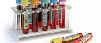
Blood cultures are done on liquid nutrient media:
- bile broth,
- sugar broth,
- liquid and semi-liquid media for growing anaerobic microbes.
How many times do you need to take a blood culture test?
To detect microorganisms, several tests of at least 3 are required, since the number of bacteria in the blood can fluctuate.
Certain types of bacteria can only be isolated by special bacteriological cultures (tank culture) of blood.
Blood culture tank - what shows
A blood culture tank shows the sensitivity to antibiotics of a particular group of bacteria. By determining the type of bacterial colony grown after culture (microbiological examination), it is possible to select effective antibiotics against this particular group of microorganisms (the so-called blood culture for sterility ).
- It is important! Before testing for blood cultures for infections - Do not take anantibiotics!
Detecting bacteria will be difficult or even impossible if the person was taking antibiotics before the blood culture test and is still infected with pathogens.
The price of biochemical analysis, and where is the best place to do it?
The listed biochemical parameters are determined in any private or public clinical diagnostic laboratory. Most laboratory sites use high-precision methods and equipment. Often, the reference values for each analysis are indicated on the study form in accordance with the age and gender of the patient.
Any laboratory, public or private, has a set of reagents and personal computers. Norms for biochemical parameters can vary greatly. If there is a need to be examined again, the examination should be carried out at the same time of day and in the same laboratory department.
The price of a biochemical analysis depends on what blood parameters need to be determined. The price of one indicator is from 100 to 700 rubles.
Doctor: Olga Shishkina ✓ Article checked by doctor
Bacteremia, fungemia (bacteriological blood test)
… bacteriological research - a set of methods used to detect and establish the nature of bacteria isolated from patients, bacteria carriers or from environmental objects.
INTRODUCTION
Blood is one of the most common materials used for bacteriological research. Blood is normal ! sterile, the presence of bacteria (bacteremia) or fungi (fungemia) is a pathology.