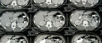
Computed tomography in modern medicine is considered the most accurate method for detecting pathology of bones and soft tissues in almost any area of the skull. CT scan of the skull allows you to determine tumor and inflammatory processes. The study is carried out on the recommendation of a doctor if indicated.
Computed tomography is one of the few research methods for diagnosing brain pathologies
CT scan of the skull: features
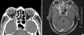
Computed tomography of the skull and brain is performed in a spiral or stepwise mode. The cut thickness is most often 1-2 mm. Devices with multiple detectors are used (multispiral tomography - MSCT).
At a minimum, two windows are used: bone and medullary. The skull is never examined separately from the brain, eye sockets, and paranasal sinuses. All changes are analyzed comprehensively.
For example, with a bone injury, it is necessary to evaluate not only the direction of the fracture line, but also the damage to the brain. Detecting intracerebral injuries is a more important task than detecting fractures.
CT diagnostic services at CELT
The administration of CELT JSC regularly updates the price list posted on the clinic’s website. However, in order to avoid possible misunderstandings, we ask you to clarify the cost of services by phone: +7
| Service name | Price in rubles |
| CT scan of the cerebral arteries and screening examination of the brain | 14 000 |
| CT pituitary gland | 7 000 |
| CT scan of the brain | 6 000 |
All services
Make an appointment through the application or by calling +7 +7 We work every day:
- Monday—Friday: 8.00—20.00
- Saturday: 8.00–18.00
- Sunday is a day off
The nearest metro and MCC stations to the clinic:
- Highway of Enthusiasts or Perovo
- Partisan
- Enthusiast Highway
Driving directions
CT Scan Basics
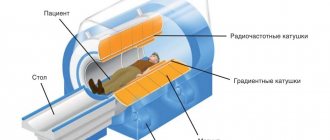
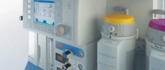
Computed tomography is based on x-rays. A computed tomograph consists of a gantry, inside of which a tube rotates, and a movable table. The table can move smoothly during spiral tomography and discretely during step-by-step tomography.
The tube rotates around the scanned object, generating X-rays. They are weakened by the object, being perceived by detectors on the opposite side. After processing the data, black and white images (slices) of the object are obtained. The greater the difference between the X-ray densities of tissues, the better they are distinguishable on CT.
How does this procedure work?
In a conventional X-ray, a small amount of radiation is directed and passed through the part of the body being examined, recording an image on a special electronic image recording plate. Bones appear white on an X-ray, soft tissues such as the heart or liver appear in shades of gray, and air appears black.
In a CT scan, numerous X-ray beams and a series of electronic X-ray detectors rotate around the body, measuring the amount of radiation absorbed throughout the body. Sometimes the examination table will move during the scan so that the x-ray beam follows a spiral path. A specialized computer program processes this large amount of data to produce three-dimensional cross-sectional images of the human body.
Improvements in detector hardware allow almost all CT scanners to acquire multiple slices per revolution.
These scanners, called multi-slice or multi-slice CT scanners, produce thinner slices in a shorter period of time, resulting in more detailed viewing capabilities.
Modern CT scanners are so fast that they can scan large parts of the body in just a few seconds.
This speed is important for all patients, but especially for children, the elderly and the seriously ill, all those who have difficulty remaining in one place for a long time, even for the short time required to obtain images.
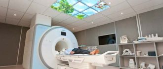
The procedure itself for a computed tomography scan of the skull bones usually takes about 10-15 minutes without contrast and a maximum of thirty minutes with contrast. A person lies down on a movable comfortable table that slides inside the CT scanner.
The patient is completely alone while the equipment is operating in the office. Medical personnel are in the next room and supervise the procedure.
The tomograph places the patient's head in a special flask, which looks like a rotating ring with built-in X-ray sensors that penetrate the human body.
This creates noise that rarely causes discomfort to patients. And if such fear is present, then you need to inform your doctor about it. Fortunately, such sensations are rare and can only be of a psychological nature.
Sometimes the doctor may ask you to hold your breath for literally seconds to ensure the patient remains completely still and to get clear results on the image.
Detectable pathology
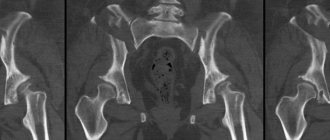
Bone trauma
It is necessary to assess the nature of the fracture (complete, incomplete, linear, depressed), the number of fragments, and the depth of their displacement. The location, direction of the fracture line, and their number are assessed. The degree of displacement of fragments in combination with the effect on the brain must be indicated.
All changes in the skull must be assessed comprehensively with the brain, eye sockets, and paranasal sinuses. For example, a fracture is often complicated by epidural hematomas, brain contusion, and parenchymal hemorrhage.
In case of wounds, a wounding object (knife, bullet, shrapnel, wad) can be detected. It is necessary to clearly assess its location, relationship to neighboring structures, direction of the wound channel, and the presence of complications.
Bone tumors
They can be benign (osteoma) and malignant (osteosarcoma, metastases). Osteomas can be compact and spongy. This is a common finding and in most cases has no clinical significance.
When malignant tumors (mainly metastases) are detected, it is necessary to assess their location and the degree of destruction of the skull.
Tumors can be limited to the spongy substance or grow through the cortical plate outward or inward, spreading into the subcutaneous soft tissues or membranes, and then into the substance of the brain.
Inflammatory lesions
Osteomyelitis most often occurs after injury or surgery. It manifests itself as local destruction of bone tissue, linear periosteal reaction, sequestration, and inflammation of adjacent soft tissues.
The inflammatory process in the paranasal sinuses can spread to the orbit and brain, leading to complications. Inflammation of the temporal bone can cause catarrhal or purulent meningitis, encephalitis.
Developmental anomalies and acquired disorders. Various deformations of the skull may be detected, including those caused by a lack of vitamin D in childhood. Anomalies in the articulation of the cervical spine with the skull are also common.
A rare, not severe pathology is basilar invagination, in which the odontoid process of the second cervical vertebra falls into the cranial cavity and can compress the stem structures and disrupt the outflow of lymph. Anomalies of the skull are often combined with abnormalities of brain development.
Postoperative changes. Milling holes, covered defects of the skull after trepanation, and metal plates may be detected. Stents are also identified to drain cerebrospinal fluid from the cranial cavity. Consequences of surgical interventions on the temporal bones, paranasal sinuses, and nasal septum are often found.
3. Main pathologies that MRI can detect
MRI is often performed if there is a suspicion of damage to the bones of the base of the skull and an oncological process. An accurate diagnosis allows specialists to prescribe competent treatment, slowing down the development of pathology. In the early stages of the disease, it is possible to cure completely.
Among the main pathologies detected by scanning, the following should be noted:
- Hypertension, characterized by increased intracranial pressure. The condition spreads to all areas of the brain. The problem manifests itself as a result of tumors, injuries and severe infectious diseases. In hypertension, MRI visualizes sparse edges of the cerebral ventricles and dilated fluid cavities.
- Cancerous and benign formations. Tumors are detected by a tomograph due to increased signals coming from abnormal formations. With timely diagnosis of a tumor, patient survival increases significantly.
- Linear fractures and cracks in bone structures.
- Intracranial damage to brain cells.
- Cystic formations in the subarachnoid space.
- Hydrocephalic syndrome.
When research is not indicated
If the patient is very restless and has motor activity, the examination may be difficult. Patients with head trauma often behave inappropriately, not understanding what is happening. They are trying to get up from the table and go somewhere.
Such patients need to be carefully monitored. If fixation does not help, anesthesia or sedation is required. Computed tomography is relatively contraindicated for children and pregnant women. This means that the study, despite the radiation exposure, can be performed for health reasons.
What you need to know when preparing for research
For a CT scan of the skull, metal objects, including jewelry, glasses, dentures, and hairpins, should be left at home or removed before the examination, as they can negatively affect the CT images. You may also be asked to remove hearing aids and removable dental work.
You need to tell your doctor about the medications you are taking and whether you have any allergic reactions to medications or, as previously known, contrast materials and dyes.
If there is a history of this, your doctor may prescribe medications (usually steroids) to reduce the risk of allergies or prevent them altogether.
The radiologist will need to know if you have asthma, diabetes, myeloma, or heart disease, kidney disease, or thyroid problems. Any of these listed conditions may increase the risk of adverse effects on a person's overall health.
How the research works
First, the patient removes all metal from the scanning area to avoid artifacts that reduce the quality of the scans. The patient is placed on the table of the device, the head is fixed in the headrest. Then the scanning begins. First, preliminary tomograms are performed in frontal and lateral projections, intended for marking. On them, the laboratory assistant marks the boundaries of the scanning area.
Scanning can be performed at an angle (with the gantry tilted) or perpendicular to the table. It is better if the sections are oriented parallel to the base of the skull. Step-by-step or spiral scanning takes several tens of seconds. During the examination, you should not move your head to avoid blurry images. The data is sent to the radiologist.
When is preparation required?
Before analyzing how the examination is carried out, it is worth mentioning preparation, because it is an important part of many diagnostic procedures. Fortunately, such a study requires preparation only when using contrast, and even then it is not difficult. The essence of preparation is a simple refusal to eat, which should be carried out approximately 8 hours before the start of the examination.
It is also worth mentioning that it is important to prepare after you come to the office. Be sure to bring loose clothing with you. The fact is that you will have to remain motionless for a long time, so you should be as comfortable as possible. Also remove all metal jewelry, and don’t forget about piercings, because all this can significantly spoil the quality of the photos.
Possible risks and complications
Without the introduction of contrast, the risks of the study are minimal. In young children, oncopathology may occur, but with an extremely low probability and with multiple studies. In pregnant women, teratogenic and mutagenic effects on the fetus are possible.
Complications associated with contrast: vein injury, contrast extravasation, allergy, anaphylaxis, renal failure.
Attention: before contrast, in order to assess renal function, you need to take a blood test for creatinine and urea.
Security Aspects
Computed tomography is a relatively safe research method. Immediate risks are associated with the administration of a contrast agent, and long-term risks are associated with radiation.
Computed tomography in children
For children, CT scans are performed according to strict indications. These are bone trauma, tumors, sinusitis, developmental anomalies. There are studies showing an increase in the incidence of cancer in patients who underwent multiple radiation studies in childhood.
Although the risk is generally very low, efforts should be made to reduce the number of radiation examinations. CT scans should only be performed as prescribed by a doctor.
For children, special protocols are used to minimize radiation exposure.
Computed tomography in pregnant women
For pregnant women, CT is also performed according to strict indications. The abdomen and chest must be shielded from radiation using lead aprons, covering the patient on all sides with them. It is necessary to select a study protocol so as to reduce the dose received.
Other Imaging Techniques
X-ray is used if CT is not possible. The sensitivity of the method is low. Fractures of the base of the skull may be missed. Vault fractures appear as linear asymmetrical lucencies.
For better visualization of individual anatomical areas, special projections are used (for example, radiography of the occipital bone according to Altschul, temporal bone according to Schuller).
Nuclear medicine (osteoscintigraphy, PET) is used to identify and stage tumors and assess their nature. The essence of the method is to inject a drug labeled with a radioactive atom into a vein, followed by assessment of its distribution in the body. Tumors and inflammatory lesions usually actively accumulate radiopharmaceuticals.
MRI shows bone less well than CT, but can identify trabecular edema. Used to evaluate soft tissue tumors and brain lesions. The method does not involve radiation, but is more expensive and time-consuming.
Indications for use
Despite the relative safety of undergoing a CT scan of the head in most situations, specialists never prescribe a referral for it unless absolutely necessary. The fact is that X-ray radiation can still have a negative effect on the body, and the procedure itself is quite expensive. For this reason, they are sent for research only in the following situations:
- the need to identify pathological processes associated with bones;
- unforeseen cases associated with injuries to the face or skull (it is worth noting that such diagnostics are suitable for studying both hidden and open injuries);
- cases of foreign objects of various types entering the area of the skull bones;
- the need for research in order to prepare for a surgical operation of any nature;
- the need to identify neoplasms, and we can talk about both benign and malignant tumors; on a computed tomography you can see their location, as well as the degree of tissue damage and size (you can also see how malignant tumors spread throughout the body, that is, metastasis);
- the need to detect teeth that have not yet erupted;
- the presence of any complex injuries, which are always accompanied by damage to blood vessels, as well as nerves and soft tissues;
- Computed tomography of this kind is also prescribed when it is important for a specialist to clarify some features of the anatomical structure of bones.
Note! Despite such a list, doctors do not always prescribe studies even in the presence of many of these indications, because computed tomography is necessary only when the doctor does not have enough information, but it can be obtained through ordinary x-rays, as well as examination, test results and oral consultation. Do not argue with your doctor about the diagnostic method, because sometimes it is really worthwhile to undergo an MRI; in some cases, an ordinary x-ray will suffice.
Skull fracture.