Preparing for an x-ray
This research method is distinguished by the fact that it does not require special preparatory measures.
You only need to come to the x-ray room at the appointed time and take an x-ray. If such a study is prescribed for the purpose of examining the digestive tract, then the following preparation methods will be required:
- If there are no deviations in the functioning of the gastrointestinal tract, then no special measures should be taken. In case of excessive flatulence or constipation, it is recommended to give a cleansing enema 2 hours before the test.
- If there is a large amount of food (liquid) in the stomach, lavage should be done.
- Before cholecystography, a radiopaque contrast agent is used, which penetrates the liver and accumulates in the gallbladder. To determine the contractility of the gallbladder, the patient is given a choleretic agent.
- To make cholegraphy more informative, a contrast agent, for example “Bilignost”, “Bilitrast”, is administered intravenously before it is performed.
- Irrigography is preceded by a contrast enema with barium sulfate. Before this, the patient should drink 30 g of castor oil, do a cleansing enema in the evening, and not have dinner.
Preparation requirements, procedure for performing skull x-rays
This type of x-ray does not require any preparatory measures. Before it is carried out, the doctor clarifies the fact that there is no pregnancy, if we are talking about a female patient, explains exactly how the procedure will take place, how many pictures will need to be taken, what is required of the patient during the process. If the procedure is prescribed for a child, the parents prepare him for diagnosis and explain in a clear manner to the child how he needs to behave. Doctors do not set any restrictions on diet or the amount of physical activity before the examination unless they are required by the patient’s general condition, regardless of the prescribed procedure.
Before starting the diagnosis, the doctor asks the patient to remove all metal jewelry and accessories from the head and neck, as they may appear on the pictures in the form of additional darkening, thereby distorting the results.
The image can be captured in different positions - the patient can lie, sit or stand, depending on which area is being examined. The body of the subject is covered with a special protective apron with lead plates. The head, if necessary, can be fixed with special belts or rollers to ensure its complete immobility while capturing the image. The doctor takes the required number of pictures. During the process, he can change the patient’s position and position.
Pictures can be taken in the following projections:
- axial;
- semi-axial;
- anterior-posterior;
- posterior-anterior;
- right lateral;
- left side.
There is also such a thing as radiography methods. It involves recording images in special projections that allow obtaining an image of a specific area. For example, the methods according to Reza, Ginzburg and Golwin differ from each other, but they all provide an overview of the optic canals and the superior orbital fissure. Images according to Schüller, Mayer and Stenvers allow you to study the condition of the temporal bones.
Most often, for a doctor to make a diagnosis, it is enough to take pictures in two projections - the front and one of the sides. The whole procedure lasts no more than 5 minutes. It is absolutely painless, and the only atypical sensation that may arise from it is a metallic taste in the mouth due to exposure to x-rays.
Types of diagnostics
Based on the area that requires X-ray examination, there are:
- diagnostics of the skull;
- diagnosis of the temporal bone;
- diagnostics of the ear area;
- jaw diagnostics, etc.
An x-ray of the skull, bones of a given section or base can reveal the following conditions:
- cracks and fracture;
- symptoms of osteosclerosis and osteoporosis;
- congenital pathologies of the bone apparatus.
Diagnostics of the temporal bone allows you to determine the condition:
- organ responsible for hearing and balance;
- temporomandibular joint;
- common carotid artery.
Based on the results of an X-ray of the temporal region, the consequences of injury, signs of acute and chronic otitis media, mastoiditis, and a tumor process are revealed.
A survey x-ray of the ear area does not help assess the condition of the small anatomical structures of the ear. An accurate picture is provided by targeted radiography, which is carried out to identify pathologies in the inner, middle and outer ear. Also, x-rays of the ear are prescribed after surgery to monitor the installation of a cochlear implant.
The research is carried out in several projections:
- straight (front and back);
- lateral (right and left);
- axial (parietal and chin);
- Towne projection (antero-posterior);
- Caldwell projection (posterior-anterior).
In most cases, direct and lateral projections are used to obtain a clear picture of the condition of the skull bones. The first allows you to evaluate the structure of the facial skeleton, cranial vault, occipital region and neck region to detect spinal cord tumors and other pathologies. X-rays in lateral projection are prescribed to determine the condition of the vault and base of the skull, cervical vertebrae, and facial skeleton.
X-ray examination of the head is divided into several types, based on the indication for the procedure:
- overview;
- sighting.
Specific studies of the temporal region are highlighted separately:
- according to Shuler;
- according to Mayer;
- according to Stenvers.
Plain radiography provides information about the structure of the cranial bones, the extent of the fracture, and the level of displacement of bone fragments after head injury. Due to the simplicity, quick implementation and low cost of the method, it is used in the diagnosis of conditions that threaten human life and health.
Sight radiographs are often prescribed in otolaryngology, ophthalmology, and dentistry. The technique allows you to obtain a focused image of a specific area of the head, which gives a complete picture of its condition. Sight radiography is carried out in the following areas:
- on the sella turcica;
- on the mastoid processes;
- on the eye socket;
- on the nasal region;
- on the cheekbone;
- on the temporomandibular joint;
- on the bones of the teeth.
Schuler X-ray is performed using an oblique projection. Thanks to this study, it is possible to study the structure of the temporomandibular joint and the pneumatic cells of the mastoid process. The method is most often used for longitudinal fractures of the cranial vault.
Mayer X-ray is prescribed to examine the middle ear and mastoid process. This method can identify a tumor or sclerosis process.
Stenvers X-ray is performed using a transverse projection to identify pathologies in the inner ear and the pyramid of the temporal bone.
X-ray of the skull: when is it needed and how is it performed?
Radiography is a quick and reliable way to determine pathologies that are located within dense tissues.
The advantages of a skull x-ray are as follows:
- high information content of images taken in various projections;
- high efficiency;
- non-invasiveness and relatively simple technology for performing the procedure;
- accessibility - today head x-rays
can be taken in almost any clinic; - low radiation exposure.
Radiography techniques
There are standard x-ray techniques:
- plain radiograph;
- targeted radiograph.
There are special images of the temporal bone:
- according to Schuller;
- according to Mayer;
- according to Stenvers.
Survey examination
This type of x-ray gives a general idea:
- about the structure of the bones of the skull;
- about the scale of fractures;
- about the degree of displacement of bone fragments.
The simplicity, quick execution and low cost of this method give it an advantage in diagnosing conditions that threaten the patient's life.
In this video you can see what an X-ray of the skull looks like when rotated. The video was provided by Vasily Vishnyakov’s channel.
Sight radiograph
This type of examination is most often used in their practice by otolaryngologists, ophthalmologists and dentists. It provides focused images of certain areas of the skull, which allows you to study them in more detail.
The following types of targeted radiography are distinguished:
- sella turcica;
- mastoid processes;
- orbits (eye sockets);
- nose;
- zygomatic bones;
- temporomandibular joints;
- teeth.
The photo shows a Schüller X-ray
According to Mayer
This method of placement gives an axial projection of the image and is used to study the condition of the middle ear and mastoid process. The antrum is the largest air-bearing cave of the process and plays an important role in conducting sound. If it is affected by a tumor or sclerosis process, the radiograph shows expansion and darkening of the walls of the antrum.
Mayer's picture: 1 - cells of the mastoid process; 2 - cave; 3 - wall of the external auditory canal; 4 - temporomandibular joint; 5 - internal auditory canal; 6 - labyrinth of the inner ear; 7 - sine; 8 - upper part of the mastoid process
According to Stenvers
This special technique is performed in a transverse projection and is intended to study the structures of the inner ear and the pyramid of the temporal bone. Determining the diameter of the internal auditory canal gives an idea of the condition of the auditory nerve (when it is inflamed, the passage is widened).
In this case, it is necessary to conduct research from both sides for comparison. A Stenvers scan is also performed if a longitudinal fracture of the temporal bone is suspected.
X-ray according to Stenvers: 1 - internal auditory canal; 2 - auditory ossicles; 3 - mastoid cells
Contraindications
The main reason why the use of skull x-rays may be limited for a patient is radiation exposure. When taking pictures, a person receives radiation exposure at a level of 0.3 mSv - 0.03 mSv. At the same time, there are standards that can be considered safe, and the number of x-rays for one patient is carefully monitored by the doctor so as not to exceed the dose and not harm health.
Particular attention is paid to pregnant women and children at an early age (up to 14 years). For them, X-rays are allowed only if there is an urgent need for examination and with a special apron on the chest and abdomen to protect the internal organs. Doctors may decide to x-ray even a newborn if they suspect a birth injury that could be fatal to the baby. The potential harm from x-rays is much less.
To ensure clear pictures, it is important to remain completely still while the device is operating. At the same time, the advisability of performing x-rays for people with mental and neurological diseases, for whom it is difficult to remain still and restrain involuntary movements, may be questionable.
Features of diagnosis and indications for testing
The brain is considered a vulnerable organ. For this reason, nature took care of its protection by surrounding it with a dense cranium. Despite the protective barrier, some injuries and certain diseases reduce this function of the skull. In such cases, a head x-ray helps to make a correct diagnosis and identify pathological changes. The mechanism of action of the diagnostic measure is associated with the penetration of X-rays into the tissues of the head. The “output” of x-ray radiation provides corresponding information recorded on a photosensitive plate.
On the film, dense tissue of the head, for example, the bone structure, has a light (white) tint, and soft tissue and the air cavity have a dark (black) tint. The diagnostic test shows conditions occurring in the following departments:
- in the lower jaw;
- in the nasal bones;
- in the eye sockets;
- in the zygomatic bone, teeth;
- in the temporomandibular joint;
- in the mastoid processes of the temporal bone, etc.
X-rays of the skull of a child or an adult are easy and do not require specific preparation. In addition, it does not require special financial expenses, so the study is accessible to every average person. Today, there is practically no alternative to x-rays for examining the skull. Despite this, medicine does not stand still, and recently digital X-ray machines are more often used, which differ from conventional ones in that they have a reduced level of radiation exposure and greater information content.
X-ray of the brain does not exist. To study the condition of this part of the head, magnetic resonance or computed tomography is performed. An X-ray examination is prescribed to diagnose the bones of the skull, when visiting a doctor with alarming symptoms or when the doctor identifies visible changes in the head area. Indications for the appointment of x-ray diagnostics are:
- the presence of trembling in the upper extremities;
- persistent headache;
- the appearance of goosebumps, darkness before the eyes;
- nose bleed;
- dizziness;
- impairment of auditory and visual function.
In addition, an X-ray of the head of a child or adult is recommended after a head injury and if a concussion is suspected. Other indications for diagnostic purposes:
- asymmetry of facial bones;
- suspicion of the formation of a cancerous tumor;
- congenital disease of the skull bones.
What does a head x-ray show:
- cystic and tumor-like neoplasm;
- symptoms of osteoporosis - destruction of bone tissue;
- congenital deformation of the skull bones;
- hernia-like neoplasm;
- increase and decrease in pressure inside the skull;
- hematoma;
- osteosclerosis;
- signs of a skull fracture;
- complications caused by the inflammatory process.
Some parents consider X-rays harmful to their newborn babies. However, in some cases you cannot do without it. In childhood (in the first month, during the first year or subsequent years), various injuries are indications for diagnosis. For example, a baby may experience damage to the skull bones while passing through the birth canal. X-ray examination of infants is also carried out in the case of the development of pathologies of the nasopharynx and sinuses (purulent processes, etc.).
Despite the effectiveness of x-ray anatomy, the diagnostic procedure has contraindications. It is not prescribed in certain cases:
- in severe general condition;
- during pregnancy and breastfeeding.
Contrast radiography is prohibited for the following conditions and diseases:
- with increased sensitivity to the contrast agent used;
- in the presence of thyroid diseases;
- during the active phase of tuberculosis;
- with severe liver and kidney disease;
- in case of decompensated diabetes mellitus.
In order not to cause harm to your health, it is important to trust a qualified doctor who specializes in such diagnostics. There is no negative effect on the body if the radiation dose is observed and there are no contraindications
Adverse reactions do not occur immediately after leaving the X-ray cabin.
Indications and contraindications
In case of traumatic brain injury, regardless of the severity, an X-ray is required - a study of the brain.
The remaining indications for the procedure are based on complaints or the presence of concomitant pathologies:
- Headaches, tremors in the limbs;
- Nausea or vomiting accompanied by migraine;
- Mental dysfunction;
- Decreased vision, development of meteosensitivity;
- Fainting, seizures, convulsive syndrome;
- Suspicion of hemorrhage in brain tissue;
- Inflammatory processes in the meninges;
- Monitoring the effectiveness of treatment for fractures and cracks;
- Decreased or lost hearing;
- Frequent nosebleeds;
- Suspicion of a neoplasm.
An X-ray of a child's head is performed only in life-threatening conditions. Children receive a large dose of radiation, which can negatively affect the body, which is in an active growth phase.
Contraindications for brain x-ray:
- Pregnancy and lactation;
- Internal bleeding;
- The presence of metal structures in the body or skull.
All contraindications (except pregnancy) are considered relative and may not be taken into account in severe cases.
Image analysis
In medical practice, it is customary to develop and check the quality of images immediately after the examination.
This makes it possible to redo the photo in case of any failures in the equipment or when using low-quality film. Description of the finished image is also carried out as quickly as possible.
Photo:
The images are analyzed by a qualified radiologist. In his conclusion, he necessarily takes into account the main dimensions of the bones, their shape and thickness, and the correct location in relation to each other.
In addition, he checks the general condition of all cranial sutures and carefully examines the vascular pattern.
The doctor also includes the general condition of the paranasal sinuses in the final description. Each parameter is individually checked against a normal value that corresponds to the patient’s age.
The clarity of the contours is checked, possible deformations that may be observed near the walls are identified.
If a tumor has been detected, the final report must indicate its overall size, current location, and structure.
The presence of a tumor is indicated by slightly increased dimensions of the sella turcica, significantly thinned walls and a double contour of the bottom.
Photo:
Deformation of the sellar walls can occur when the size of the tumor exceeds one centimeter. The main sign that the tumor is malignant is the unevenness of the structure of this neoplasm, as well as the “corrosion” of the walls. In all other cases, the walls retain a clear and clearly visible contour.
After an X-ray of the head, the doctor can observe a complete clinical picture of the condition of the skull bones.
In addition, it is highly undesirable to take any blood or urine tests within a few days after the x-ray, as this may lead to unreliable results.
This diagnosis, despite its relative simplicity, still allows for the timely identification of a large number of pathologies.
X-rays allow the doctor to get a clear picture of the condition of the bones of the skull.
Features of skull radiography in children
If children need a diagnostic procedure, doctors try to refer them not for x-rays, but for other types of examinations
tion to minimize exposure. This is due to the active growth of the child’s organs, which can be affected by small, but still certain doses of x-rays. These can be ultrasound, MRI (magnetic resonance imaging). But the problem is that it is difficult to find an alternative to x-rays. For example, MRI is only suitable for examining and studying internal organs and tissues.
An X-ray of the child's head is performed with the body covered with a lead collar. When carrying out such a procedure, it is important to reassure the baby and tell him that it is not painful and not scary. The child's calmness is important for obtaining a high-quality photo. When an X-ray of the baby's head is taken, sedatives may be prescribed.
When preparing for the study, parents often wonder what the consequences of an X-ray of the head of a child under one year old may be. First, you should know that the doctor is recommending a diagnostic procedure for good reasons. This may be a suspected fracture of the skull bones, cracks, or birth injuries.
In order to reduce the radiation exposure to the child’s body, it is worth choosing a clinic with modern equipment. Radiation doses must be recorded in the child’s medical record. This kind of fixation is especially necessary when an X-ray of the baby’s head is taken.
In custody
When referring a patient for an x-ray, doctors may hear the patient ask how harmful such a procedure is. It is worth knowing that an x-ray examination is a diagnostic procedure that will help identify diseases and injuries, thereby bringing more benefit than possible harm.
Of course, when conducting research, it is worth knowing how often you can do a head x-ray. In medical practice, it is believed that the accumulated X-rays over a person’s entire life should not exceed 150 millisieverts. For example, the radiation dose from head X-ray ranges from 0.5-1 mSv. Therefore, in world practice it is believed that for preventive purposes, examinations can be carried out annually, and to monitor the dynamics of the disease, during the treatment of diseases, if indicated, more often.
Interpretation of CT and MRI of the head
The resulting images are printed on film, after which they are placed on a table with internal lighting.
Next, the doctor compares the photo with indicators of the structure and anatomical state of the brain and skull of a healthy person. At the same time, the contours of the brain, shadows and areas of clearing, the presence of fluid accumulations and foreign bodies are assessed. Based on this, as well as the patient’s complaints and symptoms, the specialist interprets the computer or magnetic resonance imaging scan. Some features of deciphering CT and MRI images:
- Signs of the presence of head tumors on CT images are usually divided into direct (darkened area on the image) and indirect (cerebral edema). Contrast is used to identify a number of other features or denser areas of brain tissue. The latter makes it possible to distinguish a tumor from a cyst, because neoplasms are always brighter in the images.
- The presence of a hematoma will be indicated by a light, wide stripe in the area of the inner plate of the cranial vault. In case of blood loss, the reliability of the study results is almost 99%.
- If the area in the image is darkened, this may indicate an ischemic stroke. If the spot is light, the patient has a hemorrhagic stroke.
The brain is normal in a healthy person
The brain examination ends with the preparation of a protocol.
With a properly developed brain, the signal coming from the tomograph will be the same and uniform throughout the entire examination.
Positive (normal) indicators are considered to be:
- development of the structural component of the brain in accordance with the norm and anatomy;
- standard size of the ventricular system;
- absence of focal and diffuse changes in brain tissue;
- correct location and absence of displacement of the pituitary gland, cerebellum, ventricles, subdural, subarachnoid, epidural, perivascular spaces;
- standard size of eye sockets, sinuses, ear canals;
- absence of pathological changes in the sella turcica and pituitary gland;
- normal MRI signal intensity.
What does the pathology look like?
Various results and brain norms for healthy people are presented in medical reference books. Deviation from normal indicators of brain structure indicates the presence of pathology. To determine or refute this, the doctor uses the method of comparative characteristics with samples of healthy people. The change is evidenced by the following features:
- a white or light spot that is noticeably different may indicate either a malignant or benign neoplasm;
- if the brain cavities are wider than expected, edema is possible, which can compress certain areas of the brain and provoke their atrophy;
- dark asymmetrical spots may indicate fluid leakage from the spinal cord;
- with a brain aneurysm, thinning and expansion of the vascular wall will be characteristic;
- a lesion in the white matter indicates gliosis of the brain;
- light areas in the white matter area may indicate multiple sclerosis in the patient;
- with Huntington's disease, there will be atrophied areas in the area of the caudate nuclei.
It is based on the ability of X-rays (X-rays) to pass through tissues of different densities, which partially absorb the intensity of the emitted radiation. In a matter of seconds, the detector records the intensity of the emerging X-ray rays, then the data obtained in this way is processed by specialized computer programs and converted into a black and white image.
As for older models of X-ray machines, the detector is a photosensitive film through which rays pass, highlighting certain areas of the head. The denser the structure of the tissue, the more radiation it absorbs and the lighter the given area in the image. That is, the bones in the X-ray image appear lighter, and the soft tissues of the internal organs are darker.
X-rays help the doctor identify any abnormalities or structural changes in tissue. Despite numerous advances in medicine, this diagnostic method still has no worthy analogues.
The main advantages of radiography are:
- ease of implementation;
- speed of obtaining results;
- cheapness of the method;
- no need to prepare the body for examination.
How does the skull work and what functions does it perform?
The cranium is part of the human skeleton. Essentially, it forms the bone frame of the head.
Content:
- How does the skull work and what functions does it perform?
- What does a skull x-ray show and why is it prescribed?
- Indications and contraindications for x-rays of the skull
- Preparation requirements, procedure for performing skull x-rays
- Types of skull radiography
- Features of radiography of the skull in children
- How are skull x-rays interpreted?
This part of the skeleton has its own characteristics, for example, the growth and development of the bones of the skull occurs before a person reaches the age of 30-32 years. In addition, as a person grows older, the proportions of the relationship between the brain and facial parts change, the cartilage located between the bones of the base of the skull disappears, and the fontanelles (non-ossified areas of the cranial vault connecting its parts) become overgrown.
The anatomical structure of the skull includes 23 bones, two sections - the brain and the face, while the first is significantly larger in volume than the second.
In the facial part of the skull there are paired and unpaired bones: the vomer, the ethmoid and hyoid bones, the lower jaw, the inferior nasal concha, the upper jaw, the nasal, palatine, zygomatic and lacrimal bones.
The brain part of the skull is divided into a vault and a base, and is formed by the frontal, occipital, sphenoid, parietal and temporal bones. In the area of the crown there are the parietal bones and parietal tubercles - characteristic convex parts of bone tissue. The temporal bones contain pyramidal processes containing the vestibular apparatus and auditory receptors.
All the bones of the skull are connected by sutures - fixed formations of a fibrous structure. The exception is the lower jaw - it is mobile, and is connected to the main part of the skull by ligaments and paired temporomandibular joints.
What is the purpose of the skull in the human body? First of all, it is a protective box for the brain. The skull is the bony frame of the head and determines its shape. It can be argued that the protective function is the main function of this bone structure.
In the area of the skull are the original openings of the respiratory and digestive tract, as well as the human sensory organs; facial muscles are attached to his bones, which, together with the bones, determine the facial features of a person.
Thanks to the mobility of the lower jaw, a person has the ability to perform the chewing function. The bones of the skull are part of the speech apparatus, allowing communication through articulate speech, and the bones of the jaws themselves represent the base of the teeth.
The occipital bone of the brain part of the skull connects it to the spine; it provides an opening for the transition of the brain into the spinal cord.
Respiratory and speech activity, food absorption, and the work of almost all sense organs and the brain are practically impossible if the cranium cannot fully perform its functions.
How to do a head examination
X-ray of the skull and brain is a procedure that does not require special preparation. The algorithm for its implementation consists of the following stages.
- When entering the room where the X-ray machine is installed, the patient must remove all jewelry and metal products. If there are fixed dentures, this must be reported.
- The patient receives a lead apron to protect against radiation, lies on the couch, takes the desired position, following the doctor’s instructions.
- A diagnostician from a nearby office starts the device and takes one or more pictures in a few seconds.
- After the procedure is completed, the subject leaves the X-ray room, and the doctor begins to decipher the images.
X-rays of the skull are most often performed in the lateral projection, as well as in 2 additional survey projections: direct and axial. The doctor who interprets the images must perfectly understand what x-ray anatomy of the skull is and what are the criteria for its standardization.
In addition to general X-rays of the head, targeted images of its individual segments can be taken. Let's look at an example. With an oblique projection of the temporal bone, the film displays the temporomandibular joint, the upper part of the mastoid process, its pneumatic cells, as well as anterior and posterior views of the surface of the pyramid, the temporal bone itself. Diagnostics makes it possible to identify changes in the structure of the elements of a given area, signs of inflammatory or tumor processes.
Use in pediatrics
X-ray examination is used in many areas of medicine, including pediatrics. An X-ray of a child’s head is done only as directed by the attending physician. But an X-ray of a baby’s head is prescribed in extreme cases when there is a threat to life, because this type of examination has an extremely negative effect on the developing small organism.
How often can I do it?
According to generally accepted medical standards, the maximum permissible radiation dose for an adult is 150 mSv per year, the recommended dose is 15 mSv. When prescribing X-ray-based studies, the total radiation exposure for each patient should be taken into account.
If the procedure is carried out on an old-style film device, the radiation dose per session is approximately 0.5 mSv, on a modern digital one - about 0.12 mSv. Despite such a low radiation load, it is still recommended to do x-rays no more often than once every six months. Moreover, if there is a need to diagnose a pathology, X-rays of the skull are prescribed as often as the attending physician deems necessary.
Preparation and progress
There are no special rules for preparing for x-ray examination. Before diagnosis, there is no need to restrict the use of any foods, drinks, or medications.
Before entering the X-ray unit, a person must remove all jewelry and elements made of metal, as well as dentures and glasses. After this, the subject is placed on a table or seated in a chair. Sometimes a standing position is required. Those areas of the body that are below the head are covered with a special apron that prevents the passage of X-ray radiation.
While the device is operating, the person needs to be in a stationary position. If this is not possible for any reason, the head is secured with special fasteners - bandages and fasteners. Some hospitals have the equivalent of fixatives, such as bags filled with sand.
There is no discomfort or pain during the diagnostic procedure. The time it takes to capture a photo is a few seconds.
Which organs are most sensitive to the negative effects of X-ray radiation?
One of the properties of the effect of X-rays on the human body is the formation of ionizing radiation, fluxes of photons capable of ionizing matter. The ionization process is characterized by the possibility of spontaneous decay of atoms, failures in the process of cell reproduction and cellular mutations. The following organs and tissues of the human body are sensitive to the negative effects of fluoroscopy:
- bone marrow, which is the main hematopoietic organ in humans;
- thymus, lymph nodes and spleen;
- the lens is the main element of the optical system of the human eye;
- endocrine glands, responsible for the synthesis of hormones in the body;
- skin and mucous membranes;
- organs of the human reproductive system;
- a developing embryo or fetus in a woman’s body;
- organs of children (up to 16 years).
Reasons for ordering a head x-ray
Often, an X-ray of the head is the first study that is performed to find out the reasons for the patient’s poor health. Due to the fact that the doctor receives the results immediately after the examination, he has the opportunity to quickly develop a further strategy for examining the patient or a plan for his treatment.
The main indications for examining the patient’s skull bones using X-ray:
- complaints of hand tremors;
- frequent headaches;
- dizziness;
- changes in consciousness and perception of reality;
- changes in well-being after injuries and head impacts;
- deterioration of health with a sharp change in pressure, for example, during an airplane flight;
- congenital anomalies of the structure of the skull bones;
- pronounced signs of the development of a brain hernia;
- osteoporosis;
- suspicion of destruction of the skull bones;
- benign or malignant tumor of the brain and pituitary gland;
- a pronounced imbalance of hormones in the body, which is not related to thyroid diseases;
- intracranial hypertension;
- intracranial hypotension;
- formation of brain hematomas as a result of injuries and bruises;
- monitoring the condition of the bones of the skull after fractures;
- diagnosis of brain inflammation caused by skull fractures;
- complaints about persistent ENT diseases that can be caused, for example, the paranasal sinuses can be developed with abnormalities.
An X-ray of the head is required after severe injuries. This procedure is often performed even in cases where the patient is unconscious. Because the risk of late diagnosis of injuries can be fraught with serious consequences for the life of a person and his loved ones.
In the photo obtained after the examination, the doctor will clearly notice fractures, cracks and other injuries and changes in the condition of the bones.
Preparation for an x-ray examination is simple - there are no restrictions on eating, drinking, or taking medications.
Are there any differences from skull x-rays in adults?
There are no fundamental differences between X-rays of the head of a child or an adult. Of course, it is easier for adults to explain what this procedure is, why it is needed and how to behave during the diagnostic process. It is difficult to get children to remain still and hold their breath. They may also become overly anxious, especially if they see their arms and legs being restrained with straps.
However, many years of medical practice show that X-rays can be taken for both children and adults, obtaining quite informative images.
When is it prescribed and why is skull radiography important?
Various head injuries and diseases can threaten not only a person’s health, but also their life. Therefore, in most cases, when pathological manifestations or injuries occur, radiography of the skull bones is prescribed. This diagnostic procedure is capable of:
- identify fractures of the cranial vault, facial part, and lower jaw;
- detect congenital anomalies and malformations of the skull;
- establish changes in bone structures;
- diagnose sinusitis, sinusitis;
- identify osteoporosis (destruction of bone tissue);
- help in examining the sinuses, sella turcica, and orbits.
It is also worth understanding why skull x-rays are taken. X-rays are not used to examine the brain. To do this, the doctor will send you to various types of tomography, for example, MRI or CT. The reasons for referral for an x-ray may be patient complaints or visible clinical manifestations (indications).
Indications and contraindications for radiography
Indications for a skull x-ray may include:
- headache;
- asymmetry of the skull;
- suspicion of tumors of the maxillofacial area;
- neoplasms in the cranial cavity;
- traumatic brain injury;
- pathology of the sella turcica and pituitary gland;
- pathology of the temporomandibular joint.
This diagnostic method is used for metabolic and endocrine diseases, injuries of the nasal septum, and also provides a referral for an X-ray in case of a concussion. A referral for the procedure is issued by a neurologist, rheumatologist, surgeon, oncologist, and endocrinologist.
Contraindications to this type of diagnosis are pregnancy and breastfeeding. In some cases, x-rays may be performed in this category of patients, because the negative consequences of an incorrect diagnosis and, accordingly, treatment may be greater than from the effects of radiation diagnostics.
Regions of the skull examined
Depending on the clinical manifestations, diagnosis may include various types of studies, which will determine what projections are needed for skull radiography.
Using radiography, you can examine the sella turcica to exclude or confirm osteoporosis and pituitary tumors. X-rays of the temporomandibular joints will help diagnose arthrosis. Examinations of the lower jaw, nasal bones, eye sockets, cheek bones, and processes of the temporal bone are also performed.
Features of the study
X-ray of the skull is painless for the patient. The study is safe and the diagnostic value is extremely high. X-rays are taken in two projections or in one, depending on what kind of image the doctor needs to obtain.
X-ray examination does not require special preparation - patients need to remove metal objects: earrings, hair clips and other jewelry. Dentures containing metal are also removed.
X-ray of the skull in a direct projection can be done faster, but a two-projection study will not take much time.
In a typical procedure, the doctor receives results the same day or the next, and some digital machines display the image on a screen. They are usually used for urgent diagnostic purposes, when it is necessary to determine damage to the brain or skull bones as soon as possible.
Head X-rays are not performed on women during pregnancy; they are usually postponed until later. If there is a need for vital signs, then x-rays are performed for this category of patients.
Before X-rays are given to children, they are told the rules of the procedure and asked to sit quietly for a while, without moving.
What does a skull x-ray show?
X-ray scanning allows the doctor to detect:
- fractures (complete and incomplete) of the cranial bones, determine their nature and possible complications;
- congenital malformations of bone structures and the presence of postpartum pathologies;
- primary tumor neoplasms in the sphenoid sinus of the skull; the presence of bone metastases or malignant myeloma;
- foci of inflammation in the paranasal sinuses;
- the presence of neoplasms and cystic cavities;
- curvatures in the nasal septum;
- fractures in large bones of the brain and facial area and evaluate their nature;
- pathologies and damage in the internal bone plate of the skull and secondary changes in the cranial bones.
Craniography - what is it? Craniography is an x-ray method for diagnosing pathologies of the head skeleton using ionizing radiation. Modern digital X-ray equipment allows you to obtain an image of the diagnosed field on a display, paper, or can be saved in the memory of a magnetic-optical display, displaying the exact localization of pathologies. In the diagnosis of pathologies of the cranial skeleton, a survey or targeted scanning method is used. A targeted photo is taken to detect:
- fractures or pathological processes in the zygomatic bones and lower jaw;
- in the nasal bones, forming the bony pyramid;
- in the bed of the sphenoid bone and orbits;
- in the temporomandibular joints and mastoid processes of the temporal bones.
The properties of radiation diagnostics can be seen on targeted images:
- pathologies of cranial bones caused by calcification;
- calcification of tumor areas;
- localized areas of blood collection
- consequences of intracranial hypertension
- pathologies in the nasal adnexal cavities
- widening or enlargement of the cranial bones caused by acromegaly;
- deforming osteodystrophy of bone structures (Paget's disease)
- foreign objects and foci of inflammation.
Skull x-ray technique
How is a skull x-ray done and is any preparation needed? The study does not require any special preparation. You just need to remove metal accessories from your head and neck. If you have removable metal dentures, they should be removed. The patient is seated on a chair or placed on a special tripod in the form of a table. The study is carried out in 5 projections. To ensure immobility, the head is fixed in the desired position. For active patients, psychotropic medications are prescribed to reduce emotional stress.
Indications and contraindications
The feasibility of diagnosing the skull using generating X-ray radiation is due to the clinical signs of manifestation with symptoms in the form of -
- severe headaches of unknown cause;
- dizziness and fainting;
- signs of hormonal imbalance;
- nosebleeds;
- pain in the jaw;
- decreased vision and hearing;
- facial asymmetry.
The method is effective in determining the severity of traumatic brain injuries and possible tumor processes in the pituitary gland.
It is not advisable to expose patients to X-rays with a general severe condition of the body, with underlying diseases that cause impairment of respiratory and circulatory functions. Pregnant and nursing mothers cannot be examined with x-rays.
Head trauma and various brain diseases are the most dangerous. They can be identified by taking an X-ray of the brain. Although this formulation can hardly be considered correct.
It is important to understand that (in the classical sense) only includes the study of the skull bones. X-ray examination of the brain is not carried out; there are other methods for this: computed tomography, magnetic resonance imaging
Many will say that x-rays are harmful to the body, but thanks to it, more than one thousand people have been saved. The x-ray shows various injuries to the skull bones, pathologies, if any. The main thing is to diagnose deviations from the norm in time and prescribe the necessary treatment.
X-ray of the head
The human brain is an extremely vulnerable organ. Therefore, nature, in the course of evolution, took care of it, placing it under reliable protection - the skull. However, in certain cases - injuries or the occurrence of multiple diseases affecting the bones, the skull may lose its protective qualities.
To avoid the negative consequences of organ damage and to draw up an appropriate therapeutic course in a timely manner, an X-ray of the head is often prescribed.
This method has long become indispensable in the diagnosis of multiple bone diseases and is widely used in traumatology, orthopedics, oncology and other branches of medicine.
What is the survey based on?
Like all other studies of this profile, head x-rays are based on the ability of x-rays to pass through body tissue.
Moreover, tissues of different density do not reflect radiation equally, and this is recorded on the photosensitive plate in the form of areas with different color intensities.
On film or on the screen of the device, the image of the organ being examined is presented according to the principle of a negative, and dense tissue formations, such as bones, are displayed in lighter shades, while soft or hollow ones are displayed in darker shades.
With the help of such illumination, it is easy for a doctor to differentiate deviations and changes in tissue structure. Radiography (taking pictures using X-rays) is a simple and cheap examination method, which, even with numerous advances in medicine, has not found a worthy analogue. Therefore, it is one of the first prescribed for most pathological manifestations.
What can be diagnosed during a head examination?
A head x-ray is used primarily to examine the bones of the skull, but if it is necessary to visualize soft tissue, a CT or MRI will most likely be recommended. The reasons for which craniography (x-ray of the skull) is prescribed are conventionally divided into two groups - patient complaints and manifestations determined by the doctor.
So, the patient receives a referral for the procedure:
- with tremor (shaking) of the limbs;
- when darkening, flickering “flies” in the eyes;
- dizziness, loss of consciousness;
- presence of headaches;
- visual or hearing impairment;
- nosebleeds;
- pain during chewing.
Impaired hearing function is one of the reasons for prescribing a head x-ray
An X-ray of the skull will be required for head injuries, asymmetry, congenital anomalies of the facial bones, as well as for suspected cancer and the presence of endocrine disorders. X-rays allow you to determine and examine:
- cysts of various parts of the skull;
- signs of osteoporosis (bone destruction);
- congenital deformations of the skull, fractures, concussion;
- neoplasms of the pituitary gland;
- cerebral hernias, hematomas, osteosclerosis;
- intracranial hyper- and hypotension.
During the procedure, multiple neoplasms can be detected, because x-rays of the skull and benign tumors of bone tissue - osteoma, and soft meninges - meningioma are shown. In addition, the images will show the consequences of inflammatory processes - calcification.
In some cases, X-ray examination helps to establish the causes of pathologies that seem completely unrelated to the skull, for example, aseptic necrosis (tissue death). Despite the fact that the disease is localized on the heads of the femur, its development itself occurs due to improper blood circulation and has a direct connection with the vessels of the brain.
Methods of conducting X-ray examination
In modern medicine, two types of radiographic diagnostics are used, which are used based on the characteristics of pathological processes. Depending on the feasibility, the doctor prescribes a general examination or a targeted examination, although in some cases he may recommend one first and then the second for specifics.
Survey examination
A plain radiograph is most often prescribed for head injuries. In this case, not only an X-ray of the brain is taken, but also of all the bones of the skull. The overview image will show, if available:
- congenital anomalies of the skull bones;
- fractures, displacements, cracks;
- hematomas caused by a blow or bruise;
- intracranial hypertension.
The review technique has this name because it is prescribed not to a specific area of the skull, but to its entire area in order to identify all possible disorders.
This technique is prescribed when there is a suspicion of the presence of a pathological process in any part of the skull. This significantly helps to narrow the search and thoroughly examine the required part of the bone tissue.
The method is used to study the vessels of the brain, nasal, bones, orbits, jaws and other individual segments of the skull.
When conducting a targeted radiograph, you can qualitatively track all possible changes in the examined area.
In what projections are head x-rays performed?
X-rays of the head, like many other organs, are most often carried out in two projections - frontal and lateral. But sometimes, in order to more accurately determine the degree of damage and localization of pathology, it can be done in other projections that provide a better result. There are several other provisions that contribute to taking the most informative images - these are:
- right and left lateral projections;
- anteroposterior (Towne projection);
- posteroanterior (Caldwell);
- axial (along the axis of the body).
Images taken at different angles of inclination make it possible to detect the smallest deviations from the norm, both bone and brain, which are inaccessible to recognition during an examination performed in 2 projections.
Stages of craniography
X-rays of the skull are primarily carried out in two projections - lateral (sagittal) and direct (frontal). An overview image in the sagittal projection allows the specialist to assess the condition of the skull as a whole, including:
MRI of the head and spine
- cranial sutures;
- sella turcica;
- vault and base;
- facial skeleton.
To create it, the patient is positioned so that the lateral surface of the skull is parallel to the functional table.
The central ray is directed a couple of centimeters above the line connecting the external auditory canal with the superior outer orbital margin.
When positioned correctly in the image, the upper walls of the orbit, the external auditory canals and the sphenoid processes are layered on top of each other.
The clarity of the boundaries and the size of the sella turcica must be taken into account. To create a coronal photograph, the patient is positioned face down, and the correctness of this position can be checked by matching the mastoid processes.
The craniogram should clearly show the anterior parts of the parietal bones, the scales of the frontal bone, and the coronal suture. The doctor takes into account that by the age of 35 this suture ossifies and becomes invisible on photographs, just like the sagittal one.
Later, other installations are carried out if necessary.
How harmful is a head x-ray?
The dangers of the radiation field are talked about everywhere, but how dangerous is an X-ray of the skull? When examining the head using X-rays, the patient receives approximately 0.12 mSv (millisieverts).
This is no more than 4% of the radiation to which a person is exposed throughout the year, living in an area with a normal radiation field.
The same dose that would be received over the entire year - 3 mSv - can be received in just one hour of exposure to the open midday sun while relaxing on the beach.
But at the same time, doctors still do not recommend undergoing x-rays more than 6-7 times a year. In fact, there is no such thing as a “maximum permissible radiation dose.”
Since x-rays are harmful in any case and can cause the occurrence of certain pathological processes.
All appointments are carried out solely according to indications, and not a single doctor will recommend doing the procedure again.
If we are talking about a threat to human life, and x-rays are the only way to detect a dangerous disease, then it will be prescribed as many times as necessary.
In difficult situations when it is necessary to examine the head, for example, in case of severe injuries, radiography was sometimes performed even on pregnant women.
Of course, at such moments, special lead pads are used to completely cover the patient’s stomach.
A protective apron made of lead protects the body from x-ray radiation
For patients who are forced to frequently undergo such a procedure, there are general recommendations that help reduce the harmful effects of radiation. After the x-ray, you should add grape juice, apple juice and milk to your diet. And immediately after the procedure itself, you can drink a glass of red natural wine - this will help the body get rid of radiation faster.
Diagnostics of the head of children
In relation to young patients, doctors try to minimize all risks, and if an X-ray examination of a child can be replaced with an equally informative alternative, then the first one will always be refused.
Due to their small body size, children are almost completely irradiated during the procedure, and therefore receive a much greater radiation dose than adults.
In addition, their organs are actively growing, and the effect of the radiation field on dividing cells can have an extremely negative effect.
That is why X-rays are given to children only in cases where the child’s life is at stake, and it is not possible to obtain the necessary information using ultrasound or other methods.
The problem is that it is very difficult to find a worthy replacement for radiography. This is due to certain features of the bone structure, and not all bone formations of the skull can be studied using ultrasound.
And MRI, generally speaking, is not intended for examining the hard tissues of the skull.
The most common indication for head x-rays in children is trauma.
And although it is very undesirable to expose newborns to X-rays, in most cases only with their help can birth injuries to the skull be detected, which pose an even greater danger to the baby’s life.
If, nevertheless, the baby is given an x-ray, then the chest, abdomen and pelvis are covered with lead protection - a “collar” and an “apron” that do not allow harmful rays to pass through.
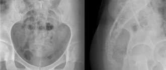
Postpartum hematomas in children (highlighted in light)
This further complicates the examination process, since it is difficult even for an adult to lie still, and almost impossible for a child.
It is necessary to calm, lay down and secure the baby correctly so that the procedure does not have to be repeated. To do this, all options are used - from the presence of relatives in the room who can hold him and calm him down to taking a sleeping pill or sedatives. For a child under one year old, this method will be the best option and a guarantee of successful imaging.
Source: https://apkhleb.ru/rentgen/golovy
X-ray diagnostics in pulmonology
Pulmonology is a branch of medicine dealing with the diagnosis, treatment and prevention of diseases of the bronchopulmonary system. Radiography is considered the leading research method using digital and classical methods. X-rays are available and allow you to obtain clear images, on the basis of which you can make a diagnosis and formulate treatment; subsequently, it is possible to dynamically monitor the course of the disease and identify residual effects.
X-rays of the lungs allow you to see various shadows that normally should not be there. Based on the characteristics of the shape, size and localization of these shadows, many pathologies can be diagnosed:
- Pneumonia of various etiologies.
- Anomalies and malformations of the respiratory system.
- Traumatic lesions.
- Infectious and inflammatory diseases of the lungs.
- Degenerative-dystrophic processes (acquired emphysema and others).
- Foreign bodies in the bronchial tree.
- Destructive pathologies (abscess, gangrene).
- Neoplasms of benign and malignant nature, sites of metastasis.
- Pleurisy.
- Fungal infections of lung tissue.
- Parasitosis.
- Tuberculosis.
X-ray image of the OGK
Indications for an x-ray are patient complaints of a prolonged cough, sputum streaked with blood, difficulty breathing, chest pain, shortness of breath, high temperature, fever, chills, changes in the blood picture. In addition, x-rays are performed to monitor the quality of treatment and identify negative consequences after the disease.
Appearance of a child's skull before the loss of baby teeth
Skull before loss of baby teeth
Children are born without teeth. In the first years of life, babies do not need them, since there are no solid foods in their diet. As more solid foods are introduced, children develop baby teeth.
From about 5 years of age, baby teeth begin to fall out, giving way to molars. Before the baby teeth fall out, the skull has a slightly different shape. The human skull gradually increases in size, while the baby teeth remain small. Molars appear immediately large and remain so for life.
An x-ray of an adult shows that there is no additional row of teeth behind the molars in the upper and lower jaw. An X-rayed skull with baby teeth often frightens unprepared people.
Baby teeth are already formed at the time of birth, but erupt gradually, each in a specific place, starting from 5-7 months. In this case, the rudiments of permanent teeth (molars) are located in the upper and lower jaws of the baby. When they finish forming, they push out the primary teeth. This is how the replacement of baby teeth with molars begins.