Carrying out sonography, or ultrasound of the hip joints in newborns, significantly speeds up the detection of congenital pathologies. The diagnostic procedure has many advantages over other studies: no harmful radiation is used, and the child’s complete immobility is not required. Using ultrasound, you can detect any destructive changes not only in the hip joint, but also in the periarticular structures.
Sonography, carried out immediately after the birth of a child, allows us to identify the most minor anomalies, which in the future can cause the development of serious diseases. Timely treatment will help the newborn to fully grow and develop.
Benefits of Sonography
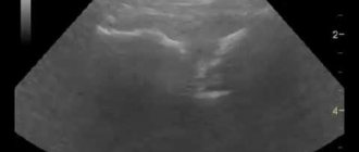
When conducting an ultrasound examination, high-frequency acoustic waves are reflected from surfaces encountered in their path. All tissues in the human body have their own special structure. They differ in density, contain a certain amount of liquid, and therefore differ significantly in the speed of reflection. These values are read by special sensors and then visualized on the monitor in the form of images. An experienced diagnostician, when interpreting an ultrasound of the hip joints in infants, identifies ligaments, tendons, bone, and cartilaginous tissues, and then assesses their condition. Ultrasound has many advantages over other methods of studying the hip joint in young children:
- there is no aggressive and extremely undesirable radiation when examining newborns;
- one of the parents is present during the diagnostic procedure, hold and calm the baby;
- there is no need for the child to be in a stationary position, so the doctor can assess the state of the joint in dynamics.
3% of newborns are diagnosed with congenital dysplasia of varying degrees. This is the name for defective one- or two-sided development of the hip joints, which is characterized by a decrease in the functional activity of the joint. The disease is highly treatable, especially if diagnosed early. The resulting images make it possible to determine the shape and degree of dysplasia, and differentiate it from other congenital pathologies. What can be diagnosed using ultrasound:
- improper development of the acetabulum;
- underdevelopment of the cartilaginous rims surrounded by the acetabulum;
- broken ligament structure;
- degrees of dysplasia - preluxations, subluxations, complete dislocations.
If any form of dysplasia is detected, doctors immediately begin therapy. To assess its effectiveness, frequent ultrasound examinations of the hip joints in infants are required. This is another advantage of the diagnostic technique. Unlike radiography, sonography can be used to constantly monitor the baby’s condition and the rate of tissue regeneration. The diagnostic procedure has only one contraindication - the child’s hypersensitivity to the ingredients of the gel used to conduct acoustic signals.
X-ray examination is also less informative due to the structural features of the hip joint in newborns. There is still little bone tissue in it, but a lot of cartilaginous tissue. And in the images obtained during radiography, only the pathological state of the bone structures is clearly visible.
How is an X-ray examination of the hip joint performed in children?
X-rays of children's hip joints are performed using a standard digital or analogue machine. No special preparation required. In some cases, a cleansing enema may be performed before the x-ray so that the contents of the intestines do not distort the picture.
X-rays are taken in the supine position, in two states - with the legs straightened and with the knees apart. Both hip joints must be included in the image to allow comparison behavior.
Together with all the preparation of the equipment, the procedure takes about ten minutes, of which the child will be exposed to x-ray radiation for about a second.
During the X-ray itself, it is important that the child does not move. To do this, the mother or laboratory assistant holds him by the knees.
Indications and contraindications
To determine normal values or deviations from it, ultrasound of the hip joint in newborns is indicated for all children under 6 months. It is often carried out within a few days after the birth of the child, especially if during pregnancy the woman’s body was affected by external or internal unfavorable factors. Here are the main indications for ultrasound of a newborn:
- a premature baby, the likelihood of developing dysplasia is quite high. The risk group also includes children from multiple pregnancies;
- gluteal or pelvic position of the fetus before childbirth, causing congenital dislocation of the joint;
- difficult pregnancy, especially complicated by severe toxicosis or deficiency of vitamins and microelements;
- a woman taking drugs belonging to various clinical and pharmacological groups during pregnancy - antibiotics, diuretics, cytostatics, immunomodulators, antivirals;
- Acute viral, bacterial or mycotic infections suffered by a woman during pregnancy. Intestinal or respiratory pathologies are especially dangerous in the 2nd and 3rd trimesters, when the fetal hip joint begins to form and foci of ossification nuclei of the articular heads form.
Any of these factors can cause the development of dysplasia. An ultrasound is performed either directly in the maternity hospital, or a few days after the mother and baby are discharged.
After birth, the child is examined by a pediatrician 1-2 times a month to monitor his development. He may prescribe an ultrasound examination if one of the signs of impaired articulation formation is detected: asymmetrical arrangement of skin folds in the groin or buttocks, shortening of the leg, specific clicking during abduction of the child’s hip.
Video: Hip dysplasia – School of Dr. Komarovsky
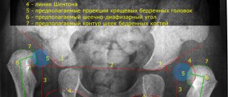
The acetabular angle or index is a radiographic term for measuring the deformity of the hip joint. The concept was first introduced by scientists Kleinberg and Liebermann in 1936. Normally, the acetabular index of the hip joint in newborns is less than 28 degrees. The indicator changes with age. By the end of the first year of life it drops to 22 degrees or less. Deviations from generally accepted standards indicate the presence of pathology in the child: dysplasia, dislocation, subluxation. Timely detection of the disease will prevent its further development and maintain the health of the joint.
Carrying out a diagnostic procedure
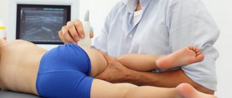
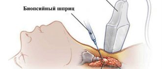
During an ultrasound, the child is placed on his side so that the examined joint is on top and bent at an angle of 20°. A gel is applied to the hip joint to facilitate the sliding of the sensor and improve the conductivity of acoustic waves. During manipulations, all articular and periarticular structures are displayed on the screen. The doctor takes pictures in the positions necessary to assess the condition of the tissues. Usually there are five of them: in the initial position, when bending and extending the leg, when abducting and bringing it to the body.
To prevent the procedure from being interrupted, pediatricians who prescribe a referral for sonography recommend not feeding the baby immediately before the examination. The child must be well-fed so as not to be capricious, but the process of regurgitation can interfere with diagnosis.
Pathology correction methods
A pediatric orthopedist deals with correction of hip dysplasia. After the most informative diagnosis and understanding of the degree of pathology in a particular patient, it is important to prescribe an appropriate treatment method.
The basic principle of any type of treatment is early initiation. Mild forms such as subluxation can be treated conservatively. The wide swaddling technique, exercise therapy, massage, and Pavlik stirrups are used. All techniques involve long-term holding of the legs in abduction and flexion positions and active movements within the permitted range.
For more complex or advanced forms, surgical treatment is used. It consists of correcting defective components of the hip joint - either the socket itself or the head (the operation never concerns the neck of the femur).
Decoding the results obtained
A child is born with a hip joint that is not yet fully formed. It contains a lot of cartilaginous tissue that will ossify as the baby grows. Until this happens, almost all violations can be easily corrected. Certain evaluation criteria for the functional activity of a joint make it possible to establish its correct and incorrect state. Ultrasound of the hip joints in infants determines the norm and abnormalities by the angles formed by the edges of the acetabulum and imaginary lines drawn along the lower points of the iliac bones. At birth, the normal parameters of the acetabular angle are from 25° to 29°. As they grow, by 12 months these values decrease and are: for boys - 18°, for girls - 20°.
A slight deviation of the angle parameters by approximately 1-2° is allowed. This may indicate both individual characteristics of the structure of the pelvis and developing dysplasia. In such cases, no therapy is carried out, and the child is under the supervision of doctors. He is scheduled for sonography at approximately 5 and 10 months, which are considered the age points. The degree of dysplasia is determined by the parameters of alpha and beta angles:
- alpha - angles formed by crossing lines drawn from the median wall of the iliac bones and lines drawn from the lower parts of the iliac bones to the upper edges of the acetabulum;
- beta - the angle formed by the upper edges of the acetabulum and the centers of the cartilaginous plates.
The parameters obtained after measuring the angles become an evaluation criterion for the normal or pathological structure of the joint. Below is a breakdown of the angle values obtained during an ultrasound examination of the hip joint in an infant:
- normal parameters - alpha angle is more than 60°, beta angle is more than 55°,
- pre-dislocation state - alpha angle values - 43-49°, beta angle - 70-77°;
- subluxation - alpha angle - less than 43°, beta angle - more than 70°;
- dislocation - the alpha angle is less than 43°, the beta angle is more than 70°, while the bone surface is strongly concave and the cartilaginous protrusion is deformed.
| Form of pathological condition | Sonography results obtained |
| Normal (correctly formed joint) | Expansion and shortening of the cartilaginous plate |
| Articulation formation too slow (preluxation) | Fixed ossification, which requires constant monitoring of the condition |
| Subluxation | Displaced head of the femur, preservation of the cartilaginous structure unchanged |
| Dislocation (dysplasia) | Underdevelopment of the hip joint, failure to cover the head of the femur with a cartilaginous protrusion |
In the case of ultrasonographic diagnosis of preluxation, treatment is not carried out, since the obtained values may indicate immaturity of the hip joint. As the joint grows, it forms correctly and functions actively. If a subluxation or dislocation is detected, doctors immediately begin therapy for the newborn. The normal values obtained during ultrasound of the hip joints in newborns do not cancel the monthly examination of the baby by a pediatrician, and sometimes by an orthopedist. Specialists constantly monitor the condition of the child’s musculoskeletal system and prescribe diagnostic measures if they suspect the slightest deviation from the norm.
General recommendations
Many mothers who are faced with diagnoses such as immaturity of the hip joints in a newborn note that there should be no reason to panic. In the vast majority of cases, only a systematic visit to an orthopedist is required for monitoring. The joints mature on their own.
Most mothers note that massage prescribed by the doctor, special therapeutic exercises and calcium supplements helped cope with the problem identified by ultrasound at an early age. Mothers try to give it to infants with breast milk, that is, they increase the dose of calcium in their own diet, and begin to specifically take calcium supplements.
Children are recommended to swaddle widely and wear diapers one size larger than required, so that their legs are more often spread apart.
For more information about the ultrasound method of hip joints for newborns and infants, see the following video.
medical reviewer, psychosomatics specialist, mother of 4 children
Before we find out how to decipher an ultrasound of the hip joint in a baby, let’s remember the content of the latest materials. The previous article highlighted when children of different ages, from newborns to babies older than one year, need to have an ultrasound of the hip joints. Also considered:
- how to prepare for the procedure;
- how it goes;
- what are the contraindications;
- what are the pros and cons of the study.
But this information cannot be called exhaustive without a detailed consideration of the interpretation of the results, existing standards and possible pathologies of the hip joints.