- What is a chest x-ray?
- In what areas is chest radiography used?
- How should you prepare for research?
- What does the diagnostic equipment look like?
- What is the basis for the research?
- How is the research conducted?
- What should you expect during and after the procedure?
- Who reviews the X-ray results and where can they be obtained?
- What are the benefits and risks of having a chest x-ray?
- Limitations of chest x-ray
What is a chest x-ray?
Among all x-ray examinations, chest x-ray is the most commonly used.
This procedure is used to take pictures of the lungs and airways, heart, blood vessels, and bones of the chest and spine. X-ray testing is a non-invasive diagnostic technique that helps doctors detect and treat various diseases. In this case, certain parts of the body are exposed to a small dose of ionizing radiation, which allows them to be photographed.
X-ray examination is the oldest imaging method and is most often used in diagnosis.
Up
Internal organs are the basis of life
The human body is a complex mechanism consisting of a huge number of cells that form tissues.
From their individual groups, organs are obtained, which are usually called internal, since the location of the organs in humans is internal. Many of them are known to almost everyone. And in most cases, until someone gets sick somewhere, people, as a rule, do not think about what is inside them. Nevertheless, even if the layout of human organs is only superficially familiar, when a disease occurs, this knowledge will greatly simplify the explanation to the doctor. Also, the latter’s recommendations will become more clear.
The concept of a system means a specific group of organs that is related anatomically and embryologically, and also performs a single function.
In turn, the apparatus, whose organs are closely interconnected, does not have the kinship inherent in the system.
They are found in several specific cavities.
So, in the chest, located within the boundaries of the chest and upper diaphragm, there are three others. This is a pelicard with a heart and two pleurals on either side with lungs.
The abdominal cavity contains the kidneys, stomach, most of the intestines, liver, pancreas and other organs. It is the torso located below the diaphragm. It includes the abdominal and pelvic cavities themselves.
The abdominal cavity is divided into the retroperitoneal space and the peritoneal cavity. The pelvic region contains the excretory and reproductive systems.
To understand in even more detail the location of human organs, the photo below serves as an addition to the above. It shows cavities on one side, and the main organs that are located in them on the other.
They are arranged into two categories: hollow or tubular (for example, the intestines or stomach) and parenchymal or solid (for example, the pancreas or liver).
The former have several layers in their tubes, which are also called shells. The inside is lined with a mucous membrane, which plays a mainly protective function. Most organs have folds on it with projections and depressions. But there are also completely smooth mucous membranes.
Next comes the submucosa, which consists of connective tissue and is mobile.
In addition to them, there is a muscular layer with circular and longitudinal layers separated by connective tissue.
The human body contains smooth and striated muscles. Smooth - prevail in the respiratory tube and genitourinary organs. In the digestive tube, striated muscles are located in the upper and lower sections.
In some groups of organs there is another membrane where blood vessels and nerves pass.
All components of the digestive system and lungs have a serous membrane, which is formed by connective tissue. It is smooth, allowing the insides to slide easily against each other.
Parenchymal organs, unlike the previous ones, do not have a cavity. They contain functional (parenchyma) and connective (stroma) tissue. The cells that perform the main tasks form the parenchyma, and the soft skeleton of the organ is formed by the stroma.
The photo shows the location of human internal organs with a description.
We can say that they are the basis of life. It is difficult to live without lower or upper limbs, but it is still possible. But without a heart or liver a person cannot live at all.
Thus, there are organs that are vital, and there are those without which life is difficult, but nevertheless possible.
Moreover, some of the first components have a paired structure, and without one of them, the entire function passes to the remaining part (for example, the kidneys).
Some structures are able to regenerate (this applies to the liver).
Nature has endowed the human body with a very complex system, which it must carefully treat and take care of what is given to it within the allotted time.
Many people neglect the most basic things that can keep the body in order. Because of this, it becomes unusable ahead of time. Illnesses appear and a person dies when he has not yet done all the things he should have done.
All organs are collected into separate systems, which helps in classifying and systematizing the human structure. This makes it easier to study the structures and their functions in the body. The following systems are distinguished:
- The musculoskeletal system is responsible for moving and taking the body into any possible position in space. The system consists of a bone skeleton, ligaments, tendons, and muscles.
- The cardiovascular system is responsible for transporting blood throughout the body. This provides the tissues with oxygen and nutrients.
- The digestive tract absorbs vitamins, microelements, proteins, fats and carbohydrates from food. This is necessary to generate energy, without which it is impossible to perform any actions.
- The organs of the respiratory system remove carbon dioxide and saturate the blood with oxygen, which is distributed throughout the body.
- The nervous system is central and peripheral, responsible for the functioning of the entire organism, collecting information from the outside world, processing it.
- Endocrine glands are responsible for maintaining homeostasis within a person.
- The genital organs are responsible for reproduction, the urinary organs are responsible for the removal of biological fluids.
The skin is also separately distinguished, which protects the insides from unfavorable external factors and is responsible for the aesthetic function.
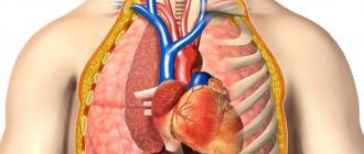
The chest cavity contains a number of vital formations. One of the most important is the heart. It is located almost in the middle of the chest, localized behind the middle third of the sternum. The size of the heart is equal to the size of a hand clenched into a fist.
Muscle tissue is very powerful; the cells are connected to each other by bridges, forming something like a canvas. This structure ensures electrical conduction and contraction of the heart. The organ provides blood circulation, receiving venous blood from the vessels, saturating it with oxygen, turning it into arterial blood. The latter, through heart contractions, ensures the delivery of oxygen and nutrients to all human systems and organs.
Also located in the chest are the bronchi and lungs. The latter are a paired organ; they occupy most of the space of a given cavity. Each lung consists of large lobes: the left one has 2, the right one has 3.
The lobe is divided into smaller formations, the structure of which contains alveoli - special bubbles that carry out gas exchange. The alveoli saturate the blood with oxygen and ensure the elimination of carbon dioxide. These structures are formed by branching of the bronchi.
The latter are large trunks that penetrate the lungs through the so-called gates, where they begin to divide into smaller formations. The bronchi, in turn, are the airways in humans.
Another organ located in the chest is the trachea. It originates from the larynx, from where it extends lower and passes into the bronchi.
The esophagus runs parallel, which has several anatomical bends; it itself is a muscular tube that provides passage for the bolus of food for further digestion in the stomach.
In addition, in addition to the structures listed above, there are large vessels - the aorta, pulmonary arteries and veins. Also in the chest are lymph nodes, nerve trunks and another gland - the thymus, or thymus.
The latter is an organ of the immune system that gradually atrophies with age. In persons over 16-18 years of age, only remnants of the thymus are present.
The organs of the abdominal cavity ensure the digestion of food and the formation of feces from its remains. They are separated from the chest by the diaphragm. The organs of the thoracic cavity are as follows:
- The stomach is a hollow formation that originates from the esophagus. The stomach is responsible for the absorption of amino acids; it contains juice, which, in addition to its digestive function, disinfects incoming processed foods.
- Then there is a transition to the small intestine, consisting of 3 sections - duodenum, jejunum and ileum. These organs are involved in the digestion of food bolus, absorption of amino acids and carbohydrates. Bile also begins to form in the small intestine.
- Next is the large intestine. Its sections are as follows: cecum with appendix, transverse colon, descending and sigmoid colon. The thick section ends with the rectum. The final absorption of nutrients and water absorption occurs in this organ. Fecal masses are formed from food gruel, which are eliminated from the body through the anus, which ends in the rectum.
- Also in the abdominal cavity are the liver, pancreas and spleen. These structures are responsible for metabolism, hematopoiesis, and bile exchange. The liver is located under the right costal arch, the pancreas is located under the left. The spleen is adjacent to the pancreas below.
- In the lateral sections of the abdominal cavity there are kidneys, which are paired formations. Above them are the secretory glands - the adrenal glands, which are very small in size. The ureters extend from the kidneys and pass into the bladder. The main function is the formation of urine, which enters the bladder and is discharged out.
DETAILS: Temperature in a cat with chronic kidney disease
In addition, the abdominal cavity also contains large and small blood vessels, lymph nodes, nerve trunks and plexuses, and there is also an omentum located here, which ensures the maintenance of all formations in their places. It also protects internal structures from traumatic effects.
The brain provides a person with mental activity, distinguishing him from other living organisms. It is essentially a mass of nervous tissue. It consists of two cerebral hemispheres, the pons and the cerebellum.
- The cerebral hemispheres are necessary to control all thought processes and provide a person with conscious control of all movements
- At the back of the brain is the cerebellum. It is thanks to him that a person is able to control the balance of the entire body. The cerebellum controls muscle reflexes. Even such an important action as withdrawing your hand from a hot surface so as not to damage the skin is controlled by the cerebellum.
- The pons lies below the cerebellum at the base of the skull. Its function is very simple - to receive nerve impulses and transmit them
- The other bridge is oblong, located slightly lower and connects to the spinal cord. Its function is to receive and transmit signals from other departments
- The rectum is a similar organ in both men and women. This is the final part of the intestine. Digestive products are removed through it. The length of the rectum should be about fifteen centimeters
- The bladder differs in location, female and male placement in the cavity. In women, it is in contact with the walls of the vagina, as well as the uterus; in men, it is adjacent to the seminal vesicles and streams that remove the seed, as well as to the rectum
female pelvic (genital) organs
- The vagina is a hollow tubular organ that extends from the genital slit to the uterus. It is about 10 centimeters long and is adjacent to the cervix, the organ passes through the genitourinary diaphragm
- The uterus is an organ made up of muscles. It is pear-shaped and is located behind the bladder but in front of the rectum. The organ is usually divided into: fundus, body and neck. Performs reproductive function
- The ovary is a paired, egg-shaped organ. This is a female gland that produces hormones. The maturation of eggs occurs in them. The ovary is connected to the uterus by the fallopian tubes
male pelvic (genital) organs
- The seminal vesicle is located behind the bladder and looks like a paired organ. This is a secretory male organ. Its size is approximately five centimeters in diameter. It consists of bubbles connected to each other. The function of the organ is to produce seed for fertilization
- The prostate gland is an organ consisting of muscles and glands. It is located directly on the urogenital diaphragm. The base of the organ is the urinary and seminal canal
In what areas is chest radiography used?
Chest X-rays are used to evaluate the condition of the lungs, heart, and chest wall. As a rule, first of all, this study is prescribed to diagnose the following symptoms:
- Dyspnea
- Severe or long-lasting cough
- Chest pain
- Chest injury
- Fever
Chest X-ray is used to diagnose and monitor the following conditions:
- Pneumonia
- Heart failure and other heart diseases
- Emphysema
- Lungs' cancer
- Position of the central catheter or endotracheal tube
- Other diseases
Up
Types of GC structure
Normosthenic and hypersthenic forms of the chest
Normal and pathological forms of HA allow doctors to evaluate the characteristics of breathing, the position of organs, and the normal functioning of the chest cavity. Among the natural forms of HA, there are 3 types:
- Normosthenic structure. The HA is cone-shaped, slightly truncated. The ribs are angled and the shoulders are at a 90-degree angle to the neck.
- Asthenic structure. The HA is narrow, elongated and flat, the ribs and fossae under and above the clavicles are well defined. The muscles are poorly developed, unlike the other 2 types.
- Hypersthenic structure. The chest is barrel-shaped, cylindrical. The diameters of the rib arcs are equal, and there is a small distance between them. The muscles are strong, there are no pits under or above the collarbones.
In addition to normal forms of GC, there are pathological ones. They are usually caused by various diseases of the skeletal or respiratory system. Can be observed in both childhood and adulthood:
- Emphysematous GC. Develops against the background of chronic emphysema. The diameter increases, the pits and ribs are more pronounced, almost flat.
- Paralytic. It appears against the background of long-term lung diseases, when the paired organ shrinks. The distance between the ribs on different sides can vary significantly. Because of this, the blades move asynchronously.
- Rachitic, keel-shaped form. Develops in adults and adolescents who suffered from rickets in childhood. The sides towards the front are squeezed inward, like a chicken.
- Funnel-shaped. It looks like they made a narrow hole in the center of the chest. A boat-shaped HA is formed in a similar way, but the depression is more like a boat. The causes of these pathologies cannot be determined, but it is assumed that they are formed in diseases of the bone marrow and bones.
Pathological forms of the chest affect body functions, interfere with normal breathing and impair heart function. It may be impossible to get rid of complex HA curvatures; in simpler cases, surgery can help.
How should you prepare for research?
In most cases, chest x-rays do not require any preparation.
During the examination, you will need to remove some or all of your clothing and wear a special hospital gown. In addition, you should remove all jewelry, glasses, dentures, and any metal or clothing that could interfere with the x-ray image.
Women should inform their doctor and radiologist of any possibility of pregnancy. As a rule, X-ray examinations are not performed during pregnancy to avoid exposure of the fetus to radiation. If x-rays are necessary, special precautions should be taken to protect the developing child.
Up
Contraindications to CT
CT scanning of the chest organs is not allowed in children under 14 years of age, as well as in pregnant women. Doctors also consider other conditions as absolute contraindications to the use of this diagnostic method:
- mental disorders in which the patient cannot control the activity of his muscles and is unable to remain calm;
- serious condition of the patient;
- severe allergy to iodine if contrast is planned to be used;
- insufficient renal function (relevant for CT with contrast);
- severe diabetes mellitus or thyroid pathology (the main contraindications for the use of contrast).
It is important to inform your doctor in advance about any existing health problems before the procedure to avoid complications!
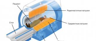
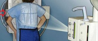
What does the diagnostic equipment look like?
Chest X-ray equipment typically includes a wall-mounted box-like machine that holds the X-ray film or matrix to take digital images, and an X-ray tube that is positioned 1.5 meters posteriorly.
In some cases, the X-ray tube is suspended above the patient's table. A drawer under the table contains X-ray film or a photographic plate for obtaining digital images.
A portable (portable) X-ray machine is a compact device that allows you to examine a patient directly in the intensive care unit or in a hospital bed. In this case, the X-ray tube is attached to a flexible arm, which is placed above the patient's body, while the photographic plate or X-ray film holder is placed behind the patient's body.
Up
Splanchnology
The study and location of human organs are considered by anatomy in a special section called splanchnology, the study of the internal organs. We are talking about structures that are located in body cavities.
First of all, these are the organs of the human abdominal cavity involved in digestion, the location of which is as follows.
Next comes the genitourinary, urinary and reproductive systems. The section also studies the endocrine glands located next to these systems.
The internal organs also include the brain. The head is located in the cranium, and the spinal canal is located in the spinal canal. But within the scope of this section, these structures are not studied.
All organs appear as systems that function in full interaction with the entire body. There are respiratory, urinary, digestive, endocrine, reproductive, nervous and other systems.
What is the basis for the research?
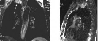
X-rays are similar to other forms of radiation such as light or radio waves. It has the ability to pass through most objects, including the human body. When used for diagnostic purposes, an X-ray machine produces a small beam of radiation that passes through the body and creates an image on photographic film or a special matrix for obtaining digital images.
X-rays are absorbed differently by different organs and parts of the body. Dense structures, such as bones, absorb radiation strongly, while soft tissue structures (muscles, fatty tissue and internal organs) transmit X-rays to a greater extent. As a result, on an x-ray, bone tissue appears white, air and air spaces appear black, and soft formations appear various shades of gray.
In a chest x-ray, most of the radiation is absorbed by the ribs, which appear white or light gray on the x-ray. Lung tissue absorbs X-rays weakly, and therefore the X-ray image appears dark in color.
Until recently, X-ray images were stored as copies on film, similar to photographic negatives. Nowadays, most images are available as digital files that are stored electronically. Such images are readily available and are used for comparison with the results of subsequent examinations to assess the effectiveness of treatment.
Up
Abdominal composition
The abdominal cavity is occupied by organs responsible for the digestive tract process.
There is also the pancreas, as well as the liver and kidneys. Next to them is the spleen, stomach, just below the kidneys and genitals, namely:
- The stomach is part of the body of the digestive system. It continues with the esophagus and is separated by a valve. In appearance it resembles a bag. The walls of the stomach produce mucus, and juice breaks down food.
- The gastrointestinal tract includes the intestines. It is large in size and length. The intestines begin after the outlet of the stomach. Body parts include intestines of different types. Thin - or in other words - duodenum, which gradually turns into a thick one, and then into a straight one. The intestines perform an important function - digest crushed foods and rid the body of food debris.
- The largest gland in size is the liver . She is involved in digestion. One of the main tasks of the organ is to carry out metabolism. The liver is located under the diaphragm and has two lobes. Thanks to a passing vein, the liver is connected to the duodenum.
- The spleen is a part of the body located above the diaphragm. It is responsible for important functions, among them hematopoiesis and protection of the body. Depending on the flow and accumulation of blood, the size of the spleen changes.
- Kidneys are organs that come in two pairs. They occupy the lumbar zone and are responsible for the regulation of homeostasis and the release of urine. The shape of the buds resembles beans. The organs are located in a fibrous capsule. Parts of the body consist of parenchyma; they include systems responsible for the accumulation and excretion of urine. The outside of the kidney is covered with a dense sheath. The outer layer of the cortical component makes up the parenchyma, and the inner part includes the medulla. The small calyces of the kidneys serve to collect urine, then the general collection of urine occurs by draining the fluid. The end of the renal pelvis is near the ureter.
- Above them are the adrenal glands . They belong to paired endocrine glands. Parts of the body regulate metabolism and also prepare the human body for stressful conditions. The adrenal glands have a medulla where adrenaline is stored. Thanks to the action of this hormone, heart contractions become faster and larger. Blood pressure begins to rise, the pupils become dilated, and glycogen quickly turns into glucose.
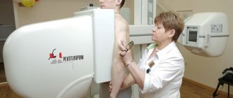
How is a chest x-ray performed?
As a rule, it is necessary to obtain two images of the chest organs: in frontal and lateral projection. At this time, the patient is positioned opposite the photographic plate holder.
A radiologist (a doctor who specializes in X-ray examinations) or a nurse presses the patient's shoulders and pelvis against the surface of the device where the photographic plate is located. For the second image, the patient is positioned sideways with his arms up.
If the patient is unable to stand, he is placed on a special table. In this case, you should remain as still as possible, and during the photo itself, hold your breath for a few seconds, which reduces the likelihood of blurring the image. When the X-ray machine is operating, the doctor moves towards the wall or leaves the treatment room into an adjacent room.
After the examination is completed, the radiologist asks the patient to wait until the images are analyzed, as additional series of images may be required.
A chest x-ray generally takes about 15 minutes. Additional imaging may be needed to evaluate changes in the chest after a few days, weeks, or months.
Up
What should you expect during and after the study?
A chest x-ray itself is painless.
The cool temperature in the treatment room and the cold surface of the photographic plate may cause some discomfort to the patient. The inconvenience is caused by the need to stand still, especially if you have arthritis, injuries to the chest wall and upper or lower extremities. The doctor or physician's assistant helps the patient find the most comfortable position, which also ensures high-quality images.
Up
Who reviews the X-ray results and where can they be obtained?
The images are analyzed by a radiologist: a doctor who specializes in performing x-ray examinations and interpreting their results. After examining the images, the radiologist draws up and signs a report, which is sent to the attending physician. In some cases, the report can be collected from the radiology department itself. Chest x-ray results can be obtained fairly quickly.
A follow-up examination is often required, the exact reason for which will be explained to the patient by the attending physician. In some cases, additional examination is carried out when doubtful results are obtained that require clarification during repeated images or the use of special imaging techniques. Dynamic observation allows timely identification of any pathological abnormalities that arise over time. In some situations, repeated examination allows us to talk about the effectiveness of treatment or stabilization of tissue condition over time.
Up
Benefits and Risks of Chest X-ray
Advantages:
- After completion of the examination, no radiation remains in the patient’s body.
- When used for diagnostic purposes, X-rays do not cause any side effects.
- X-ray equipment is relatively inexpensive and is available in most emergency departments, diagnostic centers, clinics and other institutions, making X-rays convenient for both patients and physicians.
- Since X-ray examination is quick and easy, it is particularly useful for the diagnosis and treatment of emergency conditions.
Risks:
- With excessive exposure to X-ray radiation on the body, there is always an extremely small risk of developing malignant tumors. However, the benefits of accurate diagnosis significantly outweigh this risk.
- The effective dose of radiation for bone x-rays varies.
- A woman should always tell her doctor or radiologist about the possibility of pregnancy.
A few words about reducing the effects of radiation on the body
During an x-ray examination, the doctor takes special measures to minimize radiation exposure to the body while trying to obtain the best quality image. Experts from international radiological safety councils regularly review radiology standards and produce new technical recommendations for radiologists.
State-of-the-art X-ray machines allow you to control the dose of X-ray radiation and provide filtration, which minimizes beam scattering. In this case, the patient’s organs and systems that are not examined receive a minimal dose of radiation.
Up
Helpful information
Every person is unique and inimitable. In this case, various anomalies are often encountered - for example, doubling of an organ, changes in its shape and size. It is surprising that this often goes unnoticed and does not affect health in any way.
It is also important that when any of the paired organs is removed, the other of this pair can take over its functions. And this almost always happens. At the same time, the person himself will feel the same as before.
The potential and endurance of the body are amazing; it is fragile and strong at the same time. Biological scientists and doctors have to find out the answers to a large number of mysteries of the human body. Work in this area is ongoing.
As you can see, the structure of the human body is simple and complex at the same time. Researchers still cannot fully unravel all the mysteries of the body. A person is able to carry out higher nervous activity thanks to the cerebral cortex, which is inaccessible to other biological species.
For these reasons, it is important for people to have at least a general understanding of their structure, which will help throughout their life, especially when checking their own health.
What are the limitations of chest x-ray?
Chest X-ray is an extremely useful diagnostic tool but has some limitations.
Since routine x-ray examination does not reveal some conditions of internal organs, it does not always allow an accurate diagnosis. For example, chest x-rays do not always diagnose malignant tumors of small diameter. In addition, a blood clot in the lungs, which appears with pulmonary thromboembolism, cannot be seen on an x-ray.
Therefore, to clarify the results of chest x-ray, in some cases it is necessary to use other imaging methods.
Up