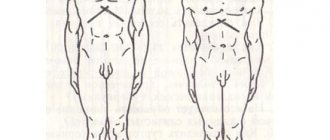
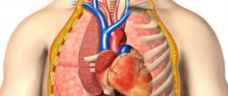
Organs of the chest schematically Magnetic resonance imaging allows you to study the structure of the internal organs. The method provides comprehensive information about the condition of soft tissues and is less indicative of bones. MRI diagnostics are prescribed to detect diseases of the chest organs. Scanning in most cases is not the method of choice for examining the lungs, but it allows for good visualization of the trachea, bronchi, pleura, heart, thymus, large blood and lymphatic vessels. What a chest MRI shows often determines the final diagnosis. The method is used as an alternative and addition to CT, radiography or ultrasound examination of the heart.
What does a chest MRI show?
Magnetic resonance imaging has high resolution characteristics; the procedure is prescribed when other imaging methods are ineffective.
Evaluates the nature of the tumor, size, and condition of the outer lining of the heart. This study reveals:
- inflammatory processes;
- hernias in the thoracic spine;
- tumors: size, degree of spread to surrounding tissues;
- myocardial infarction, its perfusion;
- morphological changes in bone and soft tissue in the sternum;
- dynamics of blood flow in blood vessels and cardiac chambers;
- determines the functional state of the heart and valves.
Helps clarify the results of radiography and computed tomography. If questionable results are obtained, additional photographs are taken.
The procedure is recommended for suspected thrombosis, cardiac aneurysms, oncopathology, myocardial ischemia, and pneumonia.
Repeated diagnostics are prescribed to evaluate the effectiveness of the recommended therapy.
What pathologies are visible in the photographs?
Chest MRI shows the following diseases:
- Injuries of the skeletal system and soft tissues.
- Neoplasms of a malignant or benign nature.
- Lesions of the pleura.
- For heart diseases.
The image of the lymph nodes allows us to determine their enlargement and inflammatory processes in this area. An image of organs makes it possible to study the spread of malignant metastases and the exact location of the tumor and its size.
Advantages and disadvantages of MRI
The advantage of the diagnostic method is safety and non-invasiveness. There is no radiation exposure, as magnetic radio waves are used.
The procedure is painless and suitable for small children. Visualizes the structures and functionality of internal organs.
If the pathological focus is localized behind a bone formation, magnetic resonance imaging will help detect it.
Allergies to contrast materials used in MRI are less common compared to iodine contrast in CT and radiography.
If the safety protocol is followed, the procedure does not pose a threat or risk to the patient. Flaws:
- price;
- prolonged stay in a stationary position causes discomfort in patients;
- not suitable for patients with metal structures in the body, insulin pumps, pacemakers, defibrillators;
- sedation can cause an overdose; to prevent it, assistants monitor the condition of the patient;
- high doses of contrast agents are fraught with nephrogenic systemic fibrosis for patients with renal dysfunction;
- MRI does not always distinguish cancerous tumors from edema fluids.
Magnetic resonance imaging is contraindicated for people with a serious condition, mental illness, pacemakers, ferromagnetic implants, and pregnant women during the 1st trimester.
Lung pathologies.
Advantages and disadvantages of the procedure
When scanning MRI of the chest organs, radiation from electromagnetic fields is used, which are completely harmless to human life. The high quality of the images makes it possible to see the beginning changes in the structure of the tissues being studied. The scanning is painless, non-invasive (without violating the integrity of the skin). Detects pathologies of soft tissues and skeletal structures that are not detected using X-rays or computer diagnostics.
The main disadvantage is the high cost of the study. It is impossible to perform the procedure on patients suffering from claustrophobia (fear of closed spaces). In this case, an open-type device is used, but due to the low power of this type of tomograph, the images are obtained with distortions. People weighing over 150 kg are not able to undergo an MRI. The need to remain motionless for a long time makes the procedure difficult for children and people suffering from mental illness.
Indications and contraindications
The use of MRI of the chest organs occurs when necessary:
- Confirm diseases of the chest (bone structures).
- Assess the speed of blood flow in the vessels of the heart after a heart attack.
- Clarify the diagnosis after CT or X-ray.
- Determine the degree of damage to the pleural cavity.
- Monitoring the dynamics of the development of the tumor process.
- Identify the development of an aneurysm at the initial stage.
- Suspicion of lung cancer (people with a smoking history of more than 15 years).
Contraindications to MRI: pregnancy (first trimester); implants; fixed dental prostheses; tattoos if the paint contains iron-containing particles; a pacemaker or other electronic device, metal wires in the body; mental disorders; the weight of the subject is more than 150 kg. The use of a contrast agent should be excluded if the patient under study has renal failure.
Preparation process
No specific preparation is required. The patient can consume water and food as usual.
Before the examination, you are asked to remove metal jewelry, piercings, and leave on hearing aids and mobile devices. This will allow you to get an accurate result, without errors.
When using a contrast agent, it is recommended to report allergic reactions, somatic diseases, and recent surgical interventions.
Women warn about pregnancy. Patients with kidney dysfunction are required to undergo a blood test first.
The doctor asks which groups of drugs have been used over the past 7-14 days.
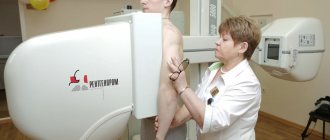
How the research works
Examination of the chest and mediastinal organs is recommended for patients in hospitals and outpatient clinics:
- the person is placed on a movable table, belts and special bolsters ensure immobility;
- if a procedure with contrast is indicated, a catheter is inserted into the vein, to which a bottle with saline solution is connected;
- medical workers leave the room, the table moves into the tomograph;
- communication with medical personnel remains throughout the duration of the study;
- contrast material is administered after the initial series of images is obtained;
- When the ring of the device begins to rotate, noise occurs. For protection, use headphones or play soft music.
Contrast enhancement expands diagnostic capabilities. After administration of the drug, you feel warmth and a rush of blood.
The diagnostician takes numerous sequential images. Once completed, the patient is asked to remain horizontal until the images are processed. The duration of the procedure is 40-60 minutes.
If the procedure was not accompanied by sedation, the patient can immediately return to their normal lifestyle.
Sedation requires recovery time. The image is interpreted by a radiological specialist.
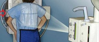
Carrying out a scan
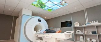
The design of an MRI machine has 3 main components: a moving table, a magnet and a tube. The latter can be of three options: completely closed (not suitable for those who suffer from a fear of closed spaces and overweight people), partially open or enclosing only a magnet.
Omniscan contrast agent for MRI
In the adjacent room behind durable glass there is a diagnostic examination room and computer equipment that receives information from the tomograph. The radiologist constantly monitors the results of the study, and if necessary, may ask the patient to take a more correct form and try not to change it, and may also take additional pictures.
Scanning occurs as follows:
- The patient must remove all jewelry and other body items and undress down to his underwear. He will be given a disposable medical shirt.
- The patient should sit comfortably on the table, his head and limbs will be secured with special belts that protect him from accidental movements that could spoil the diagnostic result.
- MRI of the thoracic spine and mediastinal organs is performed with the patient lying on his back. Depending on the type of device, the patient is either pushed into the tunnel together with the table, or a tomographic device is placed over the scanned area.
- During the process, the patient should be as immobilized as possible. You are allowed to talk to your doctor during the procedure. For this purpose, a microphone is installed in the camera of the device.
- When the tomograph ring rotates, a slight noise appears. The patient can protect their ears by using earplugs. And in some clinics, in order to calm and distract the patient, they play soft music during the procedure.
- There is no recovery period required after scanning. Immediately after receiving the transcript of the results, you can go home. There is no need to change your daily routine or limit yourself in food.
Diagnostic manipulation can last from 15 to 60 minutes. This depends on the age of the patient, the tasks set and the type of tomograph.
During the procedure, there is usually no discomfort or pain in the area being examined.
Can it be done for newborns?
Magnetic resonance imaging works thanks to a magnetic field, which is safe for people of different age groups.
To diagnose a newborn, an incubator magnetically compatible system is used. MRI reveals mediastinal tumors, intracellular compactions, emphysema, stenosis, and malformations.
Diagnosis of the child is carried out with the preliminary use of sedation: it is difficult for the baby to remain in a motionless position for a long time.
It is advisable to maintain the ability to breathe independently. Anesthesia is used in extreme cases due to the risk of adverse effects on lung tissue.
When using contrast agents, it is recommended to refrain from breastfeeding for 1-2 days.
Complications
Magnetic resonance imaging is well tolerated and complications are rare. More often it occurs:
- local increase in body temperature;
- metallic taste in the mouth - caused by a contrast agent;
- deterioration of the psycho-emotional state of a patient with mental disorders;
- tachycardia;
- cough with chronic respiratory pathologies;
- exacerbation of central nervous system diseases;
- contrast agents cause allergies, nausea, pain, hematomas at the injection site;
- sedation is fraught with overdose.
In patients with renal dysfunction, in isolated cases tumors occurred in the extremities when a high dose of contrast was administered.
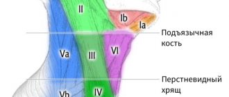
Indications
The costosternal joints connect the ribs to the sternum. Their ligaments go to the anterior and posterior surfaces of the sternum and connect to the opposite and neighboring ligaments. The cause of pain in the chest is often costosternal syndrome (found under the names costochondritis, cartilage dystrophy, arthrosis), “sliding” rib syndrome, Tietze syndrome.
Costochondritis is an inflammation of the cartilage tissue at the junction of the ribs and sternum. Muscular, osteochondral structures are involved in the pathological process, and its main symptoms are pain in the sternum, which intensifies with coughing, movements, deep breaths, and, less commonly, swelling. Added to these are psychopathological factors - increased anxiety with disruption of normal lung function, depression. Symptoms of costochondritis are similar to angina pectoris, ischemic heart disease and other cardiovascular diseases.
To differentiate diseases, to understand what exactly causes pain and what treatment method to prescribe, an examination is carried out. Therefore, if alarming symptoms appear, you cannot postpone a visit to the doctor.
Price
The cost of the procedure depends on a number of factors: the clinic that conducts the study, the doctor’s qualifications, the need to use contrast material, and the patient’s weight. Price from 2500 to 5500 rubles.
For a mobile nervous system, preliminary use of sedatives is recommended. Patients with excess body weight and a fear of closed spaces are recommended to go to a clinic that uses an open type of tomograph.
If you are hypersensitive to loud sounds, use earplugs or special headphones. Immediately after completing the procedure and receiving the results, the patient returns to his usual lifestyle.
Where to do magnetic resonance imaging of the chest cavity in Moscow and St. Petersburg
MRI of the lungs in Moscow and St. Petersburg is offered by several dozen private diagnostic centers where the most innovative high-resolution equipment is installed.
The resource’s specialists have developed a convenient search system that allows you to optimize the criteria for choosing a medical center according to dozens of necessary indicators:
- Location;
- Equipment Features;
- The need for contrast;
- Offering discounts;
- Working hours;
- Metro stations;
- Magnet power of tomograph.
You can get a free consultation in advance to resolve questions about the speed of obtaining results, the qualifications of specialists, and the individual characteristics of scanning.
There are alternative examination methods that are informative in determining a number of lung diseases:
- Ultrasound examination (ultrasound);
- Bronchoscopy;
- Virtual examination of the bronchi (MSCT).
A referral for testing is issued by the attending physician, who will select the optimal diagnosis for each patient individually.
MRI price in St. Petersburg and Moscow
You can find out the price of a chest cavity examination in St. Petersburg and MSC directly on the website, by calling the indicated numbers, or by using the institution selection form. The cost of thoracic MRI differs in different clinics, which depends on many indicators - equipment costs, room rental, and the qualifications of radiology doctors. “Unified Consultative Center for CT and MRI” is pleased to offer clients the optimal choice of medical institution depending on requirements, preferences, price range, and operating hours.