Fluorography is a non-invasive examination of the chest organs (OGC, OGP) using X-ray radiation.
This type of diagnosis is used for general medical examination of the population and is carried out as part of medical examinations for work, for the army, etc.
If you have a compulsory health insurance policy (CHI), this study is carried out free of charge.
The procedure carries radiation exposure. However, it is lower in comparison with other x-ray methods. Digital FOG irradiates less than film.
The study is not recommended for pregnant women and children under 14 years of age. Can be used during lactation.
FOG determines all the same changes in the chest organs as radiography of this area, only in slightly worse quality, therefore it is used as the first and widespread stage of the examination. The most important objective of the method is to identify clinically silent forms of pulmonary tuberculosis or cancer.
What it is
Fluorography is a study primarily of the respiratory tract. It is used as a quick method for identifying various diseases of the chest organs.
The name screening implies the ability to detect pathologies at the stage when a person already has a disease, but it is still so underdeveloped. Firstly, it does not produce clinical symptoms. Secondly, it can be identified and treated as effectively and painlessly as possible.
In addition, this is the primary allocation of contingents so as not to waste more expensive medical resources when not necessary. This is why screening was invented.
This method corresponds to the strategy of modern medicine - a focus on prevention and earlier detection of pathology. Particular attention is paid to the lungs with the current high level of smoking. A bad habit reduces resistance to infectious diseases and causes tumors.
The method itself is X-ray (X-rays pass through the chest), which implies a radiation dose, albeit a very low one.
More modern equipment is the digital FOG mode. It has positive qualities:
- not so long to wait for results;
- softer load;
- it is more convenient to store the conclusion on a computer;
- can be downloaded to a flash drive;
- Available to receive via the Internet online, print;
- it's cheaper for a clinic.
Projection diagnostics of the lungs
When performing an X-ray of the lungs, it is possible to do the study in two projections. Naturally, the harm from radiation is higher than with one photo.
However, with the help of a plain chest x-ray in two projections, a person’s life can be saved, because not all diseases are visible with a direct projection.
X-ray of the OGK in two projections is performed for diagnosis:
- pneumonia;
- pulmonary tuberculosis;
- cancerous tumors;
- pleurisy;
- the presence of abscesses, cysts;
- airiness of the lung;
- pneumothorax;
- heart sizes.
Lateral projection
Diagnosis of the chest in two projections is carried out in a direct and lateral view. The direct view is also called the anteroposterior view, a name based on the way the X-rays pass through the patient's chest cavity. When examining the lateral position, it makes no difference whether the patient is placed on the right or left side of the screen.
The image in the lateral projection is secondary - it helps to better see those organs that were closer to the screen.
Targeted lateral projection is extremely important for determining the volume of pneumonia and localizing the source of inflammation, as well as for determining the location of tumors in the lungs.
How to pass
No specific preparation is required. You can eat before FOG.
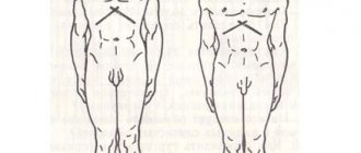
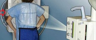
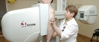
The procedure is as follows. Clothes and jewelry above the waist are removed, and lead pads are placed on the points of the thyroid and gonads. The subject stands on the step and positions his chin. Afterwards, according to the employee, patients need to take a deep breath and not breathe. Radiation is supplied from the rear side. After the photo, you can exhale and remove the pads.
Photo: aiming rays at a man.
the field is set, the shoulder blades spread, the chin rises
For immobilized, older people, the study is carried out in a supine position. There are also portable devices that can send to an address if a person is unable to transport.
What reveals
As already noted, FOG (otherwise FLG, FOGK) is a screening method effectively used to identify respiratory tract diseases. These include:
- pulmonary tuberculosis;
- cancer and other neoplastic processes;
- pulmonary sarcoidosis.
Of course, the results of fluorography can reveal a much wider spectrum of pathology. However, they usually manifest themselves in a more pronounced clinical picture and are less dangerous for the person being examined or in epidemiological terms.
Therefore, if a patient independently presents with symptoms of a chest disease or is undergoing diagnostics in a hospital department, without having had fluorography in the last year, he will be prescribed a chest x-ray. If necessary, CT scan of this area or fluoroscopy.
Such diagnostic methods will differ in accuracy. But they take longer, are more expensive and carry a higher radiation dose.
It is also important to understand that if, based on the results of FOG, the doctor identifies signs of an illness, he will still be prescribed a second, more informative method to clarify the picture in the image. On a fluorogram, lesions from approximately 5 mm can be correctly identified, and on a radiograph – from 2 mm.
Also, a complete additional examination will include an appropriate consultation with examination, blood tests, ECG (cardiogram), etc. According to the patient’s indications, a hospital will be organized according to its specialization.
Why is fluorography of the lungs done and what is the procedure?
Examination of the lungs allows early detection of pathological changes in lung tissue and can show signs of heart failure. A fluorogram shows the internal organs in the thoracic region and their connections. X-rays pass through the human body and transfer images of internal organs onto film.
What diseases does it detect?
Using fluorography of the lungs, the following diseases can be identified:
- pneumonia;
- malignant tumor;
- sclerosis;
- musculoskeletal abnormalities;
- fibrosis;
- cardiovascular diseases (enlargement, displacement of the heart);
- hernia;
- tuberculosis;
- cysts;
- abscesses.
Fluorography will also help detect foreign objects in the lungs or respiratory tract.
Types of fluorography
In addition to the traditional method of film fluorography, modern digital fluorography has appeared with less radiation exposure. A single dose of film fluorography of the lungs is 0.5 m3v (millisievert). Digital - 0.05 m3v.
The digital method is used for prevention if there are no complaints of pain and discomfort in the chest area. The device will not see cysts smaller than 5 mm, as well as a small amount of liquid and gas in the chest cavity. Computer FLG is designed to exclude serious diseases.
Radiation exposure
If digital inspection is used, its values (EDE - effective equivalent dose) will be from 0.03 to 0.060 mSv (up to 0.002 mSv), with film inspection - 0.15–0.25 mSv (per projection). For comparison: OGK radiography – 0.15–0.4 mSv, fluoroscopy values (within 5 minutes) – 2.5–3.5 mSv, CT – 6–11 mSv.
On average, a person receives about 2.4 mSv of natural background radiation per year (more in mountainous areas, less in plains). If we take into account the radiation received during everyday human activities, the contribution of fluorography once every one or two years will not be so significant.
Some countries are considering a screening program to detect early forms of lung cancer using more accurate low-dose CT scans.
Preparation and diagnostic techniques
It is advisable not to drink caffeinated drinks before the procedure.
Fluorography does not require special preparation. On the eve of the procedure, it is recommended to refrain from smoking for two to three hours, and also limit yourself to a light breakfast. It is advisable not to drink caffeinated drinks.
The procedure takes place in a special mobile or stationary room. The examination is carried out on a naked body to the waist in a special booth. The patient leans his chest against the device so that his shoulders touch the device.
You need to hold your breath for a few seconds. At the end of the procedure, take a deep breath to restore breathing.
Sometimes a person may be asked to wear a special heavy apron, which will make it possible to prevent irradiation of the internal organs of the abdominal cavity.
Is it dangerous
This method is designed to be done en masse. Therefore, it should not be dangerous, be minimally harmful, and highly informative.
FOG is not recommended for pregnant women before childbirth (in practice, as an exception, it is possible after the 36th week of gestation) and for children under 14 years of age inclusive. Therefore, if a woman is planning to have a baby, it is better to check her lungs and get a document in advance.
There are no contraindications to the medical procedure during lactation, so a mother can safely undergo a female preventive examination while breastfeeding her newborn baby; there is nothing wrong with that. Breastfeeding is not a contraindication: you can feed.
When is it necessary to pass
The current order of the Ministry of Health of the Russian Federation regulates the timing and methods of conducting medical examinations of the population according to age and profession.
For children under 14 years of age inclusive, tuberculin diagnostics (Mantoux test, Diaskintest) are used. This means that they are injected intradermally with a purified drug containing antigens of the tuberculosis bacillus, after which the doctor will look at the response. If there is the same pathogen in the body or a previous encounter with it, an immune reaction will occur, the severity of which is used to judge the possible presence of the disease.
Children aged 15–17 years also undergo tuberculin diagnostics, but FOG is also performed according to indications. Children's medical examinations are carried out once a year. Adults need to undergo FOG.
Contingents who need to do FOG once every year:
- children;
- adults with an incidence rate of over 40 per 100,000 population;
- receiving inpatient social services;
- suffering from diabetes mellitus, chronic pathology of the respiratory, digestive, urinary systems or undergoing immunosuppressive or radiation treatment;
- BUM;
- refugees;
- health workers and social service providers.
Persons who are indicated to undergo examination once every 2 years:
- adults with a tuberculosis incidence rate of less than 40 per 100,000 population.
Must be completed once every six months:
- children: not vaccinated against tuberculosis, with diabetes mellitus, nonspecific chronic diseases of the respiratory system, digestion, urinary system, reproductive system, receiving immunosuppressive or radiation treatment, immunobiological. This also includes migrant and refugee children and those living on social services;
- prisoners, remand prisoners and those released during the first two years;
- those who recovered from tuberculosis 3 years after removal from medical examination;
- under drug and psychiatric supervision;
- HIV-infected;
- maternity hospital workers.
Individual frequency decision:
- In case of contact with a patient with an open form of tuberculosis;
- Those in contact with pregnant women, newborns, children with an altered reaction to tuberculin;
- For newly diagnosed HIV infection.
In 2020 in Russia, the incidence of pulmonary tuberculosis was 53.2 per 100,000 population, and in 2020 – 48.1 per 100 thousand. Although it has been steadily decreasing over the years, the role of drug-resistant forms is increasing.
Of course, a person has the right to refuse such a diagnosis, but first of all, it is designed to identify the disease in time and protect this person and his environment, relatives. And only secondarily is it necessary for reporting.
On what terms
Fluorography is carried out free of charge, covered by the compulsory health insurance policy (CHI). The condition of the lungs can be checked in this way without a referral. To do this, you need to have a compulsory medical insurance policy and a passport with you.
Applicants entering, for example, a university, can also take the FOG for free. You can be diagnosed at a tuberculosis dispensary without the above documents.
If desired, FOG can be done not at the place of residence and registration, but in a private clinic on a paid basis, thus saving your time.
What can you suspect?
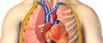
The image reveals the structures of the chest and its organs. These are the ribs, sternum, shoulder blades, spine with the spinal cord, position of the diaphragm, collarbones, lungs, heart and mediastinum, the aorta goes to the left.
Violations on the part of these organs, if they are noticeable enough for FOG, can be suspected. Then the person who has undergone the procedure will be asked to undergo further diagnostics in order to examine the pathology in detail.
If no structural changes are identified, the image is good, the conclusion after decoding will notify that no pathology of the lungs or heart has been identified.
Healthy lung tissue is low-density (retains little X-rays), but not too airy (a sign of emphysematousness). It shows unexpanded roots, the bronchial pattern is normally preserved. The results are stored in the outpatient record so that they can be monitored and studied over time.
Consumption
Pulmonary tuberculosis has a variable X-ray picture depending on its form and prevalence. This may be one focus of darkening or multiple bilateral small ones with a path to the root of the lung, where enlarged lymph nodes are sometimes identified.
Tuberculoma can be seen as a lesion with smooth ellipsoidal outlines, usually in the upper lobes, gradually spreading into the surrounding lung tissue. Also, strong darkening - calcifications - can speak in favor of infection with the tuberculosis bacillus. They indicate lime deposits in areas of subsided granuloma inflammation.
Severe and widespread forms more often give clinical manifestations. A person will have complaints with which he will consult a doctor (cough, fever).
Of course, then the patient’s fluorography data will be shown in the log book, but for the examination, an x-ray will be prescribed. The beam picture will become richer. And other analyzes will be informative.
Video with sample images:
Neoplasms
Lung cancer in the early stages of its development may also not produce obvious symptoms. Fluorography also finds such a disease. The picture will depend on its form: central, peripheral cancer. And also on the nature of growth.
Central cancer is a tumor that originates from the epithelium of the large bronchus. Therefore, it is closer to the root of the lung (from the inside), giving a shadow on the film. More often than not, the darkening is uneven and will thicken the fabric.
The mediastinum may be expanded more than it should be due to pathological lymph nodes or air exchange in the part of the lung blocked by this neoplasm may be disrupted. In the latter case, this area of the organ will also be darkened.
small shadow on the right
Peripheral cancer gives a shadow. Unlike the central one, it is located closer to the outside of the organ. This is not always noticeable in one projection in which fluorography is done (it is more informative in two).
It will look like a hearth with blurry, indistinct boundaries. However, as already mentioned, the diagnosis is clarified by other methods.
There may also be changes in the lymph nodes, but breath loss occurs only in the affected area. Therefore, the remaining zones cope well with the function of gas exchange, and symptoms do not appear for a long time. Here, the importance of screening by fluorography increases greatly.
The question will be about the possibility of surgery. The risk of cancer increases greatly if you smoke.
Metastases
Screening for respiratory diseases can also identify shadows in the lungs similar to metastatic ones, including cases where the primary focus of cancer has not been identified. Although the latter happens rarely, it serves as a reason for detailed diagnosis.
Sarcoidosis
During fluorographic examination, bilateral heavy shadows will be noted from the roots to the periphery of the lungs. Also, the changed lymph nodes of the mediastinum greatly expand it, giving rounded shadows at the base of the lungs.
In general, the picture of the disease is nonspecific. A full examination is necessary to differentiate it from tuberculosis.
Other states
Lung pathology has a corresponding picture, which can also show FOG:
- focus of darkening during pneumonia, including the consequences of the past;
- destructive and purulent diseases are also determined by the corresponding picture;
- effusion pleurisy is detected by darkening fields of most of the lung or in the sinus (lightening in the negative);
- with chronic bronchitis, emphysema, long-term and often exacerbating bronchial asthma, the diseased lungs will be more airy and, accordingly, lighter (black - in the negative).
Smokers are more susceptible to nonspecific lung pathologies.
Heart diseases can be manifested by a change in its characteristic configuration, an increase in diameter, and large size. Sometimes darkening is detected in the lower fields of the lungs or dilation of the pleural sinuses, which will indicate the presence of heart failure.
Past injuries: fractures of the ribs, collarbones can also be determined, old scars on the lungs after pneumonia, encysted chambers formed due to pleurisy and empyema.
FOG allows one to suspect many diseases, but this is not enough to differentiate them. The patient will be called for an appointment to jointly decide on further tactics.
Also, sometimes a lesion suspicious for a change in the mammary gland, mastopathy, can be identified. Then a mammogram or ultrasound of the breast, determination of hormones (sex, FSH) will be prescribed, and they may be referred to a mammologist.
Indications for the study
There are clear indications for performing a chest x-ray. If a pulmonary pathology is suspected, the doctor will prescribe an x-ray if the patient has complaints about:
- cough lasting at least a week;
- increased temperature and heat;
- sputum discharge;
- chest pain;
- wheezing in the lungs;
- shortness of breath;
- spitting up blood.
These signs primarily illustrate pulmonary problems. After a visual examination, the doctor will make a preliminary diagnosis, but can only confirm it with an x-ray.
X-ray examination helps not only to make diagnoses, but also to carry out differential diagnostics and separate one disease from another. This is extremely important, because many pulmonary pathologies have similar symptoms and it can be difficult to determine a specific diagnosis.
In addition to respiratory diseases, chest x-rays also visualize heart pathologies. Typically, diagnostics for heart diseases are performed along with electrocardiography, which will also illustrate abnormalities in the functioning of this organ.
X-ray of the chest is indicated for shortness of breath, chest pain, and rapid fatigue from the slightest physical exertion. These signs may be symptoms of chronic heart failure.
Using a chest x-ray, doctors determine the following diseases:
- heart attack and post-infarction changes in the heart;
- pulmonary embolism;
- heart defects, both congenital and acquired;
- chronic heart failure;
- cardiomyopathy;
- aortic aneurysm.
Changes in the aorta.
Aortic aneurysm. The procedure is performed for diseases of the skeletal system and spinal column. First of all, X-rays are taken if injuries are suspected, and 100% of patients who have already sustained injuries to the sternum are subject to examination.
The picture will show bruises and fractures. Most often, these can be injuries in the area of the ribs, spine and collarbone. In the image, the doctor sees not only the bone fragments themselves, but also the presence of foreign bodies and displacement of the bones in relation to each other.
If the victim has a pneumothorax and air gets into the chest cavity, this can also be seen using an x-ray.
How long to wait for results
The work schedule in the research room is structured in such a way that visitors with coupons are first accepted. Then comes the period of checking the pictures and writing forms. Thus, the reception time is reduced.
The patient learns the results of the government service and receives documents the next day when they are issued. This issue can be resolved in less than 24 hours if you need to quickly receive a description of the results and a certificate.
As an example: undergo digital fluorography, when the corresponding clinic in the city or region has the necessary apparatus and equipment to evaluate the image. This will allow you not to waste time developing the “photo”.
You can also undergo a study at a private medical institution that conducts such diagnostics, and not at your place of residence and registration. In this case, the procedure will cost some money. The price must be found in the relevant clinic.
Steadily every year
It is officially arranged, so it is necessary to undergo fluorography once every 12 months. The doctor monitors and sends for diagnosis those who missed it.
It is necessary to carry out the “flush” every year, because problems may arise during employment. They need results no older than 6 months.
Because of this, the therapist did not let me in, and I had to take a new picture. Then I went to the doctor again with the results to sign. I spent four days on everything, which can be a lot when you get a job.
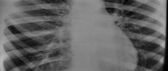
Roots of the lungs and diaphragm on x-ray
In the image, the diaphragm appears below the pulmonary field and forms a dome. The diaphragm stands high in the central part, and lowers towards the periphery, forming angles - sinuses. Normally, the dome of the diaphragm is located at the level of the fifth or sixth rib. When you take a deep breath, it flattens.
It is difficult to see the roots of the lungs during X-ray diagnostics, since they are covered by the shadow of the mediastinum. In the picture, the visible part is divided into upper, middle and lower parts.
The main shadow is given by the pulmonary artery and the smaller one by the veins, and the bronchi give contrast to the image. Externally, the root of the lung is a whole plexus of vessels and bronchi, which give the shadow in the picture.