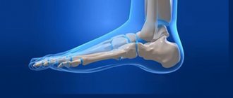
Structure of the arches of the feet
The frame of the foot is made up of bone and soft connective tissue. Its configuration includes longitudinal and transverse arches that have an arched shape with the apex directed upward. The landmarks along which these lines pass are bone formations.
The transverse arch is formed by the heads of the metatarsal bones. The support is provided by the lateral bones 1 and 5, and the central ones are located along an arc. The highest point of the transverse arch is the head of the 3rd bone.
The longitudinal arch has 2 sections: external and internal. The outer section is formed by the calcaneus, cuboid, talus, 4th, 5th metatarsal bones. The internal arch is formed by the 1st, 2nd metatarsal, talus, sphenoid and scaphoid bones.
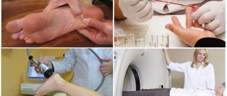
Radiology is a branch of science that studies the recognition of diseases using electromagnetic radiation. The skeleton contains a large amount of calcium, which blocks rays. The outlines of the bones on the film are white. X-ray diagnostics of flat feet helps to establish the fact of the disease and the severity of the disorders.
The study is carried out when complaining of pain in the legs or suspected flat feet during conscription. Identification of the disease can become an obstacle to entry into military educational institutions and serve as a reason for registration in the reserve.
Advantages of X-ray over other diagnostic methods
The advantage of this diagnostic method for plantar deformity is non-invasiveness and efficiency, low time costs, accessibility for various categories of patients, low radiation exposure, accurate imaging of the bone structures of the foot and joints, and the ability to determine the stage of the disease.
It is impossible to imitate disorders in the correct order of bones; x-rays record the internal structures of the foot, regardless of external signs or type of disease. With proper skill, you can obtain false data using excess weight. You can hold a heavy object or buckets of water in your hands during the study.
There are several contraindications:
- pregnancy;
- serious condition of the patient;
- presence of bleeding and pneumothorax;
- the patient's inability to remain in an upright position.
This study is sufficient to make a correct diagnosis. More expensive imaging methods - CT or MRI - will not provide the doctor with any additional information.
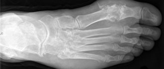
A photograph of the foot with flat feet is an important part of the diagnosis, showing the deformation of the bone structures according to the type of illness. The disease is characterized by pain in the legs, the inability to walk or stand for a long time, and the person is bothered by the knee and hip joints. If symptoms appear, you should contact a qualified professional. The doctor suggested undergoing an examination, including an x-ray of the feet. The snapshot will show existing deviations.
- Straight - the patient should place his full foot on the cassette with X-ray films, the other leg should be suspended. Used to identify the transverse form of deformation of the sole.
- Lateral – the photograph is taken in a standing position, with the other leg suspended. The ankle joint must be fixed on the film. Used to identify the longitudinal shape of changes in the ratio of bone, muscle structures, and tendons.
Diagnosis of transverse flatfoot
The transverse type of flatfoot leads to shortening and spreading of the foot in width with deviation of the first metatarsal bone and the formation of an angle between it and the main phalanx of the big toe. The transverse arch begins to rest on all the metatarsal heads.
X-ray technique
Photographs of two feet with a load are taken in a direct projection.
- The patient stands on a 24*18 cm film cassette with one leg and takes the other one back. Additional support is provided by a chair, which the subject holds with one hand.
- X-rays of the forefoot and midfoot (toes and metatarsals) are taken.
- The central beam passes through the center of the cassette.
- An x-ray of the second limb is performed.
The following angles are measured on the dried film:
- The angle between the body of the main phalanx of the big toe and the first metatarsal bone. Normally it is less than 15 degrees.
- The angle formed by the bodies of the first and second metatarsal bones. The normal value does not exceed 10 degrees.
- The angle of divergence of the metatarsal bones. Measure between bones 1 and 5, extending the lines towards the hindfoot. A value of more than 18 degrees will indicate transverse flatfoot.
Analysis of results
When assessing X-ray data of transverse flatfoot, the severity of the disease is assessed by measuring two parameters:
| Degrees of transverse flatfoot | 1 | 2 | 3 | 4 |
| The angle between the first and second metatarsal bones (in degrees) | 11-13 | 13-15 | 16-19 | Over 19 |
| the angle between the first metatarsal and the main phalanx of the big toe (in degrees) | 15-19 | 20-29 | 30-29 | More than 40 |
Flat feet on x-rays can also manifest themselves in the form of compensatory hypertrophy of the 2nd and 3rd metatarsal bones, a change in the configuration of the first metacarpophalangeal joint with the formation of exostoses.
Re: How to “correctly” take x-rays for flat feet and scoliosis?
and one more question: I was sent to take an x-ray at the clinic at my place of residence. What if, for example, instead of me, a person who was not accepted into the army because of flat feet undergoes this procedure (he said that he has one leg of the 2nd degree, the other - a 3rd degree of flat feet)? Or do they then somehow compare the photo with the foot at the military registration and enlistment office or something else?
Thanks in advance for your answer!
did not quite understand. how can I try to bring my knees together if I stand on one leg. please explain.
Diagnosis of longitudinal flatfoot
The longitudinal type of flatfoot is characterized by deformation of the longitudinal arches of the foot. The foot changes its configuration, becomes increased in length, spreads out and begins to come into contact with the support not only with the outer edge, but with the entire plane.
X-ray diagnostics of longitudinal flatfoot consists of taking photographs of two feet with a load. To perform the study, a lateral projection is used.
How to carry out the procedure
- The patient stands on a special stand, leaning on the leg being examined, and takes the other one back. To maintain balance, he holds on to the back of the chair with his hand.
- Parallel to the length of the foot under study, a standard size cassette is placed vertically at the inner edge and fixed - 18*24 cm or 24*30 cm. The long side of the film is located along the foot.
- The central X-ray beam passes from the outside to the inside of the cassette. Its location is shown in the projection of the junction of the sphenoid and scaphoid bones.
- An x-ray of the second leg is performed in a similar manner.
Evaluation of results
The following direct lines are carried out on the obtained x-ray photographs:
- the first horizontal, connecting the calcaneal tubercle with the head of the first metatarsal bone;
- the second straight line on the x-ray begins at the point of intersection of the first line with the calcaneal tubercle and rises to the scaphoid-sphenoid joint;
- the third line on the x-ray comes from the intersection of the head of the 1st metatarsal bone with the first straight line and rises to the scaphoid-sphenoid joint.
The following parameters are assessed in the triangle obtained on x-ray:
- Height of the vault. This is the length of the perpendicular drawn from the top point of the triangle to its base. The norm would be a vault height of more than 35 cm.
- Arch angle. It is located between the second and third lines in the picture. In a healthy person, this angle, measured radiographically, is in the range from 125 to 130 degrees.
In addition to these values, attention is paid to the condition of the tissues, the size of the joint spaces, and the presence of growths (exostoses).
The following table will help determine the degree of flatfoot using an x-ray.
| Degrees of flat feet | Arch height (mm) | Arch angle (degrees) | Presence of deformations |
| 1 | From 34 to 25 | 131-140 | No or initial signs of arthrosis of the talonavicular joint (stage 1) |
| 2 | From 24 to 18 | 141-155 | Arthrosis of the talonavicular joint (stage 2) with bone tissue overgrowth larger than 1 mm |
| 3 | Less than 18 | More than 155 | The appearance of signs of arthrosis of the talonavicular joint, stage 3, bone tissue growth exceeds 1 mm |
The appearance of exostoses on x-rays indicates overload of the foot joints. The initial symptoms of longitudinal flatfoot with minimal changes are considered to be a decrease in the height of the arch.
X-ray with load
Using this research method, specialists can analyze the features of the structure and anatomy of the leg joint. This method is mainly used to determine the level of flat feet in children, although this type of pathology also develops in adulthood.
The procedure does not require any special preparation; x-rays of the feet for flat feet are taken in the same projections as in a regular analysis - straight and lateral. The patient must stand on the X-ray cassette with one foot, and the machine takes two projections of the image. This method allows you to assess the level and severity of flat feet and prescribe appropriate treatment.
After the patient has completed the first course of treatment, the study procedure must be repeated. This is necessary to analyze the results of therapy.
This method allows you to observe and monitor the health status of adolescents and children. It is important to identify the pathology in time and take measures to eliminate it. Therapy for flat feet is not a complicated process; the main thing is to wear special orthopedic shoes and do special exercises.
Signs of longitudinal flatfoot
A distinctive feature of longitudinal flatfoot is the absolute contact of the foot and the floor, which results in an increase in its length, as well as its expansion.
Attention! Such longitudinal flat feet can lead to clubfoot.
In the case of the first stage of longitudinal flatfoot, there are no visible signs. Symptoms: after any physical activity, fatigue of the lower extremities is felt. Pain appears if you press on the foot. By evening time, swelling appears and the gait changes.
In the second degree of the disease, the pathology is expressed moderately. There is an increase in symptoms, and the arch is practically absent. There is a loss of muscle elasticity, and the gait ceases to be smooth. The pain is felt more and more, now it can reach the ankles and even the lower leg.
The third degree of leg deformation is the most severe. Pain now constantly accompanies the patient, plus the person may begin to worry about the lower back and periodic severe migraines. The patient cannot walk long distances, and wearing ordinary shoes becomes unbearable for him due to severe deformation of the leg.
Signs of transverse flatfoot
With transverse flatfoot, the leg is deformed as follows: the shape of the big toe changes = it becomes hammer-shaped. A thickening (bump) begins to form at its base, which is accompanied by severe pain.
Where do x-ray examinations take place, what is its price?
X-rays of the feet are performed in medical institutions engaged in radiology diagnostics. You can take pictures in the X-ray room of the clinic at your place of residence at the expense of compulsory medical insurance.
State medical institutions also conduct examinations of conscripts to identify flat feet. The district military registration and enlistment office cooperates with x-ray rooms, where they take pictures of the feet and describe them. This examination will be free for the conscript. If there are doubts about the reliability of the results, he may perform x-rays of the feet at another institution. When visiting a private clinic, diagnostics will be carried out at full cost.
If desired, any person can be examined at a non-state medical center. If there is no indication for an x-ray of the feet, the examination will not be paid for from compulsory medical insurance funds.
The average cost of a weight-bearing foot x-ray may vary depending on the region and the pricing policy of the medical institution; it ranges from 1,200 to 2,200 rubles.
Reliability of determining the degree of flatfoot from an image
X-rays of flat feet give a clear clinical picture. The photo serves as an important argument in making a diagnosis. Visualization makes it possible to evaluate all the details of the pathological process.
X-rays confirm or refute the results of plantography (a foot print painted with paint) and contourography (outlining the foot with a pencil in a standing position).
The effectiveness of X-ray examination for flat feet is 78-96%.
Contraindications for examination
Radiation exposure when performing x-rays of the feet is low. The use of the diagnostic method is not recommended:
- during pregnancy;
- in a patient in a serious and life-threatening condition.
A limitation to the examination is the inability to stand.
Flat feet are a common condition that affects approximately 40-60% of the population, regardless of age and gender. Congenital or acquired deformation of the feet causes significant discomfort and leads to the development of severe disorders of the musculoskeletal system, inflammation of the joints, and varicose veins. Effective treatment of pathology by conservative means is possible only at the initial stage, so special attention should be paid to timely diagnosis, the most accurate method of which is radiography. Let's look at how x-rays of the feet are performed for flat feet, and what indicators indicate the presence of a disorder.
The need for radiography for foot deformities
Flat feet is a disease in which the biomechanics of the foot is disrupted due to loss of shock-absorbing ability. When a person stands or walks, half the load falls on the heel. If overload occurs (for example, among athletes who run or people whose professional activities involve long periods of standing), the ligaments weaken, the arch flattens, and the body weight is partially transferred from the heel to the middle of the foot.
Due to a violation of biomechanics, the force compensation mechanism is activated. This occurs due to the spine, hip and ankle joints. Due to the increased load, the cartilage surfaces in the joints and intervertebral discs are gradually worn away. As a result, the joints become deformed, which is expressed in pain that occurs when walking in the feet, calves, lower back, and back.
Over time, the patient develops:
- arthrosis;
- varicose veins caused by a weakening of the pumping function of the calf muscles due to impaired biomechanics of the foot;
- plantar fasciitis;
- curvature of the spine of the scoliosis type.
Developed secondary diseases manifest themselves with specific symptoms. Thus, arthrosis of the knee and hip joints over time makes independent movement impossible and leads to disability of the patient. With a heel spur, a person suffers from intense pain in the heel in the morning, especially severe when he tries to step on it. Varicose veins are accompanied by swelling and a feeling of heaviness in the calves.
In most cases, foot deformity is a congenital pathology. The acquired form of the disease is less common in people whose activities involve regular heavy loads on the lower extremities.
Flat feet ranks first among congenital anomalies. Timely detection of the disease in 45% of cases is difficult due to the physiological and anatomical characteristics of the child’s body (up to 3 years). Physiological flat feet in a child is the norm; this is the stage of formation and development of the foot. Therefore, it is often confused with real pathology.
Many doctors express the opinion that diagnosis is difficult due to the presence of a fat pad on the foot in children. The subsurface space is filled with a subcutaneous fat layer - a buffer that relieves the load from the immature musculoskeletal system.
X-ray solved the problem of diagnosing flat feet at any age. Timely detection of the disease and prescription of adequate treatment increased from 35 to 68%. In adults whose flat feet are associated with improper distribution of load on the articular surfaces, after correct diagnosis and therapy, secondary changes (inflammatory, destructive processes in the tissues of the joints of the feet) sharply decreased.
Are there any contraindications
Radiation exposure during examination of patients' feet is minimal.
X-rays of the feet are contraindicated for children in the first 8-12 months of life and for pregnant women. The attending physician decides on the advisability of conducting diagnostics in patients with severe chronic diseases of internal organs. At the same time, all possible risks are taken into account.
The essence of the method
Diagnostic radiography allows you to analyze the structure, position and structural features of internal organs by passing directed x-rays through them. Due to the different densities of the tissues under study, the flow of electromagnetic waves is unevenly attenuated and scattered, which makes it possible to obtain a shadow image of the object at the output. The radiation passing through the biological medium is recorded on a special photodetector: cassette, electronic matrix. The projection of the organ or its area being studied is formed on an X-ray sensitive film or monitor screen. The object of study is the resulting photograph or digital image. An orthopedic surgeon is involved in X-ray morphometry or interpretation of the examination results.
The reliability of the method is directly related to the accuracy of the resulting projection of the body part being studied, which depends on the quality of the equipment, film and reagents, as well as diagnostic conditions. Random movements of the patient during the examination can also reduce the clarity of the image.
How do they do it?
The examination takes place in a special secure room, and the patient should not wear anything metallic. The area being examined must be freed from shoes and clothing and the instructions of the radiologist or laboratory technician must be carefully followed. There is no need to prepare for the study.
The doctor or his assistant will help you position the leg correctly, after which you will need to keep it motionless for up to 15 seconds (the total duration of the procedure is up to 10-15 minutes). If you need additional projections or obtaining images with a study load, it will take a little more time. Other parts of the body are protected by a special apron, which is removed at the end of the procedure.
Indications and contraindications for radiography
The main indication for an x-ray of the feet is a suspicion of flat feet or other structural anomalies of the bones and joints of the lower extremities in the presence of the following symptoms:
- Discomfort, pain and rapid fatigue when walking and standing for long periods of time;
- The appearance of heaviness and swelling of the legs, night cramps;
- Visible or physically palpable drooping of the arches of the feet;
- Deformation of the fingers, often accompanied by the formation of calluses that are difficult to treat;
- Protruding bones or joints;
- Widening or increasing the length of the foot.
X-rays are also necessary to establish the form and characteristics of already diagnosed diseases.
Contraindications to the study are pregnancy (all trimesters) and infancy. Children over 2-3 years old are given x-rays only in emergency or severe cases with parental consent.
The study is not performed if the patient cannot stand independently. The main limitations of radiography are associated with the additional effect of radiation on the body, which, depending on the type of equipment, is 0.1-0.6 mSv. The total radiation exposure should be no more than 1.4 mSv per year, therefore the number of studies performed is limited even in the absence of contraindications.
Research algorithm
X-rays of the feet do not require special preparation. Before the procedure, you must remove metal objects, jewelry, put away your phone and other electronic devices, and also warn the radiologist about the presence of all implants, prostheses, etc. The lower limbs should be exposed below the knee. The patient's body, with the exception of the legs, is protected with a special lead apron that prevents exposure to radiation.
In most cases, if flat feet are suspected, a load-bearing x-ray is performed to determine the deformation of the bones and joints in a state of maximum static impact on the foot. The procedure is performed standing; body position may vary depending on the required projection. The patient places the foot being examined on a special device stand - a cassette with photosensitive film, pressing the other limb in the knee. When x-raying flat feet in a direct projection, the central beam is directed strictly to the middle of the cassette; in a lateral projection, the image is taken from the side, capturing the ankle joint. The study is carried out alternately for each leg in both projections.
To obtain a reliable result, if it is necessary to repeat the examination after treatment, the patient must take the same position as the first time.
How to “correctly” take x-rays for flat feet and scoliosis?
1. I heard that when taking a photo for flat feet, you need to, without lifting your feet, try to bring your knees together so that the degree of flat feet is higher. Is it so? Also read here on the forum:
2. It is better to take x-rays for scoliosis while standing than lying down.
Who else knows what ways (tricks) to increase the degree of flat feet/scoliosis during x-rays?
Last edited by Junky; 03/06/2008 at 18:57.
Decoding the results
Normally, the limb of a healthy person has two arches: longitudinal, located along the medial edge of the foot, and transverse, located at the base of the toes. When the corresponding areas are flattened, longitudinal and transverse flat feet are formed. X-ray diagnostics includes measuring the height and angles of the arch of the foot using an X-ray in different projections, monitoring the condition of the joints and bone tissue. The study allows not only to establish the presence of the disease, but also to determine its stage. In addition to flat feet, X-rays can show manifestations of other pathologies and injuries.
X-ray for longitudinal flat feet
The lateral projection image is intended for x-ray diagnostics of longitudinal flatfoot. In the image, a triangle is determined, the apex of which is the scaphoid-sphenoid joint, and the base is the distance from the edge of the first metatarsal bone to the calcaneal tubercle. Key indicators are:
- The longitudinal angle is normally 125°-130°;
- The height of the arch of the foot, which should be at least 35 mm.
The presence of pathology can be indicated by an increase in the longitudinal angle and flattening of the arch. To determine the degree of flatfoot using an x-ray, the doctor accurately measures these indicators and evaluates the general condition of the bones and joints:
- I degree – there are no signs of limb deformity. The radiographically measured angle is 131°-140°, the height of the arch is at least 25 mm;
- II degree flatfoot on x-rays most often shows signs of arthrosis and joint deformation. The angle is 141°-155°, the height of the arch is from 17 to 24 mm;
- III degree – there are pronounced signs of deformation, the angle is greater than 155°, the height of the arch is less than 16 mm.
X-ray for transverse flatfoot
Pathological changes in the transverse arch of the foot are determined from a direct projection image. The key parameter for x-rays of transverse flatfoot is the angle of deviation between the first and second metatarsal bones, which in a healthy person does not exceed 11°. Depending on the degree of violation, its value is:
- I degree – 11-12°;
- II degree – 13-15°;
- III degree – 16-20°.
As the pathology develops, the load when walking and standing begins to be distributed not only between the 1st and 2nd metatarsal bones, but also between the 3rd and 4th, the rest of the bones become compensatory. In the stage of decompensation, all bones are located in the same plane, and the shock-absorbing properties of the transverse arch of the foot are completely lost.
Radiology methods are widely used in the diagnosis of many diseases, including flat feet. The study can be carried out in any private or public clinic on a paid or free basis. The estimated cost of the procedure is 1300-2000 rubles. depending on the conditions and type of equipment.
Article 68 (of the regulation) provides for acquired fixed deformities of the foot.
| Name of diseases | Category of suitability for military service | |||
| Flat feet and other foot deformities: | I Count | II Count | III Count | IV column |
| a) with significant impairment of functions | D | D | D | NG |
| b) with moderate dysfunction | IN | IN | B, B-IND | NG |
| c) with minor dysfunction | IN | IN | B | NG |
| d) in the presence of objective data without dysfunction | B-3 | B | SS-IND | NG, officers, midshipmen, individual. |
Point “a” includes:
- pathological equine, calcaneal, varus, hollow, planovalgus, equinovarus feet and other irreversible, pronounced curvatures of the feet acquired as a result of injuries or diseases, in which it is impossible to use shoes of the established military standard.
Important. A pathologically hollow foot is considered to be a foot that has deformation in the form of supination of the posterior and pronation of the anterior section in the presence of high internal and external arches (the so-called sharply twisted foot). The forefoot is flattened, wide and slightly adducted, there are calluses under the heads of the middle metatarsal bones and claw or hammertoe deformity. The greatest functional impairments occur with concomitant eversion-inversion components of the deformity in the form of external or internal rotation of the entire foot or its elements.
Point “b” includes:
- longitudinal grade III or transverse grade III-IV flatfoot with severe pain, exostoses, contracture of the fingers and the presence of arthrosis in the joints of the midfoot;
- absence of all toes or part of the foot at any level;
- persistent combined contracture of all toes on both feet with claw or hammertoe deformity;
- post-traumatic deformation of the calcaneus with a decrease in the Böhler angle over 10°, pain syndrome and arthrosis of the subtalar joint of stage II.
Important. With decompensated or subcompensated longitudinal flatfoot, pain in the feet occurs in a standing position and usually intensifies in the evening, when their pastiness appears. Externally, the foot is pronated, lengthened and widened in the middle part, the longitudinal arch is lowered, the navicular bone is outlined through the skin on the medial edge of the foot, the heel is valgus.
Important. Absence of a toe is considered to be its absence at the level of the metatarsophalangeal joint, as well as complete abduction or immobility of the toe.
Point “c” includes:
- moderately severe foot deformities with minor pain and static disturbances, for which standard military-style shoes can be adapted for wearing;
- longitudinal flatfoot of the third degree without valgus position of the heel bone and phenomena of deforming arthrosis in the joints of the middle foot;
- longitudinal or transverse flatfoot of the second degree with deforming arthrosis of the second stage of the joints of the midfoot;
- deforming arthrosis of the first metatarsal joint stage III with limitation of movements within plantar flexion less than 10° and dorsiflexion less than 20°;
- post-traumatic deformation of the calcaneus with a decrease in the Böhler angle from 0 to -10° and the presence of arthrosis of the subtalar joint.
Point “d” includes:
- longitudinal or transverse flatfoot of the 1st or 2nd degree with deforming arthrosis of the 1st stage of the joints of the middle foot in the absence of contracture of the toes and exostoses.
Important. A foot with increased longitudinal arches, when correctly positioned on the surface under a supporting load, is often a normal variant.
When making an expert decision, in accordance with the requirements of this article, there are special difficulties with the diagnosis and examination of such foot deformities as pathological equine, calcaneal, varus, cavus, plano-valgus, equino-varus and others acquired as a result of injuries or diseases that are irreversible There are no pronounced curvatures of the feet, which make it impossible to use shoes of the established military standard. The radiologist only needs to establish the fact and variant of foot deformities.
Quite large difficulties and discrepancies arise in X-ray diagnostics and making an expert decision when determining the degree of longitudinal and transverse flatfoot, as well as determining the stage of arthrosis of the foot joints with flatfoot.
First, let us dwell on the technique of x-ray examination of patients with suspected longitudinal and transverse flatfoot. For these purposes, radiographs of the feet are taken in a state of maximum static load on the foot, i.e. in a standing position (radiography of the feet under load).
| Figure 1. Schemes of radiography of feet under load: a - to determine longitudinal flat feet; b - to determine transverse flatfoot. |
Longitudinal flatfoot
To determine longitudinal flatfoot, lateral radiographs of the feet are taken under load (see Figure 1). Using dry radiographs, the doctor performs a graphical calculation of the longitudinal arch of the foot (see Figure 2).
| Figure 2. Scheme of graphical calculation of longitudinal flatfoot. The longitudinal arch of the foot is determined by two landmarks - the height of the arch (h) and the angle of the arch (a). They are determined on a lateral radiograph of the foot by constructing an auxiliary triangle, the vertices of which are: A - the lowest point of the heel bone; B - lower pole of the scaphoid-sphenoid joint; C - lower edge of the head of the 1st metatarsal bone. Angle ABC is the angle of the arch, and the perpendicular drawn from point B to line AC is the height of the arch (h). |
Various sources indicate that the starting point B for constructing the auxiliary triangle can be the lower pole of the talonavicular joint or the lower point of the scaphoid bone. As practice has shown, when constructing such triangles, there appears a discrepancy between the height and angle of the arch obtained from such calculations and the figures for the height and angle of the arch specified in Article 68 of the Resolution. Therefore, it should be taken into account that the main parameter for deciding the degree of longitudinal flatfoot is the height of the arch of the foot, and not the angle of the arch. This is due to the different length of the 1st metatarsal bone in each individual (in other words, different shoe sizes). As a result, patients with the same arch height but different shoe sizes will have different arch angles.
Important. Normally, the angle of the longitudinal arch is 125-130°, the height of the arch is 39 mm.
Degree of longitudinal flatfoot
- Flatfoot degree I: angle of the longitudinal internal arch 131-140°, arch height 35-25 mm;
- Flatfoot degree II: angle of the longitudinal internal arch 141-155°, arch height 24-17 mm;
- Flatfoot degree III: the angle of the longitudinal internal arch is more than 155°, the height of the arch is less than 17 mm.
Important. Longitudinal flatfoot of the 1st or 2nd degree, as well as transverse flatfoot of the 1st degree without arthrosis in the joints of the midfoot, contractures of the fingers and exostoses are not the basis for the application of this article, and do not interfere with military service, admission to military educational institutions and colleges.
Degrees of deforming arthrosis of the talonavicular joint
- Stage I arthrosis: narrowing of the joint space is less than 50%, marginal bone growths do not exceed 1 mm from the edge of the joint space.
- Stage II arthrosis: narrowing of the joint space by more than 50%, marginal bone growths exceeding 1 mm from the edge of the joint space, deformation and subchondral osteosclerosis of the articular ends of the articulating bones.
- Stage III arthrosis: the joint space is not radiologically determined, pronounced marginal bone growths, gross deformation and subchondral osteosclerosis of the articular ends of the articulating bones.
Table. Determination of the degree of longitudinal flatfoot according to the requirements of Article 68
| Norm | I degree | II degree | III degree | |
| Arch height | 39 mm | 35-25 mm | 24-17 mm | Less than 17 mm |
| Arch angle | 125-130° | 131-140° | 141-155° | More than 155° |
| Bone changes | None | Absent or stage I deforming arthrosis of the talonavicular joint with bone osteophytes no more than 1 mm | Deforming arthrosis of the talonavicular joint stage II with bone osteophytes more than 1 mm | Deforming arthrosis of the talonavicular joint stage II-III with bone osteophytes more than 1 mm |
Post-traumatic deformity of the calcaneus
Article 68 of the new Resolution introduced a requirement for the definition of post-traumatic longitudinal flatfoot as a result of injury to the calcaneus. It is recommended to take lateral radiographs of the feet under weight bearing (see Figure 1). On dry radiographs, the doctor carries out a graphic calculation using the Behler method, always of both limbs.
| Figure 3. Scheme of graphical calculation of the degree of post-traumatic displacement and the amount of flattening of the transverse arch of the foot using the Behler method. The calcaneotalar angle and the angle of the articular part of the calcaneal tuberosity (Behler angle) are formed by the intersection of two lines, one of which connects the highest point of the anterior angle of the subtalar joint and the apex of the posterior articular facet, and the other runs along the upper surface of the calcaneal tuberosity. |
Important. Normally, the calcaneal-talar angle is 140-160°, Beler’s angle is 20-40°. A decrease in the Böhler angle from 0 to 10° indicates a moderate deformation of the calcaneus. When the Böhler angle decreases by more than 10°, severe deformation of the calcaneus is indicated.
A decrease in the Beler angle indicates displacement of fragments and flattening of the longitudinal arch of the foot. Usually accompanies post-traumatic longitudinal flatfoot.
Important.
The most informative way to assess the condition of the subtalar joint is computed tomography performed in the coronal plane, perpendicular to the posterior articular facet of the calcaneus.
Transverse flatfoot
To determine transverse flatfoot, radiographs of the forefoot and midfoot are taken in a direct projection under load (see Figure 1). Using dry radiographs, the doctor performs a graphical calculation of transverse flatfoot (see Figures 4, 5).
| Figure 4. Scheme of graphical calculation of transverse flatfoot according to the requirements of Article 68: The first line is drawn along the longitudinal axis of the second metatarsal bone, the second line is drawn parallel to the longitudinal axis of the first metatarsal bone tangent to its inner surface, and the third line is drawn along the tangent to the inner surface of the main phalanx I finger The angles between the drawn straight lines are the required ones: a - angle Metatarsus varus; beta - hallux valgus angle. |
Degrees of deforming arthrosis in the first metatarsophalangeal joint
- I degree: the angle between the I-II metatarsal bones is 10-12°, and the angle of deviation of the first finger is 15-20°;
- II degree: the angle between the I-II metatarsal bones is 13-15°, and the angle of deviation of the first finger is 21-30°;
- III degree: the angle between the I-II metatarsal bones is 16-20°, and the angle of deviation of the first finger is 21-40°;
- IV degree: the angle between the I-II metatarsal bones is more than 20°, and the angle of deviation of the first toe is more than 40°.
Table. Determination of the degree of transverse flatfoot according to the requirements of Article 68
| Norm | I degree | II degree | III degree | IV degree | |
| Metatarsus varus | 10° | 10-12° | 15° | 20° | More than 20° |
| Hallux valgus | 10° | 15-20° | 30° | 40° | More than 40° |
The method presented above for determining transverse flatfoot is used for expert purposes in accordance with the requirements of Article 68 of the Decree. However, for diagnostic purposes, it is recommended to use the method of graphical calculation of transverse flatfoot, which is used at the Department of Military Traumatology and Orthopedics of the Kirov Military Medical Academy (see Figure 5).
| Figure 5. Scheme of graphical calculation of transverse flatfoot using the VMA method. Kirov: 1 - axis of the foot through the second intermetatarsal space; 2 - axis I of the metatarsal bone (drawn through the middle of the bone); 3 - line tangent to the head of the 1st finger along the outer surface; 4 - line parallel to the axis of the foot; 5 - axis V of the metatarsal bone. |
Table. Determination of the degree of transverse flatfoot using the VMA method named after. Kirov
| Corner | Norm | I degree | II degree | III degree |
| 1. Metatarsus varus a | 10° | 12-15° | 15-20° | More than 20° |
| 2. Hallux valgus b | 10° | 15-20° | 20-25° | More than 25° |
| 3. Deviation of the 5th finger | 4-5° | 7-8° | 12-15° | More than 20° |
| 4. Angle c between the I and V metatarsal bones | 15° | 20° | 25° | More than 25° |