What is avascular formation in the mammary gland and what are its consequences? The female breast consists of glandular structures and subcutaneous tissue. In the absence of pathology, the mammary gland has a homogeneous structure and smooth, clear contours. However, on an ultrasound examination of the breast, a specialist can diagnose black shadows. These are hypoechoic formations. It is important to understand that avascular formations are just a description of the diagnosed picture. This is not a diagnosis of a disease or a symptom of it.
What it is
An isoechoic formation means an area that has exactly the same density as the entire surrounding tissue of the organ. This formation can be detected only if there is a shell (see photo) - a capsule, or if there are signs of compression of this segment.
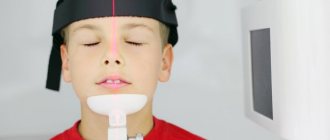
Palpation (palpation) of the thyroid gland.
Completely different symptoms are observed when the node is located inside the gland. The manifestation of other symptoms is also possible when the organ begins to produce excessive amounts of hormones. As a rule, all signs of the development of thyrotoxicosis are noted:
- tachycardia;
- fast fatiguability;
- excessive sweating;
- significant weight loss;
- sudden change of mood;
- tremor of the upper extremities.
Is it worth treating isoechoic changes in the thyroid gland?
If the formation does not have a negative effect on the patient’s hormonal background and does not cause any negative sensations, then such a node does not need treatment. The patient is advised to monitor the dynamics of the node, this will allow him to keep the condition of the defect under constant control and, if the situation worsens, take all necessary measures to eliminate the negative consequences.
But it should also be noted that if such a node was found in the thyroid gland, this could mean the following:
Carcinoma
Carcinoma on ultrasound can be completely shapeless or, on the contrary, it can have completely clear contours. Such nodes occur in a quarter of all existing cases of cancer. Most often, such nodes have uneven outlines and fully correspond to papillary carcinoma.
Cancer is found in the capsule in almost 50% of cases, having completely normal echogenicity and clear, even boundaries. In this case, the picture on the ultrasound screen is as follows:
- Wrong form of education.
- Clearly preserved contour.
- Fuzzy near the boundaries of the node.
- The presence of any hyperechoic inclusions usually indicates calcification.
- The presence of psammotous bodies inside the node.
- The presence of a convoluted structure in the vessels surrounding this section.
Benign formation
When monitoring the problematic isoechoic segment, no progression symptoms are detected. Most often, such formations occur in the following pathologies:
- nodular goiter;
- follicular adenoma;
- in nodes of adenomatous type.
Manifestation of echogenic reflections
The ultrasound method of studying internal organs is based on the quality of absorption and reflection of ultrasound in tissues. In this case, depending on the density of these structures, different images appear on the device screen. Varieties of structures differing in echo density are divided into three main types. This:
- Hyperechoic formations. Such foci have an extremely high density. Against the background of the main tissues of the organ, they stand out in a lighter shade.
- Hypoechoic. Their acoustic density is low. On ultrasound they appear as dark lesions.
- Isoechoic. Detection of such areas is difficult because they have the same echogenic density as the tissue of the organ being examined.
When describing all types of reflections, the terms formation and inclusion are used. They denote the same phenomenon, that is, a focus characterized by the ability to reflect ultrasonic signals.
How is an isoechoic inclusion detected?
The similarity of the display of such formations with the surrounding structures of the uterus largely makes it difficult to identify foci of developing pathology. Such inclusions are detected if there is a capsule (shell) or there is a feeling of squeezing of the forming formation.
For a long time, the development of such processes does not remind of itself by any symptoms, that is, the patient is not bothered by anything. Most often, isoechoic inclusions are detected by chance during a routine examination or during examination for completely other diseases.
Isoechogenic formation in the liver
If isoechoic formations were detected on an ultrasound of the liver (even if the echogenicity of the liver is increased), then this may indicate the following pathologies:
- The presence of an old hematoma that occurred due to trauma to the organ.
- Hemangioma.
- Metastases.
- The presence of a focus of hepatocellular cancer.
- Cystic formations.
To clarify the diagnosis, you need to undergo additional research methods, these may include magnetic resonance imaging (MRI), scintigraphy, angiography, and biopsy.
Isoechogenic changes in the structure of the uterus
The similarity of such formations with the structure of the uterus makes it difficult to detect on an ultrasound examination. They are detected in cases where the inclusion has a capsule, or when there is a feeling of compression of the developing formation. For a long time the process does not manifest itself with external symptoms, the patient is not bothered by anything.
If, after an ultrasound, a focus was discovered in the uterus that had an acoustic density similar to that in the surrounding tissues, then this indicates the development of the following processes:
- Uterine cancer is the presence of an adenomatous node in the mucous membrane of the organ, which appears and takes root on the myometrium.
- Presence of myomatous node.
- Presence of an adenomatous node.
- Rokitansky syndrome.
To clarify the diagnosis, you need to undergo additional research methods. Ultrasound helps to detect the presence of a tumor. Echodensity depends on the stage and degree of development of the pathology. In the initial stage, they are all visualized as isoechoic. As the process progresses (enlarges), malignant neoplasms are characterized by the appearance of hypoechoic, hyperechoic, and anechoic inclusions in the lesion.
Features of formations
A characteristic feature of echogenic inclusions is their identity with the echo density of the uterine structure. That is, these neoplasms are practically no different from other tissues of the organ. This is why isoechoicity is considered a normal manifestation of acoustic density. Healthy reproductive organs have this degree of reflectivity.
Most often, such inclusions do not pose a threat to health.
But in some cases, the detection of foci with similar density in the uterus indicates the development of both benign formations and malignant inclusions. It is the same ability to reflect the sound signal that complicates the differential diagnosis of safe isoechoic formations from such serious diseases as:
- adenomyotic node;
- myomatous node;
- uterine cancer;
- Rokitansky syndrome, which reflects the isoechoic density of the uterus, which is not completely healthy.
In the presence of a pathology of uterine development such as Rokitansky syndrome, echographic signs of other inclusions that are hyperechogenic or hypoechogenic may be found in it.
The detection of an isoechoic inclusion indicates that a tumor has been detected in the uterus. Detection of its benign or oncological nature requires a mandatory thorough examination using laboratory and instrumental methods.
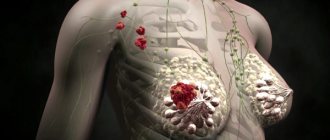
Isoechogenic formation in the mammary gland
The presence of an isoechoic formation in the mammary gland indicates the following:
- Mammary cancer.
- The presence of a normofollicular adenoma of a benign type.
- Fibroadenoma.
For an accurate diagnosis, you need to know on what day of the cycle you need to do an ultrasound of the mammary glands, and you also need to undergo additional studies.
The final diagnosis is established by a mammologist after conducting the necessary examination.
conclusions
Thus, the presence of an isoechoic formation in almost all cases means that a tumor disease of a malignant or benign nature has been detected in the body. An isoechoic formation is not necessarily cancer. In any case, ultrasound is only a screening method of examination, and to establish a more accurate diagnosis, many other additional laboratory or instrumental techniques are used.
The term “isoechoic formation” refers to a section of tissue or organ of the human body, which, during ultrasound examination, has a density similar to other tissues. This inclusion is detected only by the presence of symptoms of compression of nearby tissues or the presence of a capsule. Most often, this indicates the presence of a neoplasm, both benign and malignant.
During an ultrasound, two types of structures are revealed: hyperechoic (dense, with a high ability to reflect ultrasound, so they stand out in a light color on the screen) and hypoechoic (with a reduced density, also reflect ultrasound, but are highlighted in a dark color on the screen). Accordingly, areas that are not illuminated are isoechoic formations. Their density prevents ultrasonic waves from passing through them.
What is a hypoechoic formation?
During an ultrasound examination, all internal organs are characterized by echogenicity, or density. In the picture, the organs have a variety of colors - white, gray, black. It depends on the density of the fabric.
Ultrasound is reflected from tissues at different speeds. The denser the organ, the faster the wave reflection. In the picture this is indicated by white color and is described by hyperechogenicity. The softer the tissue or pathological neoplasm, the slower the sound comes from it. Part of the image is colored dark gray. This is hypoechogenicity.
The main symptoms of isoechoic formation
It is worth considering the fact that the neoplasm can have a malignant course, therefore, as it grows, symptoms such as compression of the esophagus, trachea, vocal cords, etc. may occur. Accordingly, the larger the node, the brighter the symptoms.
If you have such a pathology, you should pay attention to the following symptoms:
- Any difficulty breathing, symptoms may appear when changing body position.
- Hoarseness of voice.
- Difficulty swallowing.
- Feeling of a lump in the throat.
- The appearance of irregularities in the neck or a uniform increase in its size.
Any manifestations of the disease depend entirely on the location of the node and its size. If the formation is located on the outside of the organs, then there will be no symptoms of compression of the vessels, trachea and esophagus. Similar formations can be detected by palpation and ultrasound of the throat and larynx.
Completely different symptoms are observed when the node is located inside the gland. Also, the manifestation of other symptoms is possible when the organ begins to produce excessive amounts of hormones. As a rule, all signs of the development of thyrotoxicosis are noted:
- tachycardia;
- fast fatiguability;
- excessive sweating;
- significant weight loss;
- sudden change of mood;
- tremor of the upper extremities.
Isoechogenic formations can form in various tissues of the human body. But most often they are found on the thyroid gland, liver, uterus, mammary glands and ovaries. Such segments on the thyroid gland form in the form of nodes that encircle it, which can cause coughing, a feeling of suffocation, and the tumor constantly increases in size. In fifty percent of cases, such nodes are heralds of the development of cancerous tumors. Areas that have an acoustic density similar to the surrounding tissues are also found in the uterus, ovaries, and liver.
An isoechoic area of tissue is considered dangerous and malignant if:
- it has an irregular and uneven shape
- node boundaries are unclear
- there are hyperechoic inclusions, which indicates calcification
- the structure of the vessels surrounding the segment is convoluted
- psammotous bodies were detected inside the segments
In these cases, the prognosis is unfavorable; the neoplasm most likely requires surgical intervention.
Don’t be afraid, because such segments can be benign and can be treated without consequences for the body. The most important thing is to detect them in time.
Causes and symptoms of inclusions
Ultrasound makes it possible to determine the presence of a neoplasm in the uterus. Their echo density directly depends on the degree of development of the pathological process. At the initial stage, all focal pathologies in the uterus are visualized on the monitor screen as isoechoic.
If the nature of the inclusion is malignant, then as the process progresses (tumor formation increases), symptoms such as the presence of anechoic, hypoechoic or hyperechoic formations in the lesion appear.
This is how it looks on the monitor screen.
Whereas progression is not typical for benign inclusions.
Causes of isoechoic transformations in the uterus
Among the most common formations in the uterus, which initially have an isoechoic picture, the following inclusions pose a particular danger:
- Adenomyosis. It represents the detection of pathological foci in the muscular walls of the uterus, similar to the mucous membrane of the organ. Echographic signs of such changes are an increase in the size of the uterus to 13-14 cm (in length). There is an asymmetrical thickness of the uterine walls and a violation of clarity in the boundaries.
- Myomas. These inclusions in the uterine wall appear on ultrasound as thickenings with clear boundaries. In most cases, these are benign neoplasms that form from the tissues of the uterus itself. Varieties of such tumors are fibromyomas, leiomyomas, which also have an isoechoic density. An ultrasound diagnosis of fibroids is considered only an assumption and requires mandatory additional examination to exclude uterine cancer. Differentiation of such a formation is possible only after its removal. A histological examination of the changed tissues is mandatory. This makes it possible to identify the presence or absence of oncology.
- Uterine cancer. It appears as an adenomatous node on the mucous membrane of the organ. For such nodes, the form of manifestation can be both clear contours and shapeless outlines. Nodes with isoechoic signs account for more than 25% of all cases of oncological formations.
Treatment methods
Treatment of affected tissues aims to prevent their growth and development. It is important to constantly monitor the dynamics of their development. Therefore, if they do not change in size, they often do not require treatment.
An important role is played by diagnostics and constant monitoring of the condition. If the affected area constantly increases in size and unpleasant symptoms interfere with the patient’s normal life, it requires treatment. Therapy, depending on the degree of malignancy, is carried out in two forms: medication and surgical intervention. Surgery is a last resort when the cancerous nature of the tumor is confirmed.
Treatment
There are several options for treating functional cysts, but they all have their own strict indications, and not every time they can be used in pregnant women.
- Drug therapy. It is carried out only when the cyst causes clinical symptoms and/or its size exceeds 1 centimeter. To reduce the size of the formation, hormonal contraceptives and anti-inflammatory drugs are used.
- Surgical treatment is possible even for pregnant women. Often operations are performed through a tiny surgical approach (“laparoscopically”). Only in rare situations (in case of torsion or rupture of the cyst) can the classic surgical approach be used. It must be remembered that laparoscopic intervention quite often provokes the appearance of adhesions in the fallopian tubes, and this causes infertility. So the choice of treatment method in nulliparous patients should be approached with particular caution.
- Aspiration under ultrasound control of the cyst contents. The essence of the technique is to suction fluid from this formation with further administration of a drug that causes healing of the cyst bed (ethyl alcohol). This technique is most preferable in nulliparous patients. It is performed in an outpatient setting, and aspiration is tolerated quite well.
An important factor is that when undergoing any therapy, dynamic control of the size of the cyst is required (recommended by the same doctor using the same equipment).
Isoechogenic formations of the ovaries
Formed as a result of the accumulation of secretory fluid. They look like a bubble with liquid inside it. The constant presence of fluid leads to the development of a cyst and its corresponding growth. If the cyst does not grow or develops slowly, this condition only requires being under the supervision of a gynecologist. But the continuous enlargement of the cyst leads to the fact that the ovaries begin to interfere with the functioning of other organs, so in this situation, surgical intervention is inevitable and must be carried out as quickly as possible to prevent disruption of healthy parts of the body.
Isoechogenic nodule in the thyroid gland
An isoechoic thyroid nodule may indicate various disorders. This gland is a very sensitive organ; it tends to react sensitively to all adverse environmental influences, as well as to everything that happens inside the human body. It is for this reason that nodes may appear in this organ.
Such a formation can be diagnosed by palpation only if it is large. Small nodules are usually discovered by chance. If its density does not differ from the rest of the gland, then we should talk about an isoechoic node. The isoegenicity of the nodes varies depending on the stage. In the initial stages, all nodular formations have an isoechoic structure.
Isoechogenic nodes can be: In the initial stage, nodular growth is detected only by the rim surrounding it, which is formed due to increased blood flow to this area (the number of capillaries increases). With a general isoechoic background of the node, different areas are possible.
- with minor changes in structure;
- with significantly altered tissues;
- with hypoechoic inclusions.
How does such an isoechogenic neoplasm develop? In the initial stages, the echogenic structure is normal, so the follicles of these nodes can still produce hormones. But as the degenerative process progresses, the follicles die. Initially destroyed, they have limited areas, as a result of which cavities are formed. This cavity appears as a hypoechoic area on ultrasound examination.
If there is no appropriate treatment, the pathology begins to worsen. The cavities enlarge and fluid appears in them. A cyst forms.
This cyst exists for some time, then the fluid contained in it resolves. After this, scarring occurs. This indicates the onset of the next stage, when connective tissue forms at the site of the cyst. This stage can drag on for several years. How quickly this process develops depends on the individual characteristics of the organism, on the size of the growth itself and on how much the organ itself can adapt.
Further diagnostics
If an isoechoic neoplasm is detected on ultrasound during an ultrasound examination of the thyroid gland, a more in-depth diagnosis of this organ should be carried out. First, using a puncture with a thin needle and a syringe, material for a biopsy is taken. This study helps to clarify the cytomorphological structure of this node.
To determine if there is a malfunction of the organ, scintigraphy is performed. During this study, radioactive isotopes are injected into the patient's body, which allows a two-dimensional image of the gland to be obtained.
Bronchoscopy and laryngoscopy are performed if there are symptoms of compression of the trachea and larynx. It is possible to determine whether a nodule is malignant or benign using computed tomography. It also helps to determine the boundaries of the node, whether it has grown into nearby tissues and whether the vascular network is disrupted.
Recently, MRI of the thyroid gland has been increasingly used, which can accurately determine how far the process has gone, whether there are structural changes in the organ, what its shape and size are, see possible pathological transformations of its tissues, and the presence of neoplasms. The study makes it possible to see a layer-by-layer image of the gland.
If symptoms appear indicating an enlarged thyroid gland, you must consult a specialist and undergo an ultrasound examination. Usually, with timely detection, almost all isoechoic nodes are benign. But even with a malignant process in the organ, most patients are cured. The prognosis is unfavorable only in advanced cases, that is, if metastases are detected in other tissues and organs.
What is a thyroid biopsy, description of the procedure
The main task that a doctor faces during an examination is to make the correct diagnosis. And although patients are often frightened by a thyroid biopsy, the consequences of which, in their opinion, can lead to the growth of cancer, this diagnosis does more good than harm. After all, only a biopsy of the thyroid gland makes it possible to make an accurate diagnosis of nodular goiter, and to establish with one hundred percent probability whether this nodule is benign or not.
So what is meant by this procedure?
Our readers successfully use Monastic tea to treat the thyroid gland. Seeing how popular this product is, we decided to bring it to your attention. Read more here...
The full name of a thyroid biopsy or puncture is as follows: fine-needle aspiration biopsy of the thyroid gland. This name fully describes the puncture process itself. The doctor inserts a thin needle into the nodule, which is monitored by ultrasound. After the tip of the needle is inside the node, aspiration is performed. This means that the doctor performs several short suctions with a needle on the contents of the thyroid nodule. After aspiration, the doctor applies the resulting contents to a laboratory glass.
Several such punctures (injections) are performed, each time in a new node. This is necessary for a more accurate analysis of the obtained material.
The entire procedure occurs very quickly and is easily tolerated by patients. At the end of the procedure, a special patch is glued to the puncture site, after which the patient can go about his business. The only caution: do not wet the patch for two hours after the biopsy.
The resulting material is processed by a cytologist, who stains laboratory glasses with the material and further studies the material under a microscope. The purpose of the study is to determine the quality of the cells of the material being studied, and the possibility of their degeneration into a malignant tumor.
Thus, what you need to be afraid of is not a thyroid biopsy, but the possible negative consequences of refusing to do it, since in this case the diagnosis may be erroneous.
There are currently five possible research conclusions:
- A colloidal node is a positive result, since it suggests that the cells of the selected puncture are benign and cannot subsequently degenerate into a malignant tumor;
- Autoimmune thyroiditis is also a positive result, since the node was formed as a result of inflammation, as an autoimmune response of the body. The probability of node degeneration is very low;
- Follicular tumor is an intermediate result. A process of malignant transformation has been noted, but the tumor itself can be either malignant (about 15%) or benign (about 85%);
- The presence of lymphoma, papillary, anaplastic or squamous cell carcinoma, medullary carcinoma is a negative result, since these formations are malignant;
- Uninformativeness of the material suggests the need for repeated examination.
Indications
Fine-needle aspiration biopsy of the thyroid gland has become widespread due to the ability to very accurately assess the quality (up to 95%) in the study of tissue cells. But it was its wide distribution that made it possible to identify its disadvantages:
- When examining small nodes (up to 1 cm), the number of erroneous results increases, which makes such research inappropriate in such cases;
- B-cell tumors are almost impossible to determine during thyroid biopsy; for example, it is impossible to differentiate follicular carcinoma;
- The follicular variant of papillary carcinoma is determined with very low diagnostic accuracy;
- The training of personnel who perform a thyroid biopsy, as well as the analysis of the resulting material, must be at a high level. Otherwise, the number of erroneous and uninformative results increases.
- The size of nodular formations exceeds 1 cm;
- Ultrasound data indicate malignant nodes, including those smaller than 1 cm. In this case, not all thyroid nodes, or the largest ones, are biopsied, but only those in which malignant growth is suspected;
- Compression of the thyroid gland structures and subsequent surgery;
- The need for sclerotherapy with ethanol. In this case, a thyroid biopsy is performed to confirm that the nodule is benign.
Indications for the examination:
All other cases of nodular diseases of the thyroid gland can be determined using an ultrasound examination and evaluation of its results by specialists.
Contraindications and possible consequences of thyroid biopsy
The consequences of a thyroid biopsy are usually absent. Therefore, there is no need to be afraid that after it is carried out, a sharp proliferation of cancer cells will occur. The growth of the tumor after a biopsy in most cases is due to the fact that during the biopsy the size of the nodes was less than 1 cm. In this case, the possibility of error in diagnosis increases significantly. Also, the reasons for the appearance of a tumor after a biopsy of the thyroid gland and a positive result may be insufficient professionalism of the staff, which is why an erroneous result was given.
Carcinoma
Carcinoma can be completely shapeless or, on the contrary, it can have completely clear contours. Such nodes occur in a quarter of all existing cases of cancer. Most often, such nodes have uneven outlines and fully correspond to papillary carcinoma.
Cancer is found in the capsule in almost 50% of cases, having completely normal echogenicity and clear, even boundaries. In this case, the picture on the ultrasound screen is as follows:
- Wrong form of education.
- Clearly preserved contour.
- Fuzzy near the boundaries of the node.
- The presence of any hyperechoic inclusions usually indicates calcification.
- The presence of psammotous bodies inside the node.
- The presence of a convoluted structure in the vessels surrounding this section.
Isoechogenic formations are areas of human tissue that are difficult to detect during ultrasound examination, since they have an acoustic ability similar to ultrasound. The detection of such structures and segments means that a neoplasm is developing in this area of the body, which can be either benign or malignant. They may not bother a person and may not manifest themselves in any way, so they are often detected during preventive examinations, which once again emphasizes the importance of regular medical supervision.
During an ultrasound, two types of structures are revealed: hyperechoic (dense, with a high ability to reflect ultrasound, so they stand out in a light color on the screen) and hypoechoic (with a reduced density, also reflect ultrasound, but are highlighted in a dark color on the screen). Accordingly, areas that are not illuminated are isoechoic formations. Their density prevents ultrasonic waves from passing through them.
Isoechogenic formations can form in various tissues of the human body. But most often they are found on the thyroid gland, liver, uterus, mammary glands and ovaries. Such segments on the thyroid gland form in the form of nodes that encircle it, which can cause coughing, a feeling of suffocation, and the tumor constantly increases in size. In fifty percent of cases, such nodes are heralds of the development of cancerous tumors. Areas that have an acoustic density similar to the surrounding tissues are also found in the uterus, ovaries, and liver.
An isoechoic area of tissue is considered dangerous and malignant if:
- it has an irregular and uneven shape;
- the boundaries of the nodes are unclear;
- there are hyperechoic inclusions, which indicates calcification;
- the structure of the vessels surrounding the segment is convoluted;
- Psammotic bodies were identified within the segments.
In these cases, the prognosis is unfavorable; the neoplasm most likely requires surgical intervention.
Don’t be afraid, because such segments can be benign and can be treated without consequences for the body. The most important thing is to detect them in time.
Consultations: ask a question
Kremlin. Presentation of the “We Will Live” award in the category “Best Oncologist” to Dr. Skvortsov>link>
You have the opportunity to get a quick answer to your question from Dr. Skvortsov>link>
You have the opportunity to sign up for an online consultation with Dr. Skvortsov>link>
The questions are answered by the winner of the nomination “BEST ONCOLOGIST” Vitaly Aleksandrovich Skvortsov , doctor of the highest category of the State Clinical Clinical Oncology Department, kmn, oncologist surgeon, mammologist, plastic surgeon
Conclusion of breast ultrasound
QUESTION: Good afternoon! According to the results of ultrasound, in the upper inner quadrant on the left at 9 o'clock, 4 cm from the nipple, there is a hypoechoic formation with a fuzzy, uneven contour of 0.9X0.7 cm, the color circulation in which blood flow is visualized. The lymph node on the left is enlarged 1.5X0.7, the contours are clear and even. According to the ultrasound, there are signs of a space-occupying lesion in the right mammary gland (SUSP?), left-sided lymphadenopathy. Doctor, is it cancer? What should I do? Where to begin? ANSWER: Hello! Possibly cancer, but perhaps not! You need to get a mammogram, contact an oncologist, and he will refer you further! Usually a biopsy is done if possible! Immunohistochemistry is done. According to ultrasound, your tumor is small, but there is an enlarged node in the armpit; sometimes such treatment begins with chemotherapy if the diagnosis is confirmed! In general, you need to be further examined and diagnosed. It can also be a benign tumor with an enlarged lymph node. We need to figure it out!
Hypoechoic formation in the mammary gland
QUESTION: Vitaly Alexandrovich, what is a hypoechoic formation in the mammary gland? This is according to the description of breast ultrasound. ANSWER: Hello! This is a tumor and it is necessary to establish its nature: benign or malignant, then undergo the necessary treatment! To do this, you need to contact an oncologist.
Hypoechoic lymph node
QUESTION: Vitaly Alexandrovich, in the description of the ultrasound of the mammary glands it is written that in the axillary region the lymph node is hypoechoic. What does this mean? And is there any reason to worry? Thank you. ANSWER: Hello! There is reason for concern; it needs to be verified to exclude a metastatic process!
Reactive lymph nodes
QUESTION: Vitaly Alexandrovich, what are reactive lymph nodes? Thank you. ANSWER: Hello! This means that they responded to some kind of disease or infection, and are not tumorous!
Anechoic formation in the mammary gland
QUESTION: Vitaly Alexandrovich, hello! What is an anechoic formation in the mammary gland? Thank you. ANSWER: Hello! It could be a cyst or other benign tumors; usually the ultrasound doctor gives an approximate conclusion about its nature!
Breast ultrasound
QUESTION: Vitaly Aleksandrovich, according to the ultrasound description, the ultrasound shows a hypoechoic formation with an acoustic shadow with uneven edges, 14X17 mm. Could this be breast cancer? Should I do a biopsy? ANSWER: Hello! It could be cancer, and if you are over 38 years old, you need to get a mammogram and contact an oncologist, he will decide on a biopsy and further treatment!
Breast ultrasound
QUESTION: Vitaly Aleksandrovich, hello! I need your advice in the chest, they found a formation with an acoustic shadow, what should I do? Thank you. ANSWER: Hello! Normally, there are no formations in the mammary gland! In your case, you described a little about this formation! In any case, you need to contact an oncologist or mammologist, and if the ultrasound was done poorly and uninformatively, then it needs to be redone in an oncological institution in order to remove all questions about the malignancy of the origin of this formation!
Fibroadenoma
QUESTION: Good evening! Vitaly Alexandrovich, what does fibroadenoma with signs of moderate atypia mean? How dangerous is this and what to do with such a diagnosis? Thanks a lot! ANSWER: Hello! This means that fibroadenoma contains cells similar to cancer, but this conclusion seems incorrect to me. In this case, I would remove this tumor, but if you really don’t want to, then first you need to do a core biopsy!
hypoechoic formation in the mammary gland
QUESTION: Vitaly Alexandrovich, what is a hypoechoic formation in the mammary gland? ANSWER: Hello! A hypoechoic formation in the gland can be either benign or malignant! Most often it can be breast cancer, but many fibroadenomas are also hidden under this ultrasound picture; in any case, you need to contact an oncologist to perform additional examination methods!
signs of fcm of both mammary glands
QUESTION: Good afternoon! An ultrasound diagnosis was made on the right side: at 12 o’clock the dilation of the ducts was 6-8 mm with signs of deformation. Left: fibroadenoma 11*6 mm, symptom *rolling* clearly. Tell me what to do with this, and whether it is possible to do it eco-friendly. ANSWER: Hello! There is nothing to do about the expansion of the ducts, this is such a feature. The IVF procedure is a strong stimulation with hormones and usually before this procedure all fibroadenomas are removed, even as small as yours 11x6 mm, especially if it is superficial. I would remove it before the Eco procedure.
Ultrasound of breast cancer
QUESTION: Doctor, good afternoon. I did an ultrasound of the mammary glands and, according to the description, a hypoechoic formation with unclear edges is visualized in the upper outer quadrant! I was told that it is 100% a cyst! But I still worry. Is it possible to confuse a cyst and breast cancer on an ultrasound? Thank you very much for your work and answers. ANSWER: Hello! It is very difficult to confuse cysts and breast cancer. The ultrasound specialist specifically writes in conclusion that it is a cyst or a tumor!
Ultrasound - cyst or breast cancer
QUESTION: Vitaly Alexandrovich, I read your answer in which you write that it is very difficult to confuse a cyst and a tumor on an ultrasound! I got it mixed up. I was told with 100% certainty that it was a cyst! And after 6 months, when the so-called cyst began to grow, I did a biopsy and heard the protocol diagnosis of carcinoma! Or could the cyst develop into breast cancer? Thank you! ANSWER: Hello! Most likely, it was not a cyst, but a tumor. There are also cysts with cancerous changes in the center, so we always try to operate on suspicious cysts!! Each case is individual! Also in medicine there are no clear specific indications; any research method has a percentage of error!
heterogeneous formation
QUESTION: Vitaly Alexandrovich, hello. I am contacting you with the hope of understanding the results of the ultrasound. In the permanent residence, the echostructure is heterogeneous, diffuse and anechoic formations: in the NVC 8.5x5.2 mm, in the NNC up to 8.4x7.6 mm, in the VNC up to 5.4x5 mm. In the left breast, the echostructure is heterogeneous, diffuse, and anechoic formations: nvk up to 5.3 x 3.9 mm, in the vnk up to 9.4 x 4.4 mm, in nnk up to 4.1 x 3 mm and a heterogeneous ring-shaped formation at the border of the outer quadrants 8.5 x 4.9 mm with small anechoic inclusions on the periphery. Axillary lymph nodes on the right are 18x11 mm, on the left 18x7 mm, of the usual heterogeneous structure. Conclusion: Sonosigns of fcm, observation. Vitaly Alexandrovich, my question concerns a ring-shaped heterogeneous formation. What it is? The fact is that the same doctor in 2017 described its size, then it was 7.3x4.3 mm, another doctor described it on 04.2018. describes this formation as a nodular formation 8x3.7 mm, low echogenicity, with a hyperechoic center, smooth, clear contours, avascular, l/u? fibroadenoma? A trephine biopsy was performed, a fragment of breast tissue with a morphological picture of FCD and a small fragment of breast tissue. This formation is not described on mammography. I wanted to ask you if there is any reason for concern, maybe it’s worth undergoing some additional examinations, if so... which ones? Thanks in advance for your answer. Best regards, Olga. ANSWER: Hello, Olga! !These are most likely multiple small cysts on the right and left, on the left they simply describe this formation, in the center it most likely has dense contents, this happens in cysts! This doesn’t look like breast cancer, just watch it over time and do ultrasounds regularly. They write you an observation in the conclusion, which means the doctor is sure of their benign origin. For your peace of mind, you can do an MRI of the mammary glands with contrast, but this is not necessary! They are not detected on mammography and that’s good!
Formation of reduced echogenicity
QUESTION: Good afternoon! Tell me, please, what we found on the ultrasound was “a rounded formation of reduced echogenicity, its structure is close to the structures of adipose tissue, size 12.6*6.5 mm, avascular according to the central circulation, with a capsule. BI RADS 2. The ultrasound specialist said that most likely it was a fibroadenoma and nothing to worry about. But I didn’t like that the echogenicity was reduced. Please tell me what this could be? And could it be cancer? Thank you!!! ANSWER: Hello! If the ultrasound specialist said that it is a fibroadenoma or a cyst, then it is so. Breast cancer looks different. In any case, you need to observe this formation or remove it so that it does not bother you. I recommend performing a biopsy of this formation to tell what nature it is, but it is small enough and a biopsy may not work. I advise you to consult an oncologist, and he will guide you in the right direction.
Ultrasound of the mammary glands
QUESTION: Good afternoon! Did an ultrasound of the breast. In conclusion, they wrote that ECHO signs of differential FMC tissue of both mammary glands, hyperplastic lymph node in the right axillary region. Hypoechoic formation with a clear, even contour and pronounced hypervascularization, 28x11 mm. ANSWER: Hello! At first glance, nothing serious, but we need to figure it out! And in any case, this must be removed! If that's what you meant!
Ultrasound of the mammary glands
QUESTION: Good afternoon, Vitaly Alexandrovich. I will be very grateful for the answer. Mammography revealed: on the right in the outer squares (not clearly defined in the oblique projection, not visible?) an oval-shaped nodular formation, dimensions 11 * 7 mm. Macrocalcification on the left is 2 mm. On the right is a group of axillary nodes up to 7 mm. According to ultrasound, the mammary glands are symmetrical, the skin is not thickened, tissue differentiation is good. Glandular and adipose tissue is of normal structure and echogenicity, adipose tissue predominates. The retromammary space is free. the ducts are not dilated. There are no focal changes. Regional lymph nodes are not enlarged. No structural changes were detected. Please tell me whether the node needs to be removed, but how can it be determined if everything is fine on ultrasound? Thanks in advance for your answer. ANSWER: Hello! In this case, I would not remove anything and just observe in dynamics, since there are no signs of malignant growth! Most likely, this is a fibroadenoma, but it would be visible on an ultrasound, so just watch, it’s a very small formation and it could just be a lobe of the mammary gland!
Signs of breast cancer on ultrasound
QUESTION: Vitaly Alexandrovich, can you identify signs of breast cancer on ultrasound? ANSWER: Hello! I can, and an ultrasound specialist can too, but this doesn’t matter to you as a patient, what’s important to you is the doctor’s conclusion and recommendations. Signs of breast cancer on ultrasound are usually a hypoechoic formation with signs of invasive growth, according to the BIRADS classification 4 and higher.
Help me understand the ultrasound
QUESTION: The echo density of the glandular-stromal complex is increased. The milk ducts are dilated to 1.5 mm on the right and 5.5 mm on the left in the outer square, the lumen is free. In the upper outer square there is an area of reduced echogenicity measuring up to 23 mm. Thank you in advance. ANSWER: Hello! The ultrasound conclusion is issued by the doctor who performs this study, and he should help you understand it!! Based on your description, I can only say that in this case there is nothing terrible.
Ultrasound, trephine biopsy.
QUESTION: Hello, I had an ultrasound. At the border of the outer squares, a hypoechoic formation with a clear uneven contour without visible blood flow is determined with a CDV of 10.5×7×6 mm. A year ago, the ultrasound also showed the same thing. Trephine biopsy with cytology and histology - benign fibroadenoma. I deleted it this week. When removing it, the surgeon said that it was some kind of atypical fibroadenoma, without a capsule and scar color (pink). Now I'm waiting for the biopsy results. Very worried. What it might be like. How reliable is trephine biopsy today? Thank you. ANSWER: Hello! Fibroadenomas are different and look like anything, no matter what the surgeon says, wait for the planned histology! There is no point in discussing this until there is no result of planned histology!
Consultation
QUESTION: decipher the ultrasound result: at 10 o’clock closer to the oval-shaped nipple, a structure of 23 * 13 mm with uneven clear contours and calcifications (a biopsy was taken from this formation - pericanalicular fibroadenoma), deeper than it and laterally a hypoechoic zone of 21 * 13 mm with unclear contours was located . According to the results of mammography, fibrocystic mastopathy. ANSWER: In your conclusion it is written that this is a fibroadenoma, this is a benign tumor. And you better contact your local oncologist to decide on surgery.
Breast ultrasound
QUESTION: Good afternoon, tell me that this may be in VNK at 02 o’clock. the formation is 0.6x0.4 cm horizontally oriented, the contours are smooth, clear, hypoechoic, solid structure, with CDK the blood flow is avascular, calcifications are not scanned. ANSWER: Hello! It could be anything, with this conclusion you need to contact an oncologist or mammologist to rule out a malignant nature!
Breast ultrasound
QUESTION: Hello, Vitaly Alexandrovich! 10 days ago, my daughter’s ultrasound revealed fibrograndullary tissue: with a predominance of the glandular component B v/kV-rounded hypoechoic pattern with smooth contours 13mm in right m/f. Please tell me what actions we should take. Thank you in advance! ANSWER: Hello! You need to consult a mammologist! He will diagnose correctly and prescribe the necessary treatment. This could be surgery to remove the tumor before simple observation, or puncture of the cyst!
calcification
QUESTION: Good afternoon, Vitaly Alexandrovich, I had elostragrophy done, before that a month ago I had an ultrasound, there was intraductal papilloma. Today the calcification does not grow and stays there for a month, there are no changes. But I am alarmed by the words calcification. Please tell me if there is cause for concern and whether it is worth having surgery now. ANSWER: In this case, the operation is not performed; the calcification is not removed. It is necessary to observe, performing ultrasound of the mammary glands and necessarily mammography. Observation as usual.
Lipoma
QUESTION: Good afternoon! Ultrasound. The mammary glands are represented predominantly by a fatty layer with moderate fibrosis. The milk ducts are not dilated. Focal formation: on the left in the parapapillary zone in the thickness of the subcutaneous fatty tissue of increased echogenicity of an irregular shape, a formation of 11 * 6 mm (lipoma) on the right is not located. The axillary nodes on the left are 15/7 mm - unchanged, on the right 18/8 - unchanged. Supra- and infraclavicular on both sides are not located. Thanks a lot! ANSWER: Hello! In this case, an ultrasound of the mammary glands says that you have no evidence of a malignant process! You need to observe the lipoma with a mammologist at your place of residence.
Breast ultrasound
QUESTION: Hello, an ultrasound of the breast revealed a tumor that does not look like a cyst, fibroadenoma is questionable, as is nodular mastopathy. The results of the puncture: “among the erythrocytes there is an accumulation of columnar epithelium with moderate dysplasia.” The tumor increased approximately 2 times after the puncture. What could it be? Please help me decipher the puncture result. “Among the red blood cells there is an accumulation of cylindrical epithelial cells with moderate dysplasia.” ANSWER: Hello! It can be anything from a benign tumor to a malignant one. According to the biopsy, it seems that this is still a benign formation, but it is still better to finally determine what it is. Contact an oncologist at your place of residence and he will carry out all the necessary procedures, and there may even be a question about performing an operation.
Breast ultrasound
QUESTION: Hello, please tell me... An ultrasound revealed a tumor in the mammary gland measuring 1*0.8 cm, inflamed regional nodes. They took a puncture, the description was “among the erythrocytes there is an accumulation of columnar epithelial cells with moderate dysplasia.” The oncologist said the cells are dividing quickly and need to be removed urgently. What is your opinion on this matter. Thank you. ANSWER: Hello! In this case, it is necessary to perform a trephine biopsy to remove tissue from this tumor or a biopsy of a lymph node from the axillary region, if possible. If the response is benign, you just need to remove the tumor and that’s it. If the tumor turns out to be malignant, then it must be treated completely differently. I think you should communicate with your oncologist, who will choose the appropriate treatment for you.
Ultrasound of the mammary glands
QUESTION: 1st ultrasound in the upper quadrant is an isoechoic formation 8.3*4.5 mm with uneven, unclear contours, with a low-grade colorectal volume. vascularization. CONCLUSION fibroadenoma of the left breast BIRADS3. 2 Ultrasound glandular tissue with anechoic formations with clear contours and distal sound enhancement ranging in size from 3 to 10 mm in diameter in the outer quadrants. In the upper outer quadrant, a formation of reduced echogenicity is determined with clear contours measuring 8 * 4 and 7 * 5 mm, avascular with CDK, located intimately to each other, without signs of ultrasound absorption (in structure and echogenicity they correspond to lymph nodes) in the left axillary region several lymph nodes are determined with a normal echo structure and differentiation into layers with dimensions from 7*5 to 14*10 mm. CONCLUSION: sonographic signs of fibrocystic mastopathy, formations of the left breast (lymphadenomapathy?) axillary lymphadenomapathy on the left. What could it be? ANSWER: Hello! This ultrasound cannot be used to judge the problem; it is better for you to contact an oncologist for a more accurate diagnosis: You need to perform a mammo test to verify the process, otherwise this is just speculation, what it is, I think your oncologist will be able to make the correct diagnosis and, if necessary, prescribe adequate treatment!
Ultrasound of the mammary glands and diagnosis of breast cancer
QUESTION: Hello! Please help me decipher the breast ultrasound! My mother is 72 years old and has stage 1 breast cancer. ANSWER: Hello! According to this, ultrasounds describe the formation, but they describe it poorly; you cannot clearly say that it is breast cancer. Which BIRADS? Breast cancer based on what? Did you do a biopsy? Have you had a mammogram? All these data should be assessed by an oncologist and appropriate treatment prescribed.
Ultrasound after chemotherapy
QUESTION: Is it possible to immediately do an ultrasound after a chemotherapy drip? ANSWER: Hello! Of course you can if there is a need for it. Contact your doctor and he will prescribe this test for you.
Ultrasound of the mammary glands
QUESTION: Pain in the mammary gland. Ultrasound revealed a single formation, 4*3 mm, located in the upper outer quadrant, round in shape, solid structure, with smooth, clear contours, reduced echogenicity, avascular. The lymph nodes are not enlarged. Are these signs of fibroadenoma? ANSWER: Hello! This conclusion is more typical for a benign breast formation, but in this case it is better to consult an oncologist for diagnostic procedures and an accurate diagnosis.
Ultrasound of the mammary glands
QUESTION: Good afternoon! I am 47 years old. Ultrasound (Philips HD15, ultrasound scanner) revealed a formation in the upper internal quadrant: a single formation of irregular shape, with unclear uneven contours, horizontal orientation, anechoic, with mixed dorsal artifacts, containing a hyperechoic structure giving an acoustic shadow (calcification?) 0.6 cmx0 ,278 cm, avascular with CDK. After this ultrasound, the mammologist-oncologist said to observe. At a repeat ultrasound after 5 months. (13th day of MC, GE Voluson E8 device) a formation of horizontal orientation was found in the upper inner quadrant, with unclear contours 0.7 cm x 0.4 cm; with CDK, a single locus of blood flow, a structure of reduced echogenicity, can be traced. The ultrasound doctor said it’s not a cyst, we need to find out what it is. Detection of lymph nodes: no. The conclusion also says “Echosigns of fibrocystic mastopathy, space-occupying lesion (no dynamics compared to the previous study). Consultation with a mammologist in 2 weeks. Is there any cause for concern? Could this formation be a fibroadenoma or possible cancer? ANSWER: Hello! This finding requires interpretation by an oncologist and possible additional testing such as mammography and sometimes MRI. In this case, according to the description, it looks more like a benign formation, but contact an oncologist for further diagnosis.
Interpretation of breast ultrasound
QUESTION: Examination results: on the RIGHT according to CESM - hypoechoic lobule, horizontal localization, without increased blood flow in the CD mode of 0.4x0.2 cm in the i.n. square. L/u structural on both sides. What does it mean? ANSWER: Hello! This describes the normal structure of the mammary gland, where we are talking about the blood flow of the lobule. In this study there is a conclusion, which usually says what it is about.
liquid heterogeneous formations of reduced echogenicity
QUESTION: Ultrasound conclusion: In the right and left mammary glands in the subareolar areas (mainly in the right mammary gland, voluminous liquid heterogeneous formations of reduced echogenicity with clear contours, avascular 13x13mm, 8mm,7mm,6mm are determined. What can this be? ANSWER: These can be multiple breast cysts and they are not dangerous.
Cyst, fibroadenoma or breast cancer?
QUESTION: Hello! I had a mammary ultrasound and these were the results. How dangerous is this and what could happen? The structure of the mammary glands is dominated by glandular tissue with moderate diffuse fibrosis, with moderately pronounced phenomena of fatty transformation. The parenchyma layer is 11 cm, with increased echogenicity. The milk ducts are not dilated. In the left breast cancer at the base, a hypoechoic formation with smooth, partially unclear contours 23*17*19 is determined, heterogeneous in structure with an anechoic inclusion of irregular shape in the center, with blood flow loci. The nodes are not enlarged. During the ultrasound, they took a biopsy of the liquid from the anechoic inclusion; it was clear how it had collapsed. Is there a cyst, fibroadenoma or cancer, please tell me... Thank you. ANSWER: Hello! In this case, there is more evidence for a cyst with dense contents, but it is still better to contact an oncologist to perform additional examination procedures.
hypoechoic formation with calcification
QUESTION: Good afternoon. Explain. Please. Ultrasound conclusion: a hypoechoic formation with calcification in the structure, lateral acs is located in the retromamillary zone. shadows, size 5x2.2. Is this dangerous and what should I do? Thanks a lot! ANSWER: In this case, it is necessary to contact an oncologist for further examination to clarify the diagnosis.
deciphering ultrasound mf
QUESTION: Good afternoon! Ultrasound in the right mammary gland in the upper outer square at 10 o'clock, along the outer edge of the gland 0.6 mm from the surface of the skin, reveals a hypoechoic homogeneous formation of an oval shape, with smooth contours, dimensions 10.4 * 5.1 * 9.0. Regional lymph nodes are not visualized. A month ago I had a mammogram and there was nothing. Could it be cancer? ANSWER: Hello! Based on the signs that you described, most likely it is not cancer, but fibroadenoma, a benign formation.
with active vascularization with horizontal type of growth
QUESTION: Please decipher, in the vnk of the permanent residence there is a hypoechoic formation measuring 29*27 mm with uneven clear contours, with active vascularization with a horizontal type of growth. ANSWER: These are ultrasound signs of a breast tumor, which most likely has malignant signs; in this case, it is better to contact an oncologist for diagnosis and diagnosis for the correct treatment.
Fibroadenoma?
QUESTION: Hello! Ultrasound results (33 years old): in the 13 o'clock projection there is a hypoechoic formation 7.5x 3.5, a single locus of blood flow inside and 2 anechoic inclusions 4x4.2 and 4x5.2. In conclusion: fibroadenomas of the left breast are questionable. Could there be such a small fibroadenoma or fatty lobule with blood flow? ANSWER: Hello! In this case, it is most likely a fibroadenoma; the lobule looks and is described differently on ultrasound.
a hypoechoic formation with calcification in the structure is located
QUESTION: Hello, an ultrasound of the breast revealed: a hypoechoic formation with calcification in the structure, lateral ac. shadows, size 5x2.2 mm. Is it dangerous? And what would you recommend. Thanks a lot. ANSWER: Hello! You need to contact an oncologist to make an accurate diagnosis, the doctor will perform all the necessary procedures, including a biopsy of this formation, if necessary, but it seems to me that this tumor is too small for a biopsy.
hyperechoic formation with unclear smooth contours without blood flow
QUESTION: Hello doctor! Ultrasound of the breast revealed a hyperechoic formation with fuzzy, even contours without blood flow. Max40, 4mm. I am 50 years old. I made an appointment for a mammogram. I'm very worried. ANSWER: Hello! The description does not look like breast cancer, perform a mammogram and contact an oncologist at your place of residence.
irregularly shaped 3 hypoechoic formations
QUESTION: Please explain the ultrasound protocol of the permanent residence at 10 o’clock, irregular shape, 3 hypoechoic formations 12*13*14, 20*12*19, 11*21*13, distant blood flow along the periphery, retromammary space b/o, milk ducts not expanded. ANSWER: Hello! In this case, it could be cysts, but this case should be commented on by an ultrasound diagnostician, and then you need to contact an oncologist to make an accurate diagnosis, because it could be anything!
isoechoic formation of irregular shape
QUESTION: Good evening. At 18 o'clock closer to the nipple, an isoechoic formation of irregular shape with a lattice capsule with peripheral blood flow with a color circulation of 17*7 - is this cancer??? ANSWER: Hello Most likely not. Contact an oncologist at your place of residence and the doctor will give you the correct diagnosis.
hypoechoic formation without blood flow during CDK
QUESTION: Good afternoon! She was treated for T1N1M0 in 1717. In June 2020 there was lipofilling. In June of this year, a mass was found on an MRI, but in the report there was no evidence of a relapse. Now I did an ultrasound with elastography: a hypoechoic formation without blood flow with CDK. What could it be? ANSWER : Hello! These are lipogranulomas after lipofilling and this happens very often, it is not cancer, just watch and do the necessary tests regularly.
Help me decipher the ultrasound result
QUESTION: Hello, my name is Irina! I am 37 years old, a week ago I discovered bloody discharge from the mammary gland, I immediately did an ultrasound, after which I was sent to the center where they again did an ultrasound, mammography and took smears from the mammary glands, also took a puncture and a core biopsy. The biopsy results will be available in 10 days. Help me decipher the ultrasound result. Is it always oncology? Or is there hope? What could it be? ANSWER: Hello, Irina! You did not attach the ultrasound result.
Could it be cancer?
QUESTION: Good afternoon. At the end of September I discovered a small lump in my left breast. At an ultrasound and at a consultation with a mammologist, I was told that it was a cyst with smooth borders. Yesterday I had a mammogram because my breasts were a little sore. The certificate states the following: the coda, nipple, areola are not changed, the lymph nodes are not visualized, the eyeball formation is determined in the lower-inner square, a formation measuring 15/10 mm with clear, sometimes bumpy contours, hyperdense. Conclusion BIRADS-3?4??. MRI is recommended. Could it be cancer? ANSWER: Hello! It could be anything, but from the description it looks more like a benign process! Why do an MRI? You must either delete it or continue watching!
isoechoic formation with clear, even contours
QUESTION: Hello, Vitaly Aleksandrovich. Ultrasound at 3 o’clock, 2 cm from the edge of the areola, reveals an avascular isoechoic formation with clear, even contours measuring 7 by 4 mm. Is fibroadenoma dangerous? Regional lymph nodes are not changed. ANSWER: Hello! This is a benign formation, quite small and does not even require any treatment, only observation. Contact your local mammologist and he will perform all the necessary procedures to make a diagnosis and you will be monitored.
nodular formation in the right breast BI-RADS 3
QUESTION: Hello, ultrasound in the right m/f at 10 o’clock reveals a focal formation with a diameter of 5 mm * 4 mm of moderately reduced echogenicity, avascular, with unclear contours. Conclusion: nodular formation in the right mammary gland BI-RADS 3. Please tell me what this could be? ANSWER: Hello! This is most likely a breast cyst and is a benign process! But in this case, you need to undergo examination by an oncologist to make an accurate diagnosis!
Is it really cancer?
QUESTION: Examination: Acoustic access without features. Mammary glands D=S. Tissue differentiation is good. The glandular tissue does not correspond to the morphotype: it is presented in the form of a hyperplastic layer, thickened to 16 mm, with significantly increased echogenicity against the background of multiple fatty lobules. The structure of the glandular tissue is clearly heterogeneous with fragments of lobular hyperplasia with an enrichment of the ductal system. In the permanent breast, on the periphery of the parenchyma, at the border of the outer quadrants, hypoechoic tissue with an accumulation of calcifications is located Inside, with fuzzy uneven contours, a vertically oriented formation measuring 17x15 mm D. In the Color Doppler mode, vascular signals are not recorded inside. The milk ducts have a tortuous course, are condensed, the contours are uneven, and are not expanded. Cooper's ligaments are condensed and thickened. The nipples and areola are without any features. In the parva axillary region, three round hypoechoic lesions, 9-10 mm in size, with an altered echostructure and unclear contours of the lymph node are located. No enlargement of the supra, subclavian, parasternal, left axillary, or prothoracic lymph nodes was detected. Conclusion: Diffuse FCM. space-occupying lesion in the prostate with signs of an invasive type of growth. Axillary lymphadenopathy on the right. Please tell me, based on these ultrasound results, is it definitely cancer? ANSWER: Hello! In this case, this disease can be suspected, but for a more accurate diagnosis it is necessary to carry out additional examination methods. Contact an oncologist and he will carry out all the necessary procedures to make a diagnosis.
Hypoechoic formation 9/5 mm
QUESTION: Hello, I’m 33, and the ultrasound showed the following conclusion: DifDS: between fibroadenoma and the lobule of the left breast. Hypoechoic formation 9/5 mm, what to do and where to go? ANSWER: Hello! Contact a mammologist at your place of residence, and he will guide you in the right direction. Usually such a formation is not operated on, but simply observed over time, but you still need to contact a mammologist to make an accurate diagnosis.
What is VNC of the mammary gland?
QUESTION: What is VNC of the mammary gland? ANSWER: The VNK of the mammary gland is a short term for the upper outer quadrant. See picture:
1-50
51-100 101-103
Ask a Question
CATEGORIES
| Make an appointment [12] |
| Sign up for a consultation online [10] |
| Reviews about the work [6] |
| Internship of Skvortsova V.A. in the USA, MD Anderson Cancer Center [2] |
| Quota for breast reconstruction [17] Reconstruction of the mammary gland (breast) after mastectomy according to quota |
| Breast cancer [1385] All about breast cancer: diagnosis, treatment, breast restoration and reconstruction. |
| Diagnosis of breast cancer [111] |
| Breast diseases [126] |
| Mammography [25] |
| Ultrasound of the mammary glands (breasts) [103] |
| Computed tomography [35] |
| Scintigraphy (osteoscintigraphy) [18] |
| Cytological examination of breast cancer [12] |
| Biopsy and histological examination of breast cancer [45] |
| Immunohistochemical study of breast cancer [223] Immunohistochemical study |
| Her2neu [46] |
| her 2 [14] |
| ER and PR [11] |
| ki67 [64] |
| Surgical treatment of breast cancer [57] |
| Mastectomy [70] |
| Organ-preserving operations [29] |
| Preventive mastectomy [9] Preventive mastectomy |
| Breast reconstruction according to quota [46] What is included in breast reconstruction according to quota, breast correction after reconstruction, lipofilling |
| Breast reconstruction consultation [29] |
| Breast reconstruction (breast surgery) [178] Breast reconstruction |
| Breast reconstruction [109] |
| Reconstruction of the nipple and areola [9] |
| Breast lipofilling [21] |
| Mammoplasty (breast augmentation) [1] |
| Breast removal [3] |
| Chemotherapy for breast cancer [176] |
| Side effects of chemotherapy [26] |
| Taxanes (paclitaxel, docetaxel) [19] Taxanes (paclitaxel, docetaxel) |
| Paclitaxel [6] |
| Zoledronic acid [5] |
| Targeted therapy [53] |
| Herceptin [14] |
| Antihormonal endocrine therapy [208] Treatment of hormone-dependent breast cancer |
| Tamoxifen [139] Tamoxifen |
| Radiation therapy [97] Radiation therapy |
| IORT [10] Intraoperative radiotherapy |
| Cancer in situ [18] |
| Treatment of breast cancer [148] |
| Luminal A breast cancer [57] |
| Luminal In breast cancer [41] |
| Luminal In her positive breast cancer [28] |
| Her2neu positive breast cancer [14] |
| Triple negative breast cancer [64] |
| Breast cancer prognosis [20] |
| Treatment of breast cancer [34] |
| Treatment of stage 1 breast cancer [30] |
| Treatment of stage 2 breast cancer [24] |
| Treatment of stage 3 breast cancer [31] |
| Treatment of stage 4 breast cancer [41] |
| Breast cancer recurrence [8] |
| Monitoring the effectiveness of breast cancer treatment [10] |
| Medicines [32] |
| Vitamins [8] |
| Lymphorrhea, lymphostasis, lymphedema [63] Lymphostasis and lymphorrhea |
| Immunomodulators for cancer [4] |
| Keloid scar [1] Keloid scar |
| Ovariectomy for breast cancer [7] Ovariectomy |
| BRCA1, BRCA2 [6] |
| Tumor markers [38] |
| Mastopathy [5] |
| Fibroadenoma [28] Fibroadenoma |
| Breast cyst [15] |
| Injuries to the mammary gland (chest) [5] |
| Follow-up after breast cancer treatment [17] |
| Life after breast cancer treatment [116] |
| Myths and truths about breast cancer [31] |
| After breast cancer surgery [21] |
| Rehabilitation after breast cancer [12] |
| Miscellaneous [68] |
| Pregnancy and childbirth after breast cancer [1] |
Reasons for appearance
Among the factors that most often cause the development of isoechoic neoplasms are the following: heredity (if tumors were found in relatives, this increases the risk of their manifestation); poor ecology, air pollution (the entry of carcinogens and other harmful substances into the human body, the influence of radiation is especially dangerous); iodine deficiency, which affects the functioning of the thyroid gland, changing the structure of its tissues. Hypothermia, constant stress, and inflammatory processes in the body are also risk factors.
Hypoechoic formation in the uterus: examination using ultrasound
Indications for mandatory ultrasound scanning of gynecological organs are most often:
- severe pain in the lower abdominal cavity;
- disruption of the normal course of menstruation;
- uncharacteristic discharge;
- excessive leucorrhoea;
- severe disruption of the menstrual cycle;
- constant discomfort during sexual intercourse;
- inflammatory process in the uterus or ovaries;
- menopause symptoms;
- preventive examination, etc.
The procedure is often carried out when planning conception, preparing for in vitro fertilization, as well as during mandatory screenings during pregnancy.
Treatment methods
Treatment of affected tissues aims to prevent their growth and development. It is important to constantly monitor the dynamics of their development. Therefore, if they do not change in size, they often do not require treatment.
An important role is played by diagnostics and constant monitoring of the condition. If the affected area constantly increases in size and unpleasant symptoms interfere with the patient’s normal life, it requires treatment. Therapy, depending on the degree of malignancy, is carried out in two forms: medication and surgical intervention. Surgery is a last resort when the cancerous nature of the tumor is confirmed.
Isoechogenic formations of the ovaries
Formed as a result of the accumulation of secretory fluid. They look like a bubble with liquid inside it. The constant presence of fluid leads to the development of a cyst and its corresponding growth. If the cyst does not grow or develops slowly, this condition only requires being under the supervision of a gynecologist. But the continuous enlargement of the cyst leads to the fact that the ovaries begin to interfere with the functioning of other organs, so in this situation, surgical intervention is inevitable and must be carried out as quickly as possible to prevent disruption of healthy parts of the body.
Reasons for homogeneous and inhomogeneous contents
The reason for homogeneous bile in the bladder cavity is the absence of pathological formations and deposits. Transparent exudate without foreign inclusions confirms the timely evacuation of bile, the absence of congestion and the normal functioning of the gastrointestinal tract.
A common cause of changes in homogeneous bile is irregular and poor-quality nutrition, which leads to stagnation and inflammatory reactions. After a long delay in the gallbladder, the structure of homogeneous bile becomes thick, which leads to stone formation.
Factors influencing the formation of inhomogeneous bile:
- cholelithiasis (cholelithiasis) – the formation of stones in the cystic cavity;
- traumatism – injuries accompanied by bleeding can lead to the appearance of blood in the bile;
- helminthiasis – the presence of parasites in the gallbladder;
- inflammation of an infectious nature, accompanied by the formation of purulent contents;
- tumors of various etiologies.
If inhomogeneity of the bladder contents is detected, a number of additional diagnostic measures are carried out to determine an accurate diagnosis and select effective treatment tactics.