Ultrasound examination of the kidneys is not only one of the most accessible procedures, but is also a safe and informative technique. A renal ultrasound is required in many cases when various pathologies are suspected in women, men and children. Some situations require the appointment of such an examination in relation to the fetus - usually this is the third trimester; this approach makes it possible to identify diseases of the urinary system before the birth of the child.
One of the most important points is deciphering the ultrasound of the kidneys, since only after it an accurate diagnosis is established and competent treatment is prescribed.
Parameters and indicators studied
An ultrasound of the kidneys will help the doctor see such a standard set of indicators and parameters as:
- number of organs;
- location of the kidneys;
- dimensions;
- shape and contours;
- structure of the renal parenchyma;
- blood flow condition.
Quantity
Normally, a person should have two kidneys, but there are also anomalies that may be associated with
- congenital absence;
- duplication of one of the organs;
- removal of a kidney due to surgery.
Location
The kidneys are located quite high, at the level of the first and second lumbar vertebrae. Normally, the right kidney is located slightly higher than the left - this is due to the fact that it is pushed upward by the liver. A kidney that is too drooping is considered a deviation from the norm.
Location of the kidneys (dorsal view)
Dimensions
For adults, the normal kidney sizes are:
- length – 100-120 mm;
- width – 50-60 mm;
- thickness – 40-50 mm.
In children with:
- height up to 80 cm - only length and width are determined;
- height above 100 cm - all indicators are measured.
Inflammatory processes such as pyelonephritis or glomerulonephritis can increase the size of the kidneys and increase the risk of organ loss.
Shape and contours
The shape of a normal kidney is bean-shaped and has clear, even contours. In some cases, a “humpbacked” or “lobed” kidney may be the norm. Most often, these are congenital anomalies associated with abnormalities in the structure of the organ, which do not require treatment, provided the patient does not have any associated ailments.
It is also possible to identify the following features of the organ:
- uneven contours;
- changes in shapes, pelvis and cups;
- kinking of the ureter.
Anatomically, the appearance of the kidneys resembles beans with slightly rounded poles, upper and lower
Structure of the renal parenchyma
Normally, the structure should be uniformly porous. If the kidneys are affected by the disease, then this parameter in the ultrasound interpretation can be described as “increased echogenicity” or “decreased echogenicity”.
In the parenchyma there may be cysts - bubbles with fluid. They are not treated if they are small and do not change in size over time. If they cause symptoms or are unusual in appearance, a tumor may be present.
Blood flow condition
A detailed diagnosis of blood vessels is most easily obtained using Doppler.
The Doppler ultrasound method allows you to determine:
- condition of the walls of blood vessels;
- the presence of stenoses and intravascular obstructions;
- blood flow speed (normally from 50 to 150 cm/sec).
Visualization of renal blood flow. Dark colors are considered normal, bright colors are an increase in blood flow. This indicates the presence of stenosis, in which the blood flow speed can reach 200 cm/sec.
Ultrasound indicators and their standards are described in the video. Provided by the channel “Clinic of Aesthetic Gynecology”.
What factors influence deviations in renal size?
When ultrasound assessing the organs of the urinary system, it is important to take into account not only their size, but also the structure, condition of the sinus, capsule and perinephric tissue, and pay attention to the location. Enlargement of an organ does not always speak in favor of pathology, but the sonologist must conclude this in conclusion.
Diseases affecting kidney size:
- pyelonephritis, glomerulonephritis, interstitial nephritis;
- amyloidosis, nephrosclerosis;
- tumors, cysts;
- multicystic, polycystic kidney disease;
- hydronephrosis, abnormalities of structure and location.
Inflammatory pathology
Pyelonephritis is often a unilateral inflammation of the kidney tissue and pelvis. An ultrasound examination will show one kidney enlarged in size and volume. The echogenicity of the parenchyma is most often locally or diffusely reduced, and its thickness may exceed normal. The walls of the pelvis become swollen, layered, and thicken; pyeelectasis is characteristic.
Glomerulonephritis is always a bilateral process in which the glomerular apparatus of the organ is affected. The acute form of the disease has a severe course, and ultrasound reveals enlarged kidneys with diffuse changes in the parenchyma. They often have a normal ultrasound picture.
Amyloidosis and nephrosclerosis are irreversible processes that lead to renal failure and kidney shrinkage. On sonography they appear deformed, the parenchyma is often thinned and has increased echogenicity. The size of the paired organ is significantly smaller than normal, especially in the later stages.
Structural diseases
Cysts and tumors affect not only the size of the kidneys, but also their shape. Small cysts usually do not deform the organ or increase its size. Large single or multi-chamber cystic formations have the opposite effect, and with tumors such as nephroblastoma, the kidney becomes enormous.
Multicystic and polycystic disease are congenital pathologies in which almost the entire organ consists of cysts of different sizes, and when measuring it, the doctor receives parameters that exceed the norm. The only difference is that multicystic disease is a unilateral process, while polycystic disease is bilateral.
Hydronephrosis is often a congenital pathology in which the passage of urine is impaired. On ultrasound, hydronephrotic kidneys are enlarged and have an enlarged pyelocalyceal system. In the last stages of the disease, the organ shrinks and the parenchyma becomes thinner.
Thus, it is important to take into account that when deciphering an ultrasound of the kidneys, the norm depends on the patient’s age, his weight and concomitant pathology of the urinary system. When examining children, their height and age are taken into account, and the volume of the kidneys is always calculated. Despite the informative nature of the method, in the diagnosis of urinary tract diseases, sometimes preference is given to radiography or CT.
What does an ultrasound show and why is it done?
Ultrasound makes it possible to identify pathologies such as:
- formations on the kidneys (tumors benign and malignant);
- diffuse change or damage to the renal parenchyma;
- urolithiasis (kidney stones);
- nephroptosis (organ prolapse);
- inflammatory diseases, acute and chronic (pyelonephritis, as well as changes in glomerulonephritis);
- hydronephrosis;
- MKD (urolithiasis) of the kidneys;
- blockage of the ureters and expansion of the renal pelvis;
- congenital anomalies of the structure of the kidney and the blood supply system of an underdeveloped organ;
- cysts of various etiologies and localizations;
- pyeelectasis in childhood;
- kidney abscesses;
- kidney tuberculosis.
Kidney changes can be diagnosed and recognized using laboratory tests, but an ultrasound examination allows an accurate diagnosis to be made. With its help, you can observe changes in the state of organs over time, and use the results obtained during the pre- and postoperative period.
What diseases does ultrasound detect?
As it became known from statistical data, up to ninety-seven percent of kidney diseases can be detected using ultrasound. By comparing the information obtained as a result of the procedure with established standards, it becomes possible to diagnose nephroptosis, urolithiasis, dystrophy, inflammatory process in blood vessels, cyst formation, hematomas and much more.
It must be remembered that only an experienced doctor is able to correctly decipher the data, determine an accurate diagnosis, and draw up a therapeutic course.
Normal indicators
For adults and children, the ranges of normal kidney health indicators are different. There were no differences between the normal readings in men and women. Based on the special condition, the norms for pregnant women differ from the usual ones.
In adults
Normal indicators in the structure of the kidneys in adults are shown in the table:
| Height, cm | Length, mm | Length, mm | Width, mm | Width, mm | Parenchyma thickness, mm | Parenchyma thickness, mm |
| Left | Right | Left | Right | Left | Right | |
| 150 | 85 | 82 | 33 | 29 | 13 | 13 |
| 160 | 92 | 90 | 35 | 33 | 14 | 13 |
| 180 | 105 | 100 | 38 | 37 | 17 | 15 |
| 200 | 110 | 105 | 43 | 41 | 18 | 17 |
In children
The norms for children are given in the table:
| Age | Right | Right | Right | Left | Left | Left |
| Thickness, mm | Length, mm | Width, mm | Thickness, mm | Length, mm | Width, mm | |
| 1-2 months | 18,0-29,5 | 39,0 — 68,9 | 15,9-31,5 | 13,6-30,2 | 40,0-71,0 | 15,9-31,0 |
| 3-6 months | 19,1-30,3 | 45,6-70,0 | 18,2-31,8 | 19,0-30,6 | 47,0-72,0 | 17,2-31,0 |
| 1-3 years | 20,4—31,6 | 54,7—82,3 | 20,9—35,3 | 21,2—34,0 | 55,6—84,8 | 19,2—36,4 |
| up to 7 years old | 23,7—38,5 | 66,3—95,5 | 26,2—41,0 | 21,4—42,6 | 67,0—99,4 | 23,5—40,7 |
Acceptable standards for pregnant women
If the results of an ultrasound scan of pregnant women show that the organ is elongated to 2 cm or there is a slight expansion (with the pelvis and ureter), this is normal.
In children
It is quite difficult to determine exactly what size a kidney a healthy child should have. All babies develop differently, as do their internal organs. If you need to accurately determine the indicators, you should take into account your body type, height and weight.
Each age should have its own indicators:
- 0-2 months: 49 mm;
- 3 months – year: 62 mm;
- 1-5 years: 73 mm;
- 5-10 years: 85 mm;
- 10-15 years: 98 mm;
- 15-19 years: 106 mm.
In newborns, the kidney size in relation to body weight is three times larger than in a healthy adult.
Traumatic injuries
Kidney damage implies a violation of the integrity of the organ due to physical impact. It varies in severity: from minor injuries to those that pose a threat to human life.
In medicine, two types of injuries are distinguished: closed and open kidney injuries.
Closed damage
These include:
- bruise (there may be hemorrhages in the parenchyma, but there is no rupture of the hematoma);
- contusion;
- subcapsular rupture, with a hematoma present;
- crushing;
- separation of the ureter, complete or partial damage to the vascular pedicle (rupture of tissue and fibrous capsule of the kidney).
Open damage
The causes of open damage can be:
- gunshot wounds;
- knife wounds;
- possible damage to the abdominal cavity with subsequent development of peritonitis.
Photo gallery
Bruise (hematoma) of the kidney
Kidney crush
Kidney injury
Ultrasound sizes
If kidney disease is suspected, an ultrasound examination is prescribed. Minor deviations from the physiological norm are possible. If they do not exceed ten millimeters, there is no reason to panic. A difference of a centimeter or more is a reason to look for pathology of the urinary system.
When interpreting ultrasound results, you need to pay attention to the following indicators:
- correct form;
- the right kidney should be slightly smaller than the left;
- clear contours without spots;
- renal echogenicity below the parenchyma;
- the left kidney should be located above the right;
- there should be no sand or stones in the tub;
- the front and rear walls do not exceed 1.5 cm.
When deciphering, the condition of the parenchyma is additionally analyzed. Normally, it is homogeneous, without traces of changes in structure or damage. The maximum thickness is within 2.5 cm, decreasing slightly with age. If necessary, additional research methods may be prescribed to confirm the diagnosis.
Interpretation of kidney ultrasound results
To decipher the kidney ultrasound indicators, it is better to contact a specialist who will also take into account the patient’s medical history as a whole.
Special terms in the conclusion
The ultrasound report contains special terms that are incomprehensible to most patients:
- Severe pneumatosis of intestinal loops. This means that the study was difficult due to the large amount of gases in the intestines.
- Pelvis. This is a small cavity in the middle of the kidney where urine collects. Urine from the renal pelvis enters the ureter, and from there it is completely eliminated from the body.
- The fibrous capsule is the membrane that covers the outside of the kidney. Normally, it should be smooth and clearly defined.
- Echotenosis, hierechogenic inclusion, echogenic formation indicates the presence of stones or sand.
- Kidney microcalculosis means that small stones up to 5 mm or sand were found in the kidneys.
Signs of Healthy Kidneys
Signs of healthy abdominal organs:
- the shape of the kidneys is bean-shaped, the outlines of the organ are clear, there are no signs of changes in urinary outflow;
- the aortic diameter is normal, there is no aneurysm;
- The abdominal organs are normal, there is no proliferation of tissue and fluid;
- The thickness of the gallbladder is normal, the ducts are not dilated, there are no stones;
- the liver is normal, the structure is not changed.
Changes indicating pathologies
The examination may show deviations from the norm; therefore, the conclusion of the kidney ultrasound indicates the following description of the anomalies:
- the size of the organ is increased, urinary outflow is impaired, the ureters are dilated, kidney stones are present;
- the aorta is dilated, there are symptoms of an aneurysm;
- there are signs of inflammation, infection, disease;
- organs are displaced, tissue is growing, or there is fluid in the abdominal cavity;
- the walls of the gallbladder are thickened, the ducts are dilated, stones are present;
- there are signs of heatomegaly, the structure of the organ is changed.
What do the colors mean on a kidney ultrasound?
In addition to blood flow, the structure of kidney tissue can also allow it to be visualized in color, an ability called echogenicity.
Echogenicity of tissues and pathological formations on ultrasound:
| Color characteristic | Color | Formations corresponding to color characteristics |
| Anechoicity | Black | Renal pelvis, cavities filled with fluid |
| Hypoechogenicity | Dark grey | Tissues containing a lot of fluid, medulla |
| Echopositivity (normoechogenicity) | Grey | Most soft tissues of the abdominal cavity, kidney parenchyma (cortex) |
| Increased echogenicity | Light gray | Tissues with low water content, kidney capsule |
| Hyperechogenicity | Bright white | Kidney stones (formations of such density are not found in a healthy kidney) |
Characteristics of the pathology
Description of how ultrasound will show pathology in conclusion:
- If the kidney is too mobile or its position is displaced, a diagnosis of nephroptosis is made.
- A wrinkled kidney indicates nephrosclerosis.
- Hyperechoic inclusions on ultrasound (darkening, darkening) look like neoplasms in the form of sand or stones. In this case, microcalculosis is diagnosed.
- Neoplasms in the form of cysts or abscesses are diagnosed with low echogenicity.
- Seals and neoplasms in the form of tumors may indicate oncology or renal hemangioma. This pathology is usually diagnosed even when the tumor is located in the organ bed. Kidney cancer can be more accurately determined with additional cancer tests.
- Structural changes, uneven contours, enlarged kidneys or low mobility - the patient has pyelonephritis.
- Uneven contours, increased echogenicity, decreased blood flow - renal failure is diagnosed.
- The thickness of the parenchyma decreases, there is no visualization of the hydronephrotic sac - characteristic of hydronephrosis.
- If a decrease in the size of the kidneys is visible, a diagnosis of glomerulonephritis or a congenitally hypoplastic kidney is made.
- An increase in size indicates hydronephrosis, tumor processes, and blood stagnation.
- An increase in the width of the renal pelvis is inflammation or signs of diseases of the urinary system.
- A spongy kidney indicates deformation of the renal canals - Malpighian pyramids, which are affected by many cysts.
- A horseshoe-shaped kidney indicates a congenital anomaly of the fusion of the two poles of the kidney with each other. In this case, a diagnosis of pyelonephritis, nephrolithiasis, hydronephresis or arterial hypertension is made.
Preliminary preparation and ultrasound examination
The reliability of the information depends on how correctly the preparation for the procedure and the ultrasound itself were carried out. Before an ultrasound examination, it is necessary to adhere to a diet for three days, since changing the diet will facilitate the work of the kidneys and make it as easy as possible to obtain actual results. The menu should include products that will not be difficult to process. Recommended to use:
- Porridge on the water.
- Lean poultry and rabbit meat, fish fillet.
- Low-fat fermented milk products.
- Boiled eggs.
- Steamed, stewed, boiled vegetables.
- Vegetable soups or with secondary broth.
When preparing meat and fish dishes, you should avoid frying and baking; preference is given to boiling and stewing. Alcohol is strictly prohibited - as are foods whose consumption provokes flatulence, as well as heavy foods, smoked foods, chocolate, pickles and preserves.
To what extent do the results of a kidney ultrasound depend on whether food was consumed immediately before the procedure? What is of great importance here is what type of research is prescribed. If the kidney check is carried out in parallel with the abdominal cavity, the duration of fasting before the procedure should be at least 8-12 hours, which, if you follow a diet, guarantees complete processing of food in the gastrointestinal tract. If only the kidneys are to be examined and the ultrasound is scheduled for the afternoon, a light breakfast is allowed, but during the morning procedure you should refrain from it. Bladder fullness is of great importance for obtaining valid results.
Ultrasound of the organs of women and men is carried out with the patient lying on his side or on his back - this position of the body allows you to obtain the most accurate information. The skin over the organ being examined is lubricated with a special gel to avoid the appearance of air bubbles and exposure to hair. An ultrasound examination lasts from 20 to 30 minutes; health status plays an important role here.
During the session, the sonologist takes measurements of the required parameters, and also describes the characteristics of the kidneys and blood vessels. Certain moments are captured in photographs. After the study is completed, all the materials received are at the disposal of the diagnostician, it is he who gives the conclusion of the ultrasound of the kidneys, without affecting the formulation of the diagnosis, since this action is within the scope of activity of the attending physician.
Photo gallery
The photo clearly shows kidney pathologies on ultrasound images.
Coral kidney stones on ultrasound
Angiomyolipoma of the kidneys on ultrasound
Multicystic kidneys on ultrasound
Kidney tumor on ultrasound
Increased echogenicity of the renal parenchyma - is it dangerous?
According to statistics, today, against the background of general morbidity, people more often suffer from problems of the urinary system. Pathological processes in the kidneys cannot always be observed; more often they occur hidden.
Echogenicity of the kidneys can be diagnosed using ultrasound.
The technique is invasive, is completely painless and has a big advantage : using ultrasound, you can detect the slightest pathological changes even in the early stages.
This will increase the patient's chances of recovery. The diagnostic process itself takes no more than 20-25 minutes, during which time you can find out such parameters as:
the size of the organ itself, its location, neoplasms, if any.
Increased echogenicity of the kidneys may indicate:
diabetic nephropathy (enlarged kidneys, but the pyramids located in the medulla have reduced echogenicity); glomerulonephritis , which occurs in severe form, and the renal parenchyma itself diffusely increases its echogenicity. increased echogenicity of the renal sinus indicates that there are inflammatory processes, metabolic and endocrine disorders .
Kidneys whose tissue is healthy have normal echogenicity; it is homogeneous on ultrasound.
Video
The video clearly describes kidney anomalies during their development. Provided by the Petr Ivachev channel.
Do you have any questions? Specialists and readers of the HROMOSOMA website will help you ask a question
Was this article helpful?
Thank you for your opinion!
The article was useful. Please share the information with your friends.
Yes (100.00%)
No
X
Please write what is wrong and leave recommendations on the article
Cancel reply
Rate the benefit of the article: Rate the author ( 3 votes, average: 5.00 out of 5)
Discuss the article:
Video: Why kidneys are so important for the normal functioning of the human body
The kidneys are the main organ of the human excretory system, thanks to which metabolic products are removed from the body: ammonia, carbon dioxide, urea.
They are responsible for the removal of other substances, organic and inorganic: excess water, toxins, mineral salts.
All these functions are performed by the parenchyma - the tissue from which this organ consists.
The renal parenchyma consists of two layers:
- cortex , located immediately below the renal capsule. It contains the renal glomeruli, in which urine is formed. The glomeruli are covered with a huge number of vessels. There are more than a million glomeruli themselves in the outer layer of each kidney;
- medulla . Performs an equally important function in transporting urine through a complex system of pyramids and tubules into the calyces and further into the pelvis. There are up to 18 such tubules, grown directly into the outer layer.
One of the main roles of the renal parenchyma is to ensure the water and electrolyte balance of the human body. The contents - vessels, glomeruli, tubules and pyramids - form the nephron, which is the main functional unit of the excretory organ.
The thickness of the renal parenchyma is one of the main indicators of its normal functioning, since it can fluctuate under the negative influence of microbes.
But its size can also change with age, which must be taken into account when conducting an ultrasound examination.
So, in young and middle-aged people, the kidney parenchyma (normal value) is 14-26 mm.
In persons over 55 years of age, the kidney parenchyma (size and normal) is no more than 20 mm. The normal thickness of the kidney parenchyma in old age is up to 11 mm.
Parenchymal tissue has a unique ability to recover, so it is necessary to promptly treat diseases.
Dangerous consequences
Hydronephrosis
Undetected congestion in the renal pelvis in time leads to structural changes in the tissue. There are congenital and acquired disorders, the first being associated with anomalies that provoke narrowing of the ureter. With hydronephrosis, the patient experiences dull pain in the lumbar region. Renal colic often occurs, and a small amount of blood is found in the blood.
Decreased tone
This pathology is known as hypotension of the right renal pelvis. If there is a violation, urine is excreted in a normal volume and the process of urination is not difficult. As a rule, the pathology is congenital in nature and occurs due to hormonal disruptions during pregnancy, against the background of prolonged emotional stress. The development of hypotension is influenced by impaired functioning of the central nervous system and injuries of the upper urinary canals.
Stone formation
The formation of stones can lead to kidney rupture.
Stones may form in the left or right kidney from accumulated nutrients. Some types of stones grow slowly and do not affect the urinary process, while others cannot pass along with urine, as a result of which the pelvis becomes blocked. If the pathology is not treated, then the kidney ruptures.
Malignant neoplasm
In rare cases, a patient is diagnosed with a cancerous tumor or cyst of the renal pelvis. If there is a violation, there is a pathological growth of the epithelium lining the inner surface of the organ. In medicine, this pathology is called adenocarcinoma. For a long time, the neoplasm manifests itself as an inflammatory process. Vivid symptoms make themselves felt when the tumor grows into the inner walls of the pelvis.
Structural features and functions
The renal pelvis is a muscular organ that collects and moves urine further through the urinary system.
The pelvis is formed in the renal sinus, and inside them there is a mucous membrane of epithelial cells. The latter is considered two-layered because it contains the basal and superficial balls. Cells are transitional because they change depending on the fullness of the organ. The renal pelvis performs the following functions:
- Provides reliable impermeability and completely isolates accumulated urine. Normally, urine does not leave the kidneys.
- Pushes collected fluid into the ureters. This function is achieved through muscle contraction.
Indications for ultrasound of the urinary system
Any examination, even one as safe and non-traumatic as an ultrasound examination, must be carried out according to indications. For ultrasound examination, the reasons for conducting diagnostics are:
- observation for chronic diseases of the urinary system (pyelonephritis, glamerulonephritis, cysts, etc.);
- preventive examination;
- regular headaches of a migraine nature, as well as against the background of hypertension;
- swelling of the lower extremities, face;
- endocrine diseases;
- congenital pathologies of the genital organs;
- injuries and pain in the lumbar region;
- disturbance of urination (frequency, incontinence, pain during the process), suspicion of hydronephrosis;
- renal colic;
- changes in OAM data (protein, blood, mucus in urine).
Diagnosis and treatment
It is possible to identify pathology in a timely manner and prevent blockage and rupture of the kidney using diagnostic procedures such as:
- general examination of urine with diagnosis of sediment;
- bacteriological culture of flora;
- clinical blood test;
- Ultrasound;
- excretory urography using a contrast agent;
- CT and MRI.
If a pathology is detected, the attending physician will prescribe appropriate treatment.
Depending on the pathology in the renal pelvis, individual therapy is required. The inflammatory reaction is eliminated with the help of antibacterial therapy and the use of anti-inflammatory drugs. In case of congenital disorders in the urinary organs, surgery is required. Surgery is necessary for cysts, cancerous tumors and large stones. It is important to adjust your daily diet so that the disease goes away faster.
How to interpret sonography
After an ultrasound of the urinary system, we are left with a doctor’s conclusion, but often, due to medical language, replete with complex medical terms, it can be very difficult even for an adult to get to the content of the diagnosis. In this case, of course, you need to ask your doctor about everything. If for some reason you want to understand the doctor’s conclusion again on your own, our article will help you.
If your medical record contains the following conclusion:
“Echosymptom complex of an unchanged urinary system” or “no kidney pathology was detected” - congratulations, you are absolutely healthy!
If any abnormalities were detected as a result of the ultrasound examination, the doctor may use the following wording in the ultrasound protocol:
“The ultrasound symptom complex corresponds to pyelonephritis” (this could be any other disease, for example, a calculus of the right/left kidney, and so on).
An example of decoding and norms of ultrasound examination is the following passage:
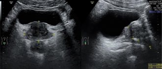
The kidneys are located in a typical position, of normal size and shape. The contours are smooth and clear. The structure of the parenchyma is homogeneous, echogenicity is average. The pyelocalyceal system is without deformation, not dilated. The sinuses are not compacted, homogeneous. No stones were identified. The adrenal glands are located typically, no changes are observed.
Abnormalities detected by ultrasound
The most informative is ultrasound diagnostics when detecting diseases such as damage to the renal vessels, nephroptosis, amyloidosis, narrowing of the ureters, organ dystrophy, abscesses, cysts, tumors, hydronephrosis, stone formation, inflammatory processes (glomerulonephritis, pyelonephritis).
When the ultrasound report indicates “severe intestinal pneumatosis,” this means that the examination was uninformative due to flatulence. In this case, the ultrasound will need to be repeated, having previously prepared, that is, by drinking carminative drugs.
Anatomical and physiological features of the kidneys in men and women
When deciphering an ultrasound of the kidneys, you also need to take into account the gender of the patient. The anatomical features of the structure of the urinary system in women and reproductive function affect some indicators. For example, during pregnancy the size of the jaw may increase.
This is due to the pressure of the uterus on the ureters. The pelvis, instead of 10 mm, expands to 17–27 mm, and remains so after childbirth. These changes become a predisposing factor for the penetration of infection and the development of the inflammatory process.