Ultrasound of the kidneys and adrenal glands is a non-invasive diagnostic method aimed at determining the function of organs and glands, their volume, structure and location. It gives good results in the diagnosis of PSP, cystic formations, neoplasms of benign and malignant nature, as well as abnormalities of organ development. As for the adrenal glands, ultrasound scanning can determine the etiology of hypertension, tumors, causes of obesity and infertility.
Ultrasound of the kidneys and adrenal glands - 3,000 rubles.
20 - 30 minutes
(duration of procedure)
Indications for ultrasound of the kidneys and retroperitoneum:
- Painful symptoms localized in the lumbar region, not caused by physical activity;
- As part of a preventive examination;
- After suffering back injuries;
- For urolithiasis (UCD);
- For any disturbances in urine excretion;
- In case of renal failure;
- In pathological conditions that precede renal failure;
- Suspicion of neoplasms of a benign or malignant nature;
- In the process of preparing for the operation to determine the tactics and volume of intervention;
- In the postoperative period to assess the effectiveness of treatment;
- To monitor the kidney after transplantation.
Types of ultrasound diagnostics
There are several types of research:
- Ultrasound echography. This is the most common method. It is based on the ability of organs and tissues with different densities and structures to reflect ultrasound waves differently. Sonography allows you to evaluate the shape, size and location of the kidneys, bladder and ureter.
- Doppler ultrasound reveals the state of blood circulation in the renal vessels.
Ultrasound is prescribed to detect urolithiasis, neoplasms, congenital pathologies and organ prolapse, to clarify the diagnosis and method of treatment.
Preparation for ultrasound of the retroperitoneum, kidneys and adrenal glands
In order to achieve the desired accuracy of a diagnostic test, the patient needs to prepare for it. All measures in this case should be aimed at minimizing the processes of gas formation, since the gas environment makes it difficult for ultrasonic waves to pass through, which does not allow achieving the desired visualization. To do this you need:
- Avoid consuming foods that cause gas three to four days before the test;
- On the recommendation of a doctor, start taking enterosorbents;
- Take your last meal no later than nine hours before diagnosis;
- An hour and a half before the procedure, drink one to one and a half liters of water or fruit drink and do not empty your bladder.
Nutrition and drinking regime before the procedure
To prepare for an ultrasound of the kidneys in women, you need to pay special attention to nutrition, since the degree of gas formation in the intestines depends on its composition. 1-2 days before the ultrasound the following should be excluded from the diet:
- legumes (beans, peas, lentils);
- fresh vegetables, fruits (cabbage, broccoli);
- dairy products, fermented milk are allowed;
- black bread, made from bran;
- baked goods, baked goods;
- carbonated drinks.
Also, in order not to cause irritation to the cells of the renal parenchyma, you should limit spicy, salty, smoked foods and marinades. You can eat your last meal 7–8 hours before the examination.
You can drink plain water if your doctor has not limited its use due to edema or heart problems. Normally, you should drink 30 ml per 1 kg of weight per day.
How is ultrasound diagnostics of the kidneys and adrenal glands performed?
Ultrasound of the kidneys and adrenal glands is a painless procedure that does not require the use of painkillers. The patient lies down on a medical couch and frees the anatomical area of the abdomen from clothing. A special medical gel is applied to it. Its use ensures close contact with the ultrasound scanner sensor and improves the passage of ultrasonic waves.
Having applied the sensor to the treated area, the diagnostician begins to move it and rotate it at different angles, achieving maximum clarity of the image that is displayed on the screen. Penetrating into the soft tissues of the body, ultrasonic waves are reflected from them, which allows the formation of visual images. Based on them, the specialist makes a conclusion about the condition of the organs and existing pathologies.
Indications for use
An ultrasound is performed only in cases where the patient experiences certain symptoms that suggest the presence of disorders in the functioning of the kidneys and adrenal glands.
The adrenal glands produce hormones, so disturbances in the hormonal cycle may be a reason for an ultrasound.
Frequent increases in blood pressure
In particular, if pigmentation or stretch marks on the skin appear on the patient’s body, this is already a reason for an ultrasound examination.
In addition, it is very important to undergo an ultrasound scan of the adrenal glands when blood pressure rises and falls for no apparent reason, the patient is constantly accompanied by weakness and loss of strength.
Excessive excess weight, which appears in a patient even if the basics of proper nutrition and an active lifestyle are observed, are also an important reason for conducting an ultrasound.
Women who cannot become pregnant are referred for such examination.
Of course, the most serious reason for an immediate ultrasound examination is the suspicion of the presence of a tumor of the kidneys or adrenal glands.
Headache
The adrenal glands and kidneys belong to the urinary system, so any pathology associated with urine excretion must be examined in order to find out the causes of the pathology and then prescribe effective treatment.
Reasons for undergoing an ultrasound may also be:
- systematic severe pain in the lumbar region;
- increased swelling of different parts of the body;
- headache;
- sudden jumps in blood pressure;
- abnormalities during urination.
All of these symptoms may characterize malfunctions of the kidneys and adrenal glands. Additionally, patients are recommended to undergo a urine test for laboratory testing.
Such an integrated approach to studying the circumstances of the pathology allows you to correctly determine the cause, establish a diagnosis and prescribe the most effective treatment for both the kidneys and adrenal glands.
How is an adrenal ultrasound performed?
During an ultrasound examination of the adrenal glands, the patient can lie on his back or stomach, lying on his back, or even standing. The area of the lower back and lower abdomen being examined should be freed from clothing. A medical gel is applied to them, which ensures tight contact with the ultrasound scanner sensor and improves the passage of ultrasonic waves.
It is important to know that during the examination, healthy adrenal glands are not visible due to the fact that the structure of their tissue coincides with the structure of the retroperitoneal tissue. As a consequence, diagnostic capabilities are limited to determining the area in which they are located... But even here, the success rate is not always 100%. Thus, it is possible to determine the desired area on the right in 90% of cases, on the left - only in half of the cases.
When starting a diagnosis, the doctor first of all finds the right kidney, determines the right hepatic lobe and the inferior vena cava. The right adrenal gland is located in the area formed by the extreme points of the above organs. After visualization and examination of the right adrenal gland, the patient is asked to lie on the right side and begins to identify the left one.
Before the examination
How to prepare for a woman's kidney ultrasound?
Basic Rules:
| Change food | The accumulation of large amounts of gas in the intestines makes it difficult to obtain a clear image. 2-3 days before the examination it is necessary to exclude from the diet:
Following these simple rules will help eliminate diagnostic errors. Recommended Products:
A routine ultrasound examination is carried out on an empty stomach, in the morning, after a fasting break for 18-19 hours. | Flatulence |
| Fighting increased gas formation | Includes taking the following medications:
| Good drug at an affordable price |
| Empty your bowels | If you are prone to constipation:
| At home |
| Fill your bladder | This measure will help to more accurately determine the condition of the urinary tract. In order to do an ultrasound of the bladder in women, you need to drink 500 ml of still clean water an hour before the study. | Do not urinate before the diagnostic procedure. |
During the examination, a special gel is used. Bring a towel with you to dry yourself after the diagnostic procedure.
Interpretation of the study of ultrasound results of the kidneys and adrenal glands
After performing an ultrasound of the kidneys and retroperitoneal space, the diagnostician begins to decipher the results. To do this, he compares the results obtained with the results that determine the norm according to the following parameters:
- The shape and size of organs - for each of them there are average indicators;
- The location of the internal organs relative to each other - due to the density of placement, if one of the organs is pathological, it can exert compression on the others;
- Tissue homogeneity, which makes it possible to detect inflammatory processes, malignant neoplasms and even microscopic injuries.
The sizes of healthy kidneys and adrenal glands are presented in our table below:
| Options | Kidneys | Right adrenal gland | Left adrenal gland |
| Length | About 11 cm (13 cm for pregnant women) | From 1 to 1.5 cm | From 0.8 to 1.5 cm |
| Width | About 6 cm | From 0.3 to 1.6 cm | From 0.8 to 1.5 cm |
| Height | From 4 to 5 cm. | From 1 to 2 cm | From 0.8 to 1.5 cm |
A little anatomy
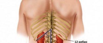
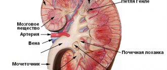
The kidneys are a paired organ of the urinary system, located in the retroperitoneal space at the lumbar level on either side of the spine.
The right kidney is located slightly lower than the left, because anatomically it is pushed downwards by the liver.
Most often, bean-shaped buds are found, but there are two more normal variants:
- “lobulated” kidney, found in children;
- A “humpbacked” kidney is formed during intrauterine development due to pressure on it from the spleen.
An important stage of the study is to determine the size and volume of the kidneys depending on age. The norms for children are presented in the table:
| Age | Length, cm | Width, cm | Thickness, cm |
| Newborns | 4–5 | 2,5–2,7 | 2–2,3 |
| 1 year | 5,5–6,5 | 3,5–3,7 | 2,5–2,7 |
| 5 years | 7,5–8,5 | 4–4,3 | 2,8–3,0 |
| 10 years | 8,5–10 | 4,5–4,8 | 3,3–3,8 |
If the size of the kidneys does not coincide with normal sizes, then the doctor pays attention to the height and weight of the child and evaluates the indicators using special tables regarding his build.
The size of the kidneys in an adult varies within the following limits:
- length 9-12 cm;
- width 4.5-6 cm;
- thickness 3.5-5.5 cm.
The most accurate indicator is the volume of the kidneys, which is closely related to the body composition of a child or adult. It is determined by the formula: length × thickness × width × 0.52.
Also, when performing an ultrasound, the doctor determines the blood flow in the renal artery, which, if disturbed, can cause arterial hypertension. In this case, it is very important to know how to prepare for a kidney ultrasound.
It should be noted that in the retroperitoneal space this paired organ is supported only by the surrounding fatty tissue, therefore, with sudden weight loss and thinning of the adipose tissue around the kidneys, they can change their position, that is, descend. Depending on the degree of prolapse, disruption of their functioning may occur.
The location of the kidneys allows them to be in close contact with many organs of the abdominal cavity, including the intestines. Anterior to them lies the large intestine, which under certain conditions can be an obstacle to examination.
What does a doctor determine during an ultrasound?
When performing an ultrasound examination of the kidneys, the doctor checks all organs of the urinary system, because many diseases are combined. For example, pyelonephritis - inflammation of the kidneys due to a bacterial infection, often occurs together with cystitis (inflammation of the bladder). In addition to these diseases, ultrasound examination can reveal
- urolithiasis;
- prolapse of the kidney – nephroptosis;
- neoplasms (tumors, cysts);
- congenital pathologies;
- pyelitis (inflammation of the renal pelvis).
Ultrasound helps the specialist decide on the choice of treatment method. For example, the need for surgical intervention or conservative treatment for urolithiasis.
Ultrasound of the kidneys of pregnant women
Preparation for ultrasound diagnostics during pregnancy does not differ from the standard one given above, with a small caveat. You should definitely consult your doctor about enemas and taking laxatives.
- For three days before the examination, a pregnant woman should follow a diet.
- A few hours before the examination, take activated carbon tablets or Espumisan in the usual dosage.
- Kidney ultrasound is performed with a full bladder. Pregnant women go to the toilet more often. Bring a bottle of still water with you to the clinic. If you can't stand it for long, drink a glass of water after going to the toilet.
To eliminate understandable inconveniences and discomfort, it is possible to perform a repeated planned ultrasound of a pregnant woman at home if the first examination did not show kidney pathology.
How to do an ultrasound
The patient is asked to undress to the waist, unbutton and lower his trousers or skirt slightly. Then he lies down on the couch. The doctor applies an echo-conducting gel to the surface of the skin. By moving the sensor in the area where the kidneys are located, he sees a two-dimensional black and white image on the monitor and reports the necessary data to the nurse.
First, the ultrasound is performed in the supine position. Then the patient is asked to turn on his stomach and on the side opposite to the side being examined . Determine the size, structure and topography of the kidneys. The doctor will ask the patient to take a strong breath and exhale as much as possible to determine the respiratory mobility of the kidneys. To exclude nephroptosis (prolapse of the kidney), the examination is continued in standing and supine positions.
When finished, the patient wipes off the applied gel with napkins or a towel and gets dressed.
[ads-pc-3]
The entire diagnostic procedure takes about 15 minutes. Dopplerography of the kidney vessels will require more time, 25-30 minutes. After completion, the patient is given a doctor’s report on the condition of the urinary system.