Yes, before the examination you can eat, drink and take medications with the exception of MRI with contrast, MRI examinations of the abdominal cavity and pelvis. At the moment, there is no data on the presence of a pathological effect of MRI on the fetus. However, due to the peculiarities of the course of pregnancy, it is recommended to perform MRI in the first trimester of pregnancy only strictly according to the indications determined by your attending physician. The duration of the examination in the tomograph depends on the type of MRI machine and the type of examination. The study, which is carried out on a modern 1.5 T MRI scanner, lasts about 15 minutes.
Is it possible to eat before an MRI and why is it forbidden to drink a lot of liquid?
Magnetic resonance imaging is one of the most effective diagnostic techniques. If a pathology or injury to the musculoskeletal system is suspected, radiography is first performed, and an MRI is prescribed for a more detailed study. The doctor evaluates the intraosseous structure, the condition of muscles and ligaments, and identifies minor damage and neoplasms. Advantages of MRI: no preparation, painlessness, information content, visualization of articular cartilage, periosteum, spongy and cortical substance, bone marrow. MRI of the lumbar region visualizes the area under study layer by layer, taking dozens of slices, which a computer program combines into one image. During MRI of the lumbar region, sections are made in the transverse and sagittal projection with a thickness of 3-4 mm. WATCH THE VIDEO ON THE TOPIC: How to prepare for the examination. Magnetic resonance imaging
Applications and types
Medicine does not stand still, and some of the latest MRI equipment allows additional examination in a vertical position, which shows any displacement of the discs and pressure on the nerve endings that a person experiences on the spine in its natural state (vertical).
Until recently, examination of the spine was carried out using X-rays, but today, thanks to magnetic resonance imaging, it is possible to see changes in the human skeleton and identify the exact cause of their appearance. The procedure helps to identify numerous dangerous diseases in the early stages, promotes timely treatment, which reduces the risk of developing severe consequences and complications.
Tags: eat, can, department, front, spine, lumbar
About the author: admin4ik
« Previous entry
Lumbar Spine - Preparing for MRI
Magnetic resonance imaging can be considered one of the most prescribed examinations of the human body. This diagnostic method is accurate and highly informative. The procedure is inherently complex, although it is quite easy to carry out. But, despite all the advantages of MRI, there are still certain limitations, contraindications and subtleties, which are individual for each patient and are determined during an individual examination by the doctor.
All iLive content is reviewed by medical experts to ensure it is as accurate and factual as possible.
Many people constantly complain of pain in the lumbar region or buttocks. The pain can be aching or sharp, and can also cause numbness in these areas of the body. From school anatomy lessons we know that the lowest part of the spine is the lumbosacral region. It consists of five separate tubular-type bones, which together create a ridge in this area of the back. These tubular bones provide support for part of the body, and at the same time they are quite flexible and allow the body to move.
WATCH THE VIDEO ON THE TOPIC: MRI of the spine
Magnetic resonance imaging is one of the types of studies aimed at establishing the boundaries of the lesion in the spine. MRI of the spine allows you to identify all pathologies, including tumors and congenital anomalies. Most often, such a study is necessary for spinal stenosis. The study allows us to identify disorders and changes in bone tissue, determine the correctness of blood flow, and new formations in the intervertebral discs. Only with the support of this procedure can a complete clinical picture be drawn. MRI, not only of the lumbosacral region, is usually prescribed in a number of cases when a detailed picture is needed: Preparing for an MRI of the lumbosacral spine does not require time. It is more important to make sure that there are no contraindications for the study. The procedure is prohibited under the following conditions: There are also restrictions for those who suffer from severe claustrophobia.
How to publish an article on our website? One of the most effective, popular and rapidly developing research methods is magnetic resonance imaging (MRI). MRI of the lumbosacral spine is the best way to thoroughly examine degenerative changes and pathologies. With the help of MRI, you can make a correct diagnosis and begin timely treatment of detected ailments and abnormal changes. A neurologist prescribes a diagnosis of the lumbar region and, according to the results obtained, analyzes the full clinical picture of the disease. Having put together the patient’s complaints and his medical history, the doctor makes a diagnosis and begins a course of therapy.
Modern medicine actively uses the achievements of other scientific disciplines. Thus, medicine managed to replenish its diagnostic arsenal with another non-invasive, non-destructive, painless, informative method of layer-by-layer study of the human body - magnetic resonance imaging or MRI.
How to prepare for research?
MRI of the spine does not require changes in diet or other restrictions: preparation consists only of eliminating various contraindications for a particular patient. Sometimes, for this purpose, the attending physician may prescribe additional types of research and tests.
Diagnosis is not made if a person has:
- hearing prosthesis;
- knitting needles;
- orthopedic structures that hold bones in a certain position;
- defibrillator;
- insulin pumps;
- braces;
- pacemaker;
- tattoos with metallic inclusions;
- titanium plates;
- vascular clips;
- artificial heart valves;
- fragments and so on.
Metal structures tend to resonate with the magnetic field of a working tomograph, thereby causing rapid heating of foreign inclusions and significant distortion of the results obtained. The possibility of conducting research is discussed privately only with specialists in the relevant field.
Contraindications also include early pregnancy. However, some unforeseen circumstances may cause an unscheduled tomography session, these include: the likelihood of miscarriage, the presence of a tumor formation in the mother, developmental pathologies in the fetus, incorrect intrauterine placement of the child.
The doctor will most likely not allow a patient who has:
- hemolytic anemia;
- severe mental condition;
- epilepsy;
- intracranial aneurysm;
- impairment of the kidneys or liver (with the introduction of a contrast solution);
- uncontrolled limb movements;
- alcohol intoxication;
- narcotic state;
- severe form of claustrophobia;
- overweight from 190–205 kg.
Eating food on the eve of the session is allowed, but, nevertheless, in the morning it is better to replace the usual products with a “lighter” breakfast: vegetable salad, unsweetened jelly or slimy porridge. The last glass of liquid should be consumed a maximum of 2 hours before the test.
Preparing for an MRI - is it possible to eat, drink, smoke, drink alcohol?
Modern research methods make it possible to accurately and very quickly recognize the disease and prescribe the correct treatment. MRI of the lumbosacral spine reveals the whole picture of what is happening inside the body and helps the doctor make an accurate diagnosis of back diseases. This method made a major breakthrough in medicine, providing a detailed analysis of the spinal column without any intervention in the patient's body. The value of the study lies in the speed of the procedure and digital processing of the data obtained. By forming error-free conclusions based on the photo, the doctor can easily understand the essence of the problem and plan further actions to eliminate the disease. The procedure takes from 45 to 60 minutes and does not require any serious preliminary preparation. Such an examination does not require preparation of the patient.
When is magnetic resonance imaging contraindicated in the lumbar region?
The method is harmless, the contraindications are:
Early pregnancy - first trimester. Cases where a person has a disease in which he cannot lie still for the required period of time. Claustrophobia only for closed type installations. Metal implants in the body; any metal inclusions that cannot be removed. Devices built into the body that support its functions. The patient’s weight, depending on the type of installation, exceeds 100 or 130 kilograms. There are more modern modifications that do not have weight restrictions.
Why do an MRI?
Currently, medicine practically cannot do without magnetic resonance imaging.
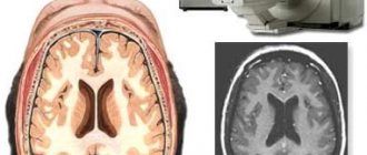
What is the purpose of MRI of the spine? Diseases of the musculoskeletal system and nervous system are very difficult to diagnose in any other way. The magnetic field created around the patient helps visualize the entire internal picture of the body, which clearly shows any changes in the organs. In addition, MRI does not expose the body to radiation, so it is not considered a harmful study. A series of images obtained from MRI scans show detailed images of all layers of organs and parts of the human body. For diseases of the musculoskeletal system, doctors most often refer for an MRI to establish an accurate diagnosis.
Why do doctors resort to this particular diagnosis? The most common are diseases of the cervical and lumbosacral spine. For example, it takes no more than an hour to conduct an MRI of the lumbar spine and interpret the results; an MRI of the cervical spine takes about 20 minutes. Compared to other methods of examining the spine, this is the fastest and most informative way to make a diagnosis.
When is it okay to not stick to a diet?
Sometimes you can eat and drink whatever you want before an MRI. This freedom is allowed to patients who are prescribed an MRI of the lumbar region. Before an MRI of the spine and vertebrae, as well as before an MRI of the brain, it doesn’t matter what you eat, the main thing is in what quantities.
Even if the study has nothing to do with the stomach, overeating is still not recommended. The effect of a magnetic field on a person is absolutely harmless, but individual increased sensitivity cannot be discounted. Fear of closed spaces can trigger a panic attack, followed by nausea and vomiting. Therefore, it is better if the examination on a magnetic resonance tomograph is carried out “lightly”.
Pay attention to exactly how they plan to have your tomography performed. It is not uncommon for an MRI of the head to be done using a special contrast agent. It is injected into the blood the day before in order to better monitor the blood flow. In this case, the last meal should be no later than 4 hours before the procedure.
Another diagnostic method that uses a contrast agent is computed tomography (CT). It differs significantly from MRI, because it is based on X-rays, although the resulting images also allow you to see the scanned organ in 3D format. Be that as it may, you should not eat before an MRI or a CT scan with contrast. As for medications, they can be taken on schedule for any study.
Contraindications
Before conducting the study, the doctor asks the patient several questions to identify any contraindications. For a study such as resonance tomography, the list of contraindications is small. However, they must be taken into account, since a person’s life may depend on it. These include the following:
- It is strictly forbidden to carry out this hardware research in the presence of various metal objects in the human body. For example, vertebral or other prostheses, pins, clips on blood vessels, etc.
- Implanted pacemakers, cardiac pacemakers, electronic pumps and neurostimulators are also a contraindication for magnetic resonance imaging, since it creates a fairly powerful magnetic field.
- Claustrophobia can also become a contraindication without the introduction of special sedatives. During the study, a person has to remain motionless for a long time in a narrow, enclosed space. This can cause worsening attacks of claustrophobia.
- Only a doctor can refer you for an MRI scan. You cannot undergo such an examination without a referral. The doctor takes into account all contraindications and accurately indicates the area that needs to be examined and why the MRI is being performed. To avoid having to do a tomography again, you should not go for an MRI first, and only then see a doctor, trying to save money. The tissues and organs shown in the images should be in the areas that the doctor needs to make a diagnosis. When choosing an area for research on your own, you can make a mistake.
- Many studies are carried out with the introduction of a contrast agent for a more informative result. In such cases, you need to take into account the fact that you may be allergic to the contrast agent.
- People who are overweight (more than 120 kilograms) cannot undergo an MRI. This is due to the fact that the device cannot withstand a heavy load. In addition, it is a narrow tube that a person with a large waist will not fit into. If an MRI of the cervical spine is necessary, then such restrictions can expand to 180 kilograms.
- There are also restrictions for pregnant women. In the first trimester of pregnancy and during breastfeeding, tomography is contraindicated for women. If there is an urgent need, a nursing mother can have an MRI, but breastfeeding is prohibited for the next two days.
What does it feel like during an MRI?
The procedure is completely painless. After all, a person cannot physically feel the influence of magnetic waves. Some may feel discomfort from being in a dark and cramped tomograph scanner, and the table is most often hard and cold. Therefore, immediately ask for a blanket and pillow, because the procedure takes almost an hour, sometimes more.
A working CT scanner typically makes a loud buzzing noise, so staff may provide special earplugs for the patient to muffle the sounds. There is also a constant connection between the patient and doctors inside the scanner. It supports the entire scanning procedure. Also, some modern tomographs even have a TV or special headphones so that the patient can pass the time faster.
Doctors warn people (whether women or men, regardless of gender) to move less while lying inside the tomograph. Any unnecessary movement may subsequently affect the MRI of the vertebral or sacral region. For medical reasons, some cannot stay in one position for a long time. You can ask for a sedative injection. You can eat and drink after the scan and lead your usual lifestyle. There is no recovery period.
Preparing for the examination
As with many other instrumental studies, little preparation is required for MRI of the spine.
First, there is usually the question of what you need to take with you to the examination. The main thing that should never be forgotten is the doctor’s direction. Any clinic has the right to refuse to conduct a study without a referral from a specialist. For the doctor who will conduct the MRI study, so that he can better understand the picture of the patient’s health status, it is necessary to bring the results of previous examinations, an outpatient card, medical history, extracts and other doctor’s reports. When deciphering what an MRI shows in the images, the doctor will rely on existing data on a particular deviation.
If you have benefits or a voluntary health insurance policy, you must take them with you to the study. Many insurance companies offer programs that cover, for example, MRI of the cervical spine, or other modern instrumental diagnostic methods.
As for the clothes that are best to wear, it is better to choose the most comfortable ones, without metal elements, inserts, sparkles and rhinestones. Before the study, the laboratory technician may ask you to remove this or that item of clothing if he believes that it will interfere with the tomography. The laboratory technician may also ask you to wear a disposable uniform provided by the clinic for greater safety.
Preparing for an MRI of the cervical spine means that piercings, jewelry and costume jewelry are absolutely not allowed during such studies. Cosmetics also need to be washed off before diagnosis, since many manufacturers use metals in the production of cosmetics. As for tattoos and permanent makeup, it is better to warn the laboratory technician about their presence on the body.
Unlike X-ray examination of the sacrum, tomography does not require emptying of the gastrointestinal tract. For x-rays, this is necessary so that gas contamination or fullness of the intestines does not interfere with seeing, for example, a vertebra and a crack on it. All layers of the human body are visible on MRI, so there is no need to cleanse the gastrointestinal tract.
Before going for the study, the question arises about how to prepare for an MRI of the spine, whether certain preparation is required. Can I eat before the test or should I stick to a strict diet for a week? What can you eat and what can you not?
Before a standard examination of the lumbosacral or cervical spine, you can eat anything, as well as take any medications, since this will not affect the MRI process or result at all.
But if the examination is planned with the introduction of contrast, then it is necessary to consult a doctor about taking medications and eating before it. In this case, you usually cannot eat before an MRI; the contrast study is performed on an empty stomach.
Preparation for an MRI of the lumbar region consists mainly of the moral awareness that you will have to spend 15 to 60 minutes in a confined space without moving, depending on the purpose of the study.
Preparation for MRI of the lumbosacral or cervical spine is identical. Both studies can be carried out absolutely at any time of the day. The exception is a contrast study, since it is best done on an empty stomach, so patients prefer to go for it in the morning.
Many patients are afraid to undergo magnetic resonance imaging for the first time, since the narrow tube of the tomograph and the incredibly loud noise are truly intimidating. In fact, this test does not cause any pain. The main difficulty is maintaining stillness. Otherwise, there are no particular difficulties in preparing yourself for an MRI of the lumbar spine.
The latest techniques used in medicine for diagnosing defects of the spinal column and its segments make it possible to recognize them in a timely manner and prescribe appropriate therapy. Preparation for MRI of the lumbosacral spine does not require special manipulations or the use of medications. This technique is the safest, as it does not cause negative effects on the patient’s body.
What does this method show?
From the images, the doctor receives information:
About the relative position of the vertebrae and discs, whether the picture corresponds to their correct anatomical dislocation. About the condition of bone tissue, cartilage, nerve fibers and blood vessels supplying the lumbar region. Pathological changes in discs and vertebrae determine the presence of hernias, which indicates the disease osteochondrosis. Is there compression of the nerve roots as a result of degenerative processes in the lumbar region.
About diseases and disorders:
Ankylosing spondylitis, neoplasms in the spinal cord and vertebrae, possible metastases, diseases associated with the spinal cord - multiple sclerosis, syringomyelia, myelitis, osteoporotic changes, rheumatoid lesions, spinal stenosis, spondylosis, changes in the physiological curvature of the lumbar region, infectious diseases.
What is MRI
To obtain the highest quality and informative image of the part of the human body being examined, magnetic resonance scanners are used. Diagnostics occurs using the radiation of electromagnetic energy and radio waves inside the tomograph.
The uniqueness of the technique allows us to study in detail not only organs, but also tissues, cartilage, intervertebral discs, and the vascular system of the body.
The safety and painlessness of the event makes it possible to carry it out for children of any age. The only contraindication may be the use of anesthesia in a child to make him completely immobile.
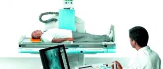
The patient is placed inside the device in a horizontal position on a special couch. This procedure may cause some discomfort. MRI scanners produce a loud, unpleasant noise, and you also need to lie still for about 30 minutes.
You must lie still during the procedure
In special cases, intravenous administration of a special colorless substance may be required to improve the overall image. The liquid has no contraindications and does not cause side effects.
Introducing the patient to the procedure
After all metal objects are left in a special room along with electronic devices, shoes and outerwear, the patient will go to the diagnostic room. Next, he will have to climb onto a horizontally located couch, next to which there is often a miniature ladder for convenience.
The person is carefully placed on the table surface, after which, in some cases, doctors fix the legs and arms to avoid unwanted movements while the tomograph is operating. If the study was to be carried out with contrast, a special solution is injected into one of the veins, allowing us to study some formations, tissues and vessels in more detail. Then the device will gradually enter the “tunnel” of the device and the scanning process will begin.
For the next 10-30 minutes, the patient will have to listen to a series of unpleasant sounds, regularly replaced by intermittent noise. This phenomenon is explained by the active operation of the magnetic device - it is absolutely safe. However, doctors may suggest special headphones or earplugs to prevent psychological discomfort.
Magnetic resonance imaging does not cause any pain in a person. However, if the patient feels unwell, this should be immediately reported to doctors through the use of a special signaling device. At the end of the set time, the sound noise stops and the couch returns to its original position. The patient should gradually rise from the surface and, excluding sudden movements, head towards the exit.
Basic rules for preparing for MRI
Preparing for an MRI of the spine involves following certain rules and restrictions:
- Before the examination, warn the specialist about the presence of pacemakers, metal and electronic implants, hearing aids, and piercings.
- It is recommended that people suffering from claustrophobia and other psychological phobias take sedatives in advance. In special cases, in case of panic attacks during scanning, the doctor may use a stronger sedative.
Using the phone is prohibited
- Lay out communication means, bank cards, keys, watches in advance. Women should remove makeup and wigs.
- Wear special disposable clothing that will exclude any influence on the MR device.
There are a number of contraindications for magnetic resonance imaging, the main ones are listed in the table.
What to do with bad habits?
Is it possible to drink alcohol before an MRI? This is a question to which neurologists answer in unison “under no circumstances.” You need to abstain from drinking alcohol for at least two days, and doctors assure that this is not just a standard prohibition, but a truly serious contraindication. There are three good reasons:
- Firstly, during the tomography the patient must be completely sane and follow the doctor’s instructions as clearly as possible. The scan lasts at least 40 minutes and the patient must lie still during this time, otherwise the image will be distorted. It is impossible to predict how a drunk person will behave in a confined space, so they simply will not let him into the manipulation room.
- Secondly, alcoholic drinks affect metabolic processes, and therefore the results of MRI. It will still not be possible to get an accurate picture as long as the blood contains at least a tiny fraction of alcohol, especially when it comes to MRI of the central nervous system.
- Thirdly, alcohol may be incompatible with a magnetic field and this can lead not only to distortion of images, but also to real complications of current pathological processes in the body.
The situation is no less strict with smokers. Nicotine has a serious effect on all nervous functions and in particular on the central nervous system and brain cells. Under its influence, the blood vessels of the brain inevitably narrow, which means the blood flow to the nervous tissues decreases. To get an objective picture, before an MRI of the brain, the doctor will certainly ask the patient not to smoke for at least several hours.
So, proper preparation for MRI is an essential stage of successful diagnosis. In order for the tomography to be justified and help identify the true cause of the disease, the patient must strictly adhere to the medical recommendations received the day before. Please note that prohibitions often apply not only to food, drinking and bad habits, but also to cosmetics, jewelry, and tattoos. Be sure to tell your doctor about the presence of implants, dentures, and any foreign bodies in the body.
Many patients are concerned about when they can eat and drink before an MRI. Magnetic resonance imaging allows you to check almost all organs and systems; it is prescribed in case of an unspecified diagnosis, if it is necessary to study the function of organs and blood flow.
Is a diet necessary before the procedure?
It is important to understand whether you should adhere to a certain diet before an MRI of the spine. Standard MR diagnostics, unlike ultrasound and x-ray examinations, does not require adherence to a special diet or a ban on taking medications.
It is strictly forbidden to drink alcohol 72 hours before the tomography. Before the procedure, you can eat a light breakfast and drink a couple of glasses of water 60-80 minutes before.
An exception is when performing an MRI with the introduction of a contrast agent into the body. On the eve of the procedure, 2-3 days before, it is recommended to start following a diet.
The main diet should consist of baked vegetables, boiled meat, fish, and dairy products. You should avoid fatty and carbohydrate foods. Completely eliminate alcohol, milk, and carbonated drinks.
You can include fish and vegetables in your diet
Research results
The results of an MRI examination of the spine are a series of pictures that are converted into a 3D image using complex computer programs. Images are taken from different angles and with different slice thicknesses, and deciphering the MRI of the spine is the job of a radiologist.
There are times when you need to undergo a magnetic resonance examination again (for example, after therapy). How often can an MRI of the spine be done? The answer is as necessary: this may be a repeat examination after the completed course of treatment (dynamic observation). In medicine, this procedure is often carried out along with such diagnostic methods as CT or radiography, but, unlike the latter, the method is completely safe.
What preparation is needed in the MRI room?
The patient and those accompanying him are prohibited from bringing the following items and devices into the MRI room:
- technical devices that interfere with the operation of the tomograph;
- removable dentures, jewelry, jewelry, hairpins;
- You need to take out keys, plastic cards, devices, money from your pockets in advance;
- change into disposable clothing, which will be offered by the laboratory assistant;
- Warn the radiologist about pregnancy, stimulants, and possible contraindications.
How is an abdominal MRI performed?
The research is considered completely harmless. However, certain restrictions still exist.
During the first trimester of pregnancy, it is better to refrain from such examination. Such a recommendation is not based on any objective data on the harm caused, but is actually reinsurance.
At other times, the examination can be carried out any number of times over an as long period as desired.
If it is necessary to conduct a computed tomography scan, then the situation is different. How often can an abdominal CT scan be done? Without harm to health, it is recommended to carry out this procedure no more than once every six months. But if treatment requires such an examination several times in a row, then this is possible
When thinking about how often an abdominal CT scan can be done, you need to remember that the patient receives an additional dose of x-ray radiation.
Attention! Although there is no harm from constantly conducting MRI, it does not always make sense to spend time on it. The fact is that to control your health, you can be examined at a certain frequency (for example, once a year), but there is little point in doing this too often.
There is a condition: before the MRI, remove metal objects from yourself - chains, watches, from your clothing pockets - keys, coins, etc.
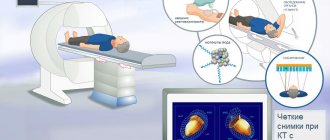
Abdominal MRI does not cause any discomfort. The subject’s task is to lie still for 30 minutes and, at the doctor’s request, hold his breath. The patient is placed in a closed capsule, which has ventilation, lighting and an intercom through which you can contact the doctor. The doctor watches him through a video camera.
- high diagnostic value compared to ultrasound and computed tomography;
- painlessness of the procedure.
Other Dietary Recommendations
In addition to following a diet, the patient needs to take some medications to normalize digestion. On the eve of the study, it is recommended to drink activated charcoal or Espumisan. Immediately before the procedure itself, antispasmodics are prescribed - No-Shpu, Spazmalgon. In some cases, a cleansing enema is performed.
When diagnosing the liver and pancreas, the doctor prescribes a special low-carbohydrate diet, which must be followed for 2 days. This is necessary to reduce the load on the organs being examined in order to obtain reliable tomography results. A low-carbohydrate diet consists of completely abstaining from fruits, cereals, confectionery and flour products. You should limit your consumption of potatoes, carbonated and sugary drinks. Patients are allowed to eat only buckwheat, kefir, green vegetables, and nuts.